Discuss stridor in an infant. Ms ent university exam pattern for 20 marks.
Reading File
Finding Sources
Finding Sources
Finding Sources
Reading File
Reading File
Reading File
Finding Sources
Reading File
Reading File
Reading File
Finding Sources
Reading File
Reading File
Reading File
Reading File
Loading Image
Here is a complete 20-mark university exam answer on stridor in an infant, written in the MS ENT long-essay format.
STRIDOR IN AN INFANT
(20 Marks - MS ENT University Examination Pattern)
DEFINITION
Stridor is a high-pitched, harsh respiratory sound produced by turbulent airflow through a partially obstructed segment of the upper airway at the level of the larynx or trachea. In infants, it is one of the most common referrals to an ENT surgeon and demands systematic evaluation because it can indicate a life-threatening airway lesion.
CLASSIFICATION
A. By Phase of Respiration
| Phase | Level of Obstruction | Example |
|---|---|---|
| Inspiratory | Supraglottic / Glottic | Laryngomalacia, vocal fold paralysis |
| Expiratory | Infraglottic / Trachea | Tracheomalacia |
| Biphasic | Glottic / Subglottic | Subglottic stenosis, subglottic hemangioma |
B. By Onset
- Congenital - present at or shortly after birth
- Acquired - develops after an initial symptom-free period
C. By Age of Presentation
- < 6 months: predominantly congenital causes (laryngomalacia, tracheomalacia, vocal cord paralysis, subglottic stenosis)
- 6-18 months: croup, subglottic hemangioma (peak growth phase)
- > 18 months: foreign body, papillomatosis, epiglottitis
CAUSES / ETIOLOGY
Supraglottic
- Laryngomalacia - most common, accounts for 60% of all neonatal laryngeal problems
- Saccular cysts and laryngoceles
- Macroglossia, micrognathia (Pierre Robin sequence)
- Epiglottitis (older infants/children)
Glottic
- Vocal fold paralysis (VFP) - second most common cause of neonatal stridor
- Laryngeal webs
- Laryngeal cleft
- Papillomatosis
Subglottic
- Subglottic stenosis - congenital or acquired (post-intubation)
- Subglottic hemangioma
- Croup (acute laryngotracheobronchitis)
Tracheal
- Tracheomalacia
- Vascular rings and slings (double aortic arch, pulmonary artery sling)
- Congenital tracheal stenosis (complete tracheal rings)
PATHOPHYSIOLOGY
Turbulent airflow arises when the airway lumen is reduced below a critical threshold. Because airway resistance is inversely proportional to the radius to the fourth power (Poiseuille's law), even small reductions in infant airway diameter produce disproportionate increases in resistance and work of breathing. The infant trachea is approximately 4 mm in diameter; thus any mucosal edema of 1 mm reduces the cross-sectional area by nearly 75%.
INDIVIDUAL CAUSES IN DETAIL
1. LARYNGOMALACIA (Most Important)
Definition: Partial or complete collapse of the supraglottic structures on inspiration due to structural and neuromuscular immaturity.
Pathophysiology:
- The epiglottis is long, narrow, and omega-shaped
- Aryepiglottic folds are short and tightly tethered
- Redundant mucosa of arytenoids prolapses into the airway on inspiration
- Underlying neuromuscular hypotonia affects the laryngeal adductor reflex (LAR), a vagal nerve-mediated reflex; dysfunction of the afferent-brainstem-efferent pathway results in poor laryngeal tone
- GOR (gastro-oesophageal reflux) is found in the majority and may be causative or self-perpetuating (high negative intrathoracic pressures promote reflux)
Classification (KJ Lee):
- Type 1: Foreshortened/tight aryepiglottic folds
- Type 2: Redundant supraglottic tissue
- Type 3: Posterior epiglottic collapse (associated with underlying neuromuscular disorders)
Clinical Features:
- High-pitched, fluttering inspiratory stridor onset within first 2 weeks of life
- Worse when supine, crying, feeding, or during intercurrent URTI
- Improves in prone position and with neck extension
- Most severe around 6-9 months, spontaneous resolution by 18 months in ~90%
- Severity increases with activity, may disappear during sleep
- Severe cases: feeding difficulties, failure to thrive, sternal/intercostal recession, pulmonary hypertension, cor pulmonale
Diagnosis:
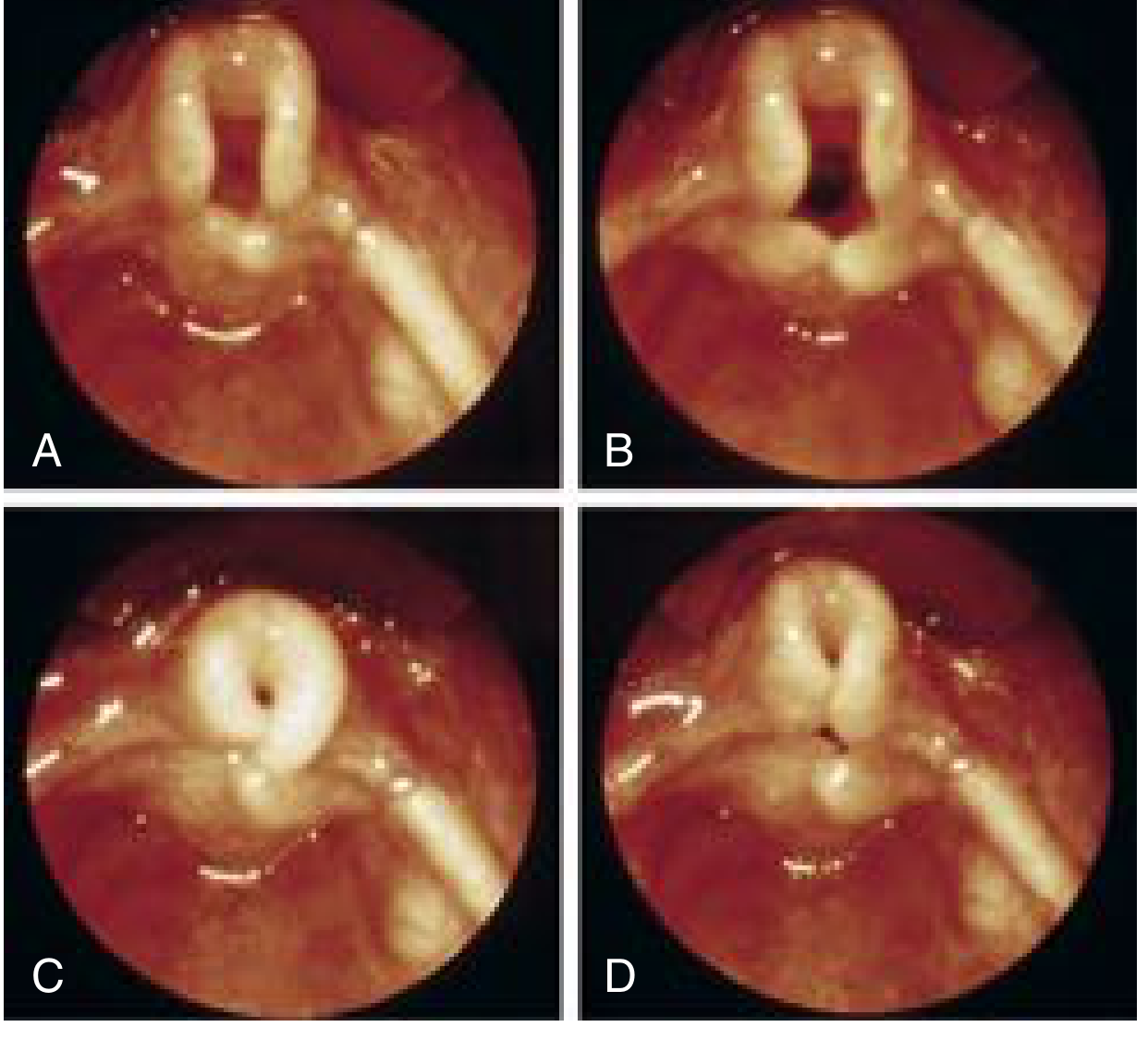
- Awake flexible fibreoptic nasopharyngoscopy (gold standard, outpatient)
- Shows: omega-shaped epiglottis, short aryepiglottic folds, prolapse of arytenoid mucosa into glottis on inspiration, normal vocal fold mobility
- MLB (microlaryngoscopy and bronchoscopy) under GA: indicated for severe stridor, failure to thrive, atypical features, or suspicion of synchronous airway lesion (10-20% incidence)
- Note: MLB requires spontaneous ventilation under light anaesthesia; beak of laryngoscope in the vallecula - deep anaesthesia masks supraglottic collapse
Treatment:
- 90% - observation with regular weight monitoring and reassurance
- Antireflux medication (PPI/H2 blocker) for 4 weeks if GOR present
- Surgical - Aryepiglottoplasty / Supraglottoplasty (endoscopic): for failure to thrive, cyanosis, apnoeic episodes, or pulmonary hypertension
- Division of each aryepiglottic fold, excision of redundant mucosa/submucosal tissue over arytenoids; cuneiform cartilages may be partially excised
- The interarytenoid "bridge" of mucosa is preserved to prevent scarring
- Minimal bleeding; no routine antibiotics or steroids; immediate improvement post-op
- Tracheotomy: rarely required for most severe cases
- Caution: CHARGE syndrome - supraglottoplasty can worsen epiglottic collapse
2. VOCAL FOLD PARALYSIS (VFP)
Frequency: Second most common cause of neonatal stridor
Causes:
- Birth trauma/forceps delivery (traction injury to vagus nerve)
- Iatrogenic left VFP after PDA ligation or interrupted aortic arch repair (left RLN passes around ductus arteriosus; incidence 1-7.4%)
- CNS anomalies: Arnold-Chiari malformation, hydrocephalus, nucleus ambiguus dysgenesis
- Cardiomegaly, TEF repair
- Idiopathic
Clinical Features:
- Unilateral VFP: weak cry, inspiratory or biphasic stridor, feeding difficulties, aspiration
- Bilateral VFP: normal voice but stridor, dyspnoea, cyanosis, apnoeic episodes; bilateral VFP may paradoxically abduct on expiration and adduct on inspiration, mimicking normal movement to the untrained eye
Diagnosis:
- Awake flexible nasolaryngoscopy (to avoid sedation artefact on cord movement)
- Laryngeal ultrasound (sensitivity 0.84, specificity 0.95)
- Laryngeal EMG under light anaesthesia to differentiate paralysis from arytenoid fixation
- Imaging (MRI/CT) to rule out cardiac and neurological causes
- Barium swallow to detect aspiration
Treatment:
- Mild/unilateral: observation; most idiopathic cases resolve spontaneously
- Severe/bilateral: tracheotomy; lateralization procedures in older children
3. SUBGLOTTIC STENOSIS (SGS)
Congenital SGS: Narrowing of the laryngeal lumen, diagnosed in first months with persistent inspiratory/biphasic stridor; mild cases present later as recurrent croup
Acquired SGS: Most commonly from prolonged endotracheal intubation in premature neonates; also from blunt neck trauma
Diagnosis: Endoscopic grading (Cotton-Myer classification):
- Grade I: <50% obstruction
- Grade II: 51-70%
- Grade III: 71-99%
- Grade IV: complete obstruction
Treatment: Severity-based - endoscopic balloon dilation, cricotracheal resection, or laryngotracheal reconstruction
4. SUBGLOTTIC HEMANGIOMA
- Benign endothelial tumours that enlarge throughout year 1; not always apparent at birth
- Cutaneous hemangiomas in a "beard distribution" are a clue to airway hemangioma
- Biphasic stridor appearing after first month of life without another explanation should raise suspicion
- Spontaneous regression by age 5 years
- Treatment: Propranolol (beta-blocker) - first line; systemic steroids, laser, or surgery for large lesions
5. VASCULAR RINGS AND SLINGS
- Double aortic arch (most common vascular ring), right-sided aortic arch, pulmonary artery sling
- Compress trachea and/or oesophagus
- Symptoms from birth; worsen with URTI; dysphagia if oesophagus compressed
- CXR may show tracheal narrowing; contrast oesophagram shows posterior indentation; CT/MRI angiography is definitive
- Treatment: surgical division of the ring
CLINICAL ASSESSMENT
History
- Age of onset: congenital causes present at/shortly after birth; hemangiomas develop after 1 month
- Duration: chronic = congenital; acute = infective, foreign body
- Positional variation: prone improves laryngomalacia; position-independent = subglottic lesion
- Feeding difficulties and failure to thrive: severity marker
- Associated symptoms: cyanosis, apnoea, voice change, cutaneous lesions
- Birth history: difficult delivery (VFP), prematurity/intubation (acquired SGS)
- Family history; syndromic features (CHARGE, Down syndrome)
Examination
- Phase and pitch of stridor
- Degree of respiratory distress (recession, use of accessory muscles, SaO2)
- Voice quality (aphonia = laryngeal web/atresia; weak cry = VFP; muffled = supraglottic)
- Weight and growth chart (failure to thrive)
- Cutaneous hemangiomas (beard distribution)
- Dysmorphic features, macroglossia, micrognathia
Investigations
- Awake flexible fibreoptic nasopharyngoscopy - first-line, dynamic assessment of supraglottis and vocal fold movement
- MLB under GA - gold standard for complete airway assessment; performed under spontaneous ventilation and light anaesthesia
- CXR: mediastinal widening (vascular anomaly), subglottic narrowing (steeple sign in croup), radiopaque foreign body
- CT/MRI neck and thorax: vascular rings, masses, extent of stenosis
- Barium swallow: oesophageal compression, aspiration
- Echocardiogram: associated cardiac anomalies, PDA
- pH monitoring / impedance study: to quantify GOR
- Laryngeal EMG: differentiate VFP from arytenoid fixation
MANAGEMENT SUMMARY TABLE
| Cause | First-line Treatment | Surgical Option |
|---|---|---|
| Laryngomalacia (mild) | Observation + antireflux | Supraglottoplasty |
| Laryngomalacia (severe) | Antireflux + supraglottoplasty | Tracheotomy (rare) |
| Vocal fold paralysis (unilateral) | Observation | Injection medialization |
| Vocal fold paralysis (bilateral) | Tracheotomy | Lateralization/arytenoidectomy |
| Subglottic stenosis (mild) | Observation | LTR / cricotracheal resection |
| Subglottic hemangioma | Propranolol | Laser / surgery |
| Vascular ring | Refer cardiac surgery | Division of ring |
| Subglottic cyst | Endoscopic marsupialization | - |
COMPLICATIONS OF UNTREATED SEVERE STRIDOR IN INFANTS
- Failure to thrive - increased work of breathing + feeding difficulties
- Pulmonary hypertension - due to chronic hypoxia
- Cor pulmonale
- Pectus excavatum - permanent chest wall deformity from chronic severe sternal recession
- Obstructive sleep apnoea
- Death - from acute airway obstruction
IMPORTANT EXAM POINTS
- Laryngomalacia: 60% of all neonatal laryngeal problems; resolves spontaneously by 18 months in 90%; surgical threshold = failure to thrive / cyanosis / apnoea / pulmonary hypertension
- VFP: second most common cause; left cord more often affected; left RLN at risk during PDA ligation
- Subglottic hemangioma: think of it with beard-distribution cutaneous hemangiomas; treat with propranolol
- MLB should be under spontaneous ventilation, light anaesthesia, laryngoscope beak in vallecula
- 10-20% of laryngomalacia cases have a synchronous second airway lesion - always screen
- GOR is found in the majority of laryngomalacia patients; may be causative or a consequence of high negative intrathoracic pressures
- Biphasic stridor = glottic or subglottic level pathology
Sources: Scott-Brown's Otorhinolaryngology Head & Neck Surgery (Vol. 2); Cummings Otolaryngology Head and Neck Surgery; KJ Lee's Essential Otolaryngology; Tintinalli's Emergency Medicine
This is a shared conversation. Sign in to Orris to start your own chat.