
Is there any relation between McBurney's point and liver
McBurney's point liver relation anatomy
McBurney's point appendix anatomy abdomen
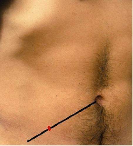
A clinical photograph of the human lower abdomen illustrating surface anatomy landmarks used in surgical evaluation. A black line represents the spino-umbilical line, extending from the umbilicus to the right anterior superior iliac spine (ASIS). A red mark is placed on this line at the junction of the lateral one-third and the medial two-thirds, identifying McBurney's point. This landmark is clinically significant as it traditionally corresponds to the base of the vermiform appendix and serves as a primary site for tenderness assessment in cases of suspected acute appendicitis. The photograph provides a clear visual guide for physical examination techniques in emergency medicine and general surgery.
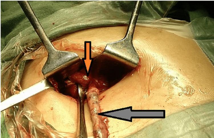
This clinical photograph captures an intraoperative view of an emergency appendicectomy through a McBurney's point incision, revealing a rare anatomical variation: a duplicated vermiform appendix. Two distinct appendices are visible within the surgical field, stabilized by metal retractors. An inferiorly positioned, elongated appendix is indicated by a gray arrow. Located superiorly, an orange arrow points to a second appendix that appears acutely inflamed and gangrenous; a small, pale, firm fecolith is visible near its base. The surrounding tissues exhibit signs of severe inflammation, including marked vascular congestion and erythema. There is visible pooling of glistening seropurulent fluid and pus in the peritoneal cavity, consistent with appendiceal perforation and localized peritonitis. This image serves as a high-level educational resource for surgical residents and medical students, illustrating the clinical presentation of appendiceal duplication and the surgical anatomy of the cecal base during complicated appendicitis.
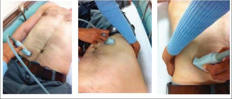
A series of three clinical photographs demonstrating the 'graded compression' technique for point-of-care ultrasound (POCUS) of the appendix. The images show a medical practitioner applying a high-frequency linear transducer to the patient's right lower quadrant (RLQ). The probe is oriented in transverse and oblique planes over McBurney's point to identify the vermiform appendix. Visually evident is the practitioner's use of their non-scanning hand to provide complementary steady pressure on the abdominal wall. This manual compression is an essential clinical maneuver designed to displace overlying bowel gas, reduce the distance between the abdominal wall and the retrocecal or pelvic structures, and assess the compressibility of the appendix. The target audience for this procedural imagery includes emergency physicians and radiologists learning to diagnose acute appendicitis through ultrasonographic findings such as increased appendiceal diameter (>6mm) or non-compressibility.
McBurney's Point and the Liver - Is There a Relation?
What is McBurney's Point?
- Named after Charles McBurney, who described it in 1889 as the point of "maximum tenderness, one half to 2 inches inside the right anterior spinous process of the ilium on a line drawn from the umbilicus."
- Deep tenderness here (McBurney's sign) is a hallmark of acute appendicitis, signaling peritoneal irritation at the appendix site.
Where is the Liver?
The Connection - Indirect and Clinical
| Aspect | Detail |
|---|---|
| Same textbook domain | McBurney's point is described in Gastrointestinal and Liver Disease texts because appendicitis is a GI condition managed by the same specialty |
| Liver disease can mimic/complicate appendicitis | Hepatic pathology (e.g., hepatomegaly, right-sided liver abscesses, Fitz-Hugh-Curtis syndrome) can occasionally cause right-sided abdominal pain that overlaps with or is confused with McBurney's point tenderness |
| Portal pyemia | A rare but serious complication of ruptured/gangrenous appendicitis is pylephlebitis (septic portal vein thrombosis), which can spread infection to the liver via the portal circulation, causing hepatic abscesses |
| Referred pain confusion | The right upper quadrant (liver/gallbladder) and right lower quadrant (McBurney's point) can overlap in clinical presentation, especially in conditions like a high-lying retrocecal appendix |
Summary
- Anatomically: No relation. McBurney's point (right iliac fossa) and the liver (right hypochondrium) are in completely different quadrants.
- Clinically: Appendicitis (diagnosed partly via McBurney's sign) can, if complicated by perforation, lead to portal vein sepsis and liver abscesses - this is the closest pathological link.
- The most well-known clinical association remains McBurney's sign = acute appendicitis, not liver disease.
During my anatomy viva our professor wants to correlate McBurney's point while my discussion topic is liver. What will be questions
Anatomy Viva: McBurney's Point vs Liver - Expected Questions & Model Answers
TIER 1 - Opening / Basic Questions
Q1. What is McBurney's point? Where exactly is it?
Q2. Which quadrant is McBurney's point in? Which quadrant is the liver in?
- McBurney's point = right lower quadrant (right iliac fossa)
- Liver = right upper quadrant (right hypochondrium + epigastrium)
- They are in opposite quadrants on the right side of the abdomen.
TIER 2 - The Real Correlation Questions
Q3. What is the vascular connection between the appendix (at McBurney's point) and the liver?
Appendix → Appendicular veins → Ileocolic vein → Superior mesenteric vein (begins near McBurney's point) → Portal vein → Liver
Q4. What is the clinical significance of this portal venous connection? (The DANGER question)
- Pyogenic liver abscesses - bacteria travel via the portal vein to the liver
- Jaundice + high spiking fevers - classic presentation of pylephlebitis
- Septicemia and high mortality if untreated
"In the past, most cases of pyogenic liver abscess were a consequence of appendicitis complicated by pyelephlebitis (portal vein inflammation) in a young patient." (Sleisenger and Fordtran's Gastrointestinal and Liver Disease)
"Other complications of appendicitis include pyelephlebitis, portal venous thrombosis, liver abscess, and bacteremia." (Robbins Pathologic Basis of Disease)
Q5. When palpating a grossly enlarged liver, where do you START palpating - and why does that connect to McBurney's point?
"To palpate for an enlarged liver one should place the hand on the right iliac fossa with the fingers pointing towards the left axilla." (S. Das - Manual on Clinical Surgery)
Q6. What surface markings of the liver are clinically relevant, and how do they contrast with McBurney's point?
| Landmark | Location |
|---|---|
| Upper border of liver | 5th intercostal space, right midclavicular line |
| Lower border (right) | Right costal margin, midclavicular line |
| Lower border (left) | Crosses epigastrium to left 5th intercostal space |
| McBurney's point | 1/3 from right ASIS to umbilicus (RIF) |
Q7. How does pain in appendicitis vs liver disease differ, and could they be confused?
- Appendicitis: pain starts periumbilically (visceral, T10 dermatome), then shifts to McBurney's point in the right iliac fossa (parietal peritoneum irritation)
- Liver/gallbladder pain: pain is in the right hypochondrium, may radiate to right shoulder tip (diaphragmatic irritation, referred via phrenic nerve C3/4/5)
- Retrocecal appendix - pain is higher up, mimicking liver/renal colic
- High-lying cecum - appendix sits near the liver, so tenderness can be in RUQ
- Acute cholecystitis - can cause RLQ radiation mimicking appendicitis
TIER 3 - Advanced / Show-off Questions
Q8. Name a complication of appendicitis that directly affects the liver and name the portal vein tributary responsible.
Q9. Which surgical incision is used at McBurney's point? Why is it NOT used for liver surgery?
Quick Summary Table for Your Viva
| Connection | Detail |
|---|---|
| Vascular | Appendicular veins → SMV (starts in RIF/McBurney's area) → Portal vein → Liver |
| Pathological | Appendicitis → Pylephlebitis → Pyogenic liver abscess |
| Clinical exam | Palpation of enlarged liver begins at the right iliac fossa (McBurney's region) |
| Quadrant contrast | McBurney's = RLQ; Liver = RUQ - same side, opposite quadrants |
| Pain distinction | Appendicitis shifts to RIF; Liver pain is RUQ ± referred to shoulder |
| Surgical | McBurney/Gridiron incision for appendix; Kocher's/subcostal for liver |