
Spinocerebellar tract
Reading File
Now let me search the medical library for spinocerebellar tract content in parallel.
Finding Sources
Finding Sources
Good results. Let me read the main sections from Neuroanatomy through Clinical Cases and Guyton & Hall in parallel.
Reading File
Reading File
Excellent content. Let me fetch the diagram image and read more detail on the tracts from Neuroanatomy.
Loading Image
Reading File
I now have comprehensive content from two major textbooks. Let me compile the full answer.
Spinocerebellar Tracts
The spinocerebellar tracts are ascending pathways that carry unconscious proprioceptive and somatosensory information from the spinal cord to the cerebellum. They are distinct from the posterior column-medial lemniscal system, which carries conscious proprioception. There are four main tracts, organized by body region and type of information conveyed.
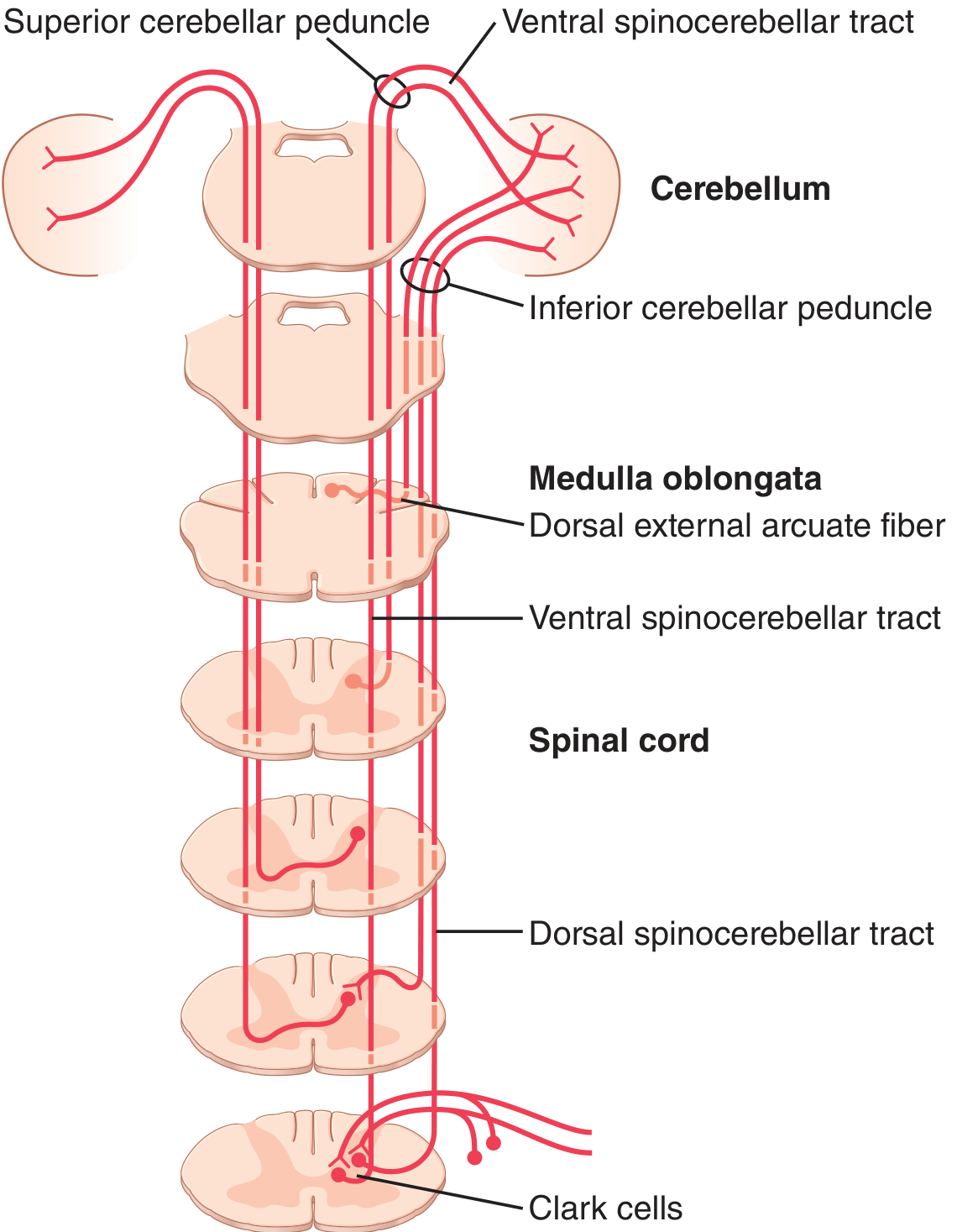
Overview: The Four Tracts
| Tract | Body Region | Cell of Origin | Peduncle |
|---|---|---|---|
| Dorsal spinocerebellar (DSCT) | Lower limb | Nucleus dorsalis of Clark (C8-L2/3) | Inferior cerebellar peduncle |
| Cuneocerebellar | Upper limb & neck | External cuneate nucleus (medulla) | Inferior cerebellar peduncle |
| Ventral spinocerebellar (VSCT) | Lower limb | Spinal border cells | Superior cerebellar peduncle |
| Rostral spinocerebellar | Upper limb | Spinal cord neurons | Superior + inferior cerebellar peduncles |
These tracts divide into two functional groups based on what information they carry:
- Afferent sensory feedback (peripheral receptors → cerebellum): DSCT + cuneocerebellar tract
- Efference copy of spinal interneuron activity (central motor signals → cerebellum): VSCT + rostral spinocerebellar tract
- Neuroanatomy through Clinical Cases, 3rd Ed.
1. Dorsal Spinocerebellar Tract (DSCT)
-
Carries: proprioceptive signals from muscle spindles, Golgi tendon organs, large tactile receptors, and joint receptors of the lower extremities and trunk
-
First-order neurons: peripheral sensory neurons enter via dorsal roots → ascend in the gracile fasciculus
-
Second-order neurons: synapse in the nucleus dorsalis of Clark (Clarke's column), a column of cells running from C8 to L2/L3 in the dorsomedial intermediate gray
-
Course: axons ascend ipsilaterally in the dorsolateral funiculus, just lateral to the lateral corticospinal tract
-
Entry to cerebellum: via the inferior cerebellar peduncle → terminates as mossy fibers in the ipsilateral cerebellar cortex (vermis and intermediate zone)
-
This pathway tells the cerebellum the momentary status of: (1) muscle contraction, (2) tension on tendons, (3) positions and rates of movement, and (4) forces acting on body surfaces
-
Guyton & Hall Textbook of Medical Physiology; Neuroanatomy through Clinical Cases, 3rd Ed.
2. Cuneocerebellar Tract (Upper-limb equivalent of DSCT)
- Carries: unconscious proprioception from the upper extremities and neck
- Course: large-diameter fibers enter the cuneate fasciculus and ascend ipsilaterally to synapse in the external (accessory/lateral) cuneate nucleus in the medulla - the upper-limb analog of Clarke's column
- Entry to cerebellum: via the inferior cerebellar peduncle → ipsilateral cerebellum
- Both DSCT and cuneocerebellar tract provide rapid feedback allowing fine adjustments to ongoing movements
3. Ventral Spinocerebellar Tract (VSCT)
- Carries: NOT primarily peripheral sensory information - instead carries an efference copy of motor signals arriving in the anterior horns from corticospinal and rubrospinal tracts, plus activity of spinal cord interneurons (the internal motor pattern generators)
- Cell of origin: spinal border cells, located along the outer edge of the central gray matter, and from interneurons of the anterior horn
- Course: crosses the midline in the spinal cord (decussates), ascends in the ventral/anterior funiculus, crosses again within the pons (double-crossing), resulting in an ipsilateral projection overall
- Entry to cerebellum: via the superior cerebellar peduncle → terminates in both sides of the cerebellum
- This tract tells the cerebellum which motor commands have arrived at the anterior horn (the "efference copy")
4. Rostral Spinocerebellar Tract
- Carries: information about activity of spinal cord interneurons for the upper extremity, analogous to VSCT for the lower limb
- Entry to cerebellum: via both superior and inferior cerebellar peduncles
Functional Significance
The spinocerebellar tracts are the fastest conducting pathways in the entire CNS - impulses can travel at up to 120 m/sec. This speed allows instantaneous appraisal by the cerebellum of changes in peripheral muscle actions, which is essential for real-time motor coordination and smooth movement.
The cerebellum uses all this incoming data to:
- Compare intended movement (from cortex via efference copy) with actual movement (from peripheral receptors via DSCT/cuneocerebellar)
- Detect and correct errors in motor execution
- Coordinate timing and intensity of descending motor signals
Since these signals do not reach the cerebral cortex, deficits from damage produce ataxia (incoordination) rather than loss of conscious proprioception.
- Guyton & Hall Textbook of Medical Physiology, p. 768-780
- Neuroanatomy through Clinical Cases, 3rd Ed., p. 736
- Eric Kandel - Principles of Neural Science, 6th Ed.
Clinical Relevance
- Friedrich's ataxia: degeneration of spinocerebellar tracts + posterior columns + corticospinal tracts → cerebellar ataxia + loss of conscious proprioception
- Lateral medullary (Wallenberg) syndrome: interruption of the ascending dorsal spinocerebellar tract → ipsilateral limb ataxia
- Subacute combined degeneration (B12 deficiency): posterior columns predominantly, but spinocerebellar tracts also involved
This is a shared conversation. Sign in to Orris to start your own chat.