
Endoscopic ultrasound
Reading File
Now let me search the medical library for endoscopic ultrasound content, in parallel with a web search and PubMed search.
Finding Sources
Searching the Web
endoscopic ultrasound EUS indications technique clinical applications 2024
Searching PubMed
"endoscopic ultrasound"[MeSH Terms] AND "clinical applications"
Good - I found rich dedicated EUS sections. Let me read the main textbook sources in parallel.
Reading File
Reading File
Reading File
Excellent content found. Let me read the indications table and get additional content on EUS staging and FNA from the textbooks.
Reading File
Reading File
Finding Sources
Now let me fetch the key EUS images and read more about EUS staging and FNA from the Clinical GI Endoscopy textbook.
Reading File
Loading Image
Loading Image
I now have excellent textbook content and images. Let me also get the chronic pancreatitis EUS image and read a bit more from the Yamada section on EUS.
Loading Image
Reading File
I now have rich content from multiple textbooks and several images. Let me compile a comprehensive answer.
Endoscopic Ultrasound (EUS)
What is EUS?
Endoscopic ultrasound combines traditional mucosal endoscopy with a separate ultrasound probe mounted on the echoendoscope tip. Because the pancreas, bile duct, and mediastinal structures lie within a few millimeters of the GI lumen, placing the transducer intraluminally allows use of higher ultrasound frequencies (7.5-20 MHz) than transcutaneous scanning - yielding far superior resolution at the cost of reduced penetration depth. This makes EUS uniquely able to characterize the five-layer wall structure of the GI tract and adjacent organs in real time.
- Bailey and Love's Short Practice of Surgery, 28th ed., p. 182
- Clinical Gastrointestinal Endoscopy Expert Consult, 3e
Instrument Types
| Type | Ultrasound Arrangement | Optics | Primary Use |
|---|---|---|---|
| Radial echoendoscope | Radially arranged (360° scan) | Forward-viewing | Diagnostic staging (esophagus, stomach) |
| Linear echoendoscope | Linearly arranged (alongside needle path) | Side-viewing | Guided biopsy (FNA/FNB), therapeutic procedures |
The linear scope has a working channel like an ERCP scope, allowing real-time needle visualization during EUS-FNA. The radial scope gives a more intuitive anatomical cross-section but cannot guide needles in real time.
- Bailey and Love's, p. 182
GI Wall Layers on EUS
EUS resolves the GI wall into five distinct layers:
| Layer | Appearance | Tissue Correlate |
|---|---|---|
| 1 | Hyperechoic | Interface/superficial mucosa |
| 2 | Hypoechoic | Deep mucosa (muscularis mucosae) |
| 3 | Hyperechoic | Submucosa |
| 4 | Hypoechoic | Muscularis propria |
| 5 | Hyperechoic | Serosa/adventitia |
Identifying which layer a lesion arises from directs both the diagnosis (e.g., lipoma from layer 3, GIST from layer 4) and the T-stage.
Indications
Diagnostic
- Staging of esophageal/gastric/rectal malignancy (T and N staging)
- Staging of hepatobiliary malignancy (pancreatic cancer, cholangiocarcinoma)
- Subepithelial lesions (SELs) - characterizing intramural masses (lipoma, GIST, carcinoid, varices, cysts)
- Diagnosis of choledochal microlithiasis / biliary sludge
- Evaluation of chronic pancreatitis - parenchymal and ductal criteria
- Idiopathic recurrent acute pancreatitis (IRAP) - 7-point checklist including bile duct stones, pancreatic masses, pancreas divisum, SOD, CP, AIP
- Suspected pancreatic masses - EUS detects small masses missed by CT
Therapeutic / Interventional
-
EUS-FNA/FNB - tissue acquisition from pancreatic masses, lymph nodes, submucosal lesions, left adrenal, portal adenopathy
-
Transgastric drainage of pancreatic pseudocysts and abscesses (EUS cystgastrostomy)
-
Coeliac plexus neurolysis/block for pain management in pancreatic cancer
-
Biliary interventions - EUS-guided biliary drainage (increasingly performed)
-
EUS-guided gastroenterostomy for gastric outlet obstruction
-
Bailey and Love's, Table 9.6, p. 182
EUS in Tumor Staging
Esophageal Cancer
EUS is the most accurate pre-operative staging modality for esophageal cancer:
- T staging accuracy: 87-92% (vs. CT: 51-74%)
- N staging sensitivity: 92-95%, specificity 50-54%
- EUS is superior to CT for primary tumor and regional nodal status
- EUS-FNA improves N-staging accuracy to >90% for celiac axis nodes
- CT or MRI should be performed before EUS to exclude distant metastases
- Post-chemoradiotherapy: a ≥50% reduction in tumor cross-sectional area correlates with treatment response, though EUS cannot reliably distinguish residual cancer from inflammation/fibrosis
EUS image - esophageal tumour with adventitial invasion:
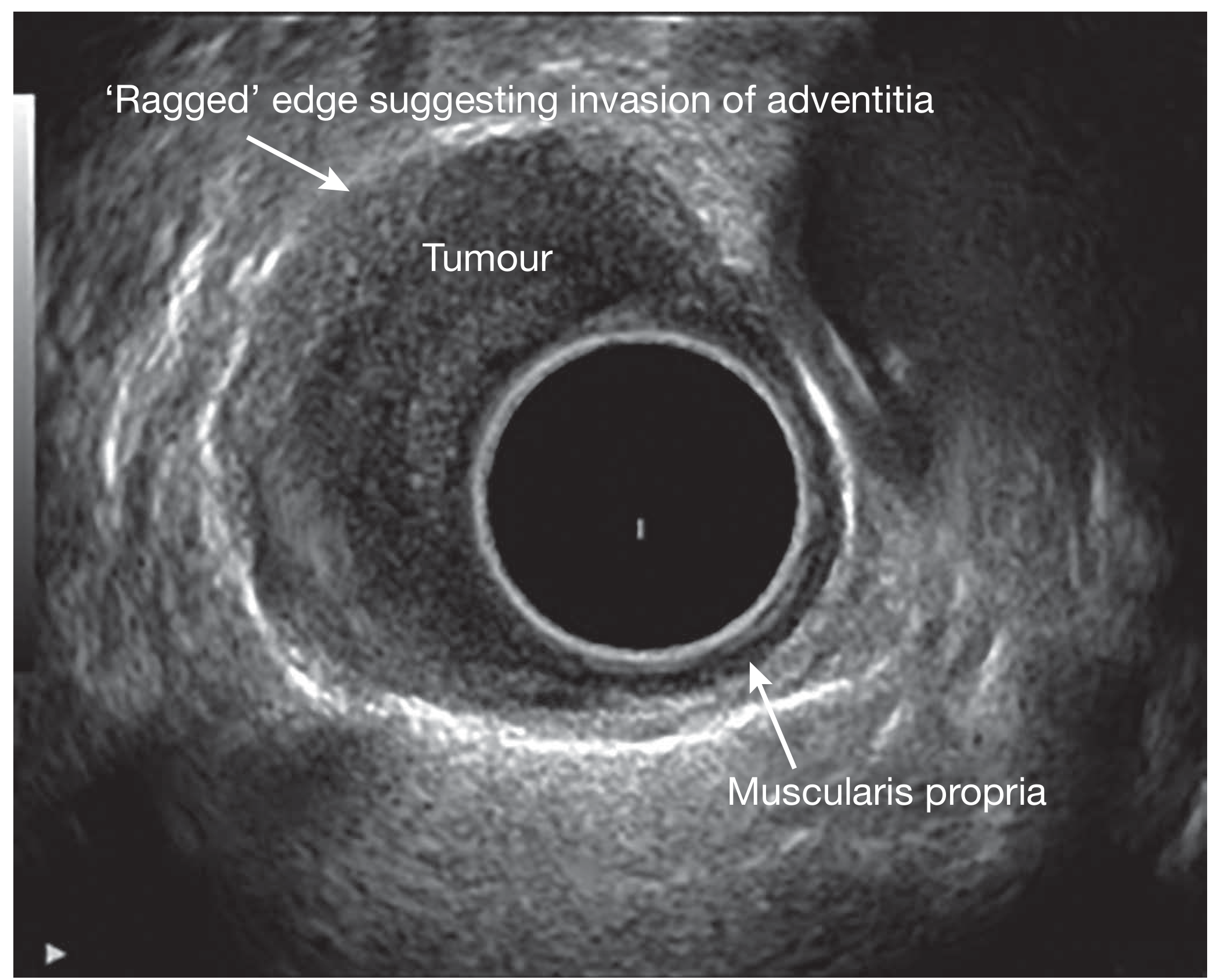
EUS image of an oesophageal tumour. The 'ragged' edge at the top indicates invasion into the adventitia (T3/T4 disease). The intact muscularis propria layer is visible at the bottom. - Bailey and Love's, Fig. 9.20
- Yamada's Textbook of Gastroenterology, 7e, p. 976
EUS-FNA (Fine Needle Aspiration)
EUS-FNA is performed using the linear echoendoscope with real-time needle visualization. A needle (typically 22G or 25G; 19G for core biopsy) is passed through the working channel and directed into the target lesion under continuous ultrasound guidance.
EUS-FNA image - pancreatic head mass:
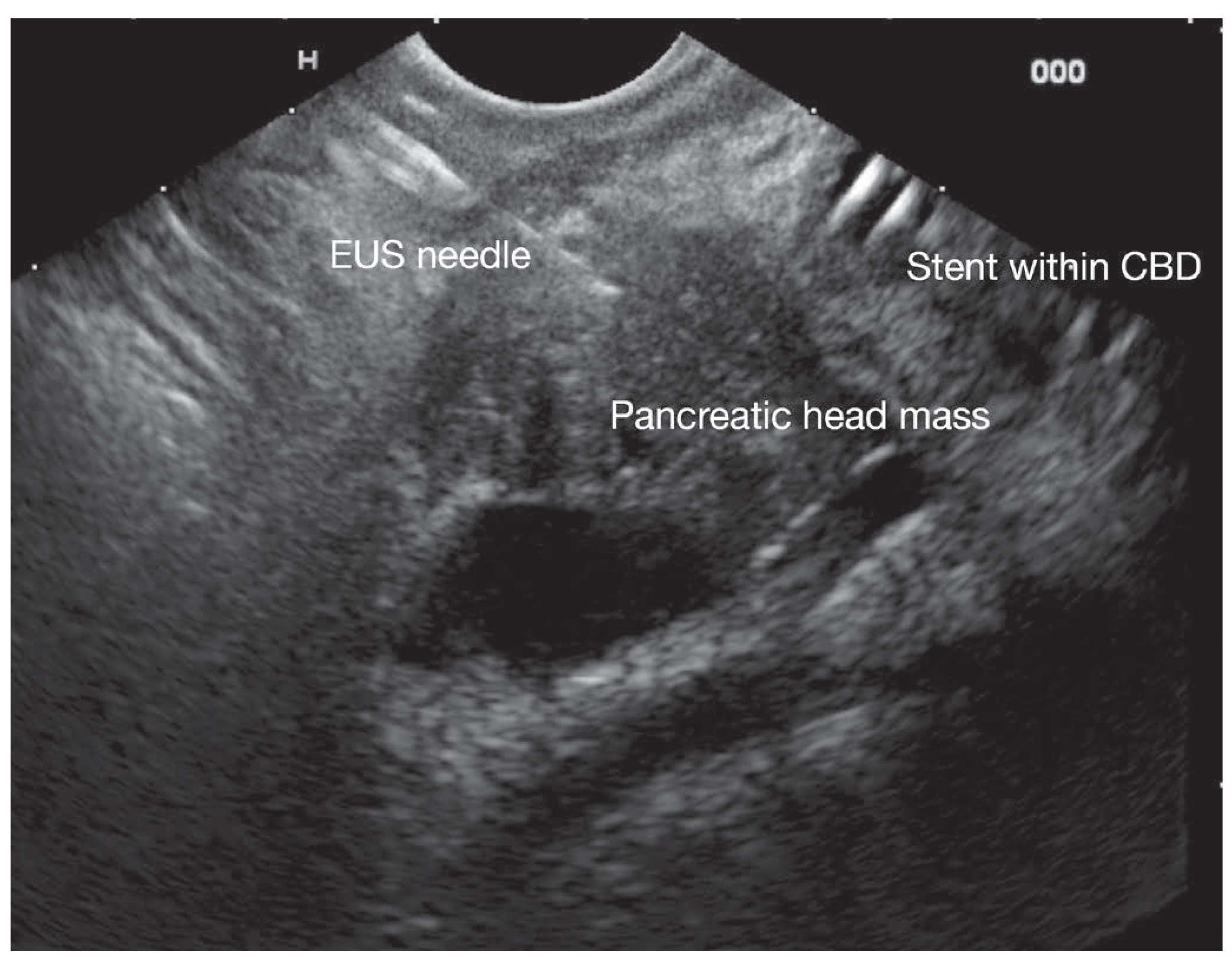
EUS-guided FNA of a pancreatic head mass. The EUS needle is clearly visible entering the mass. A biliary stent is also visible within the CBD (common bile duct). - Bailey and Love's, Fig. 9.21
Key EUS-FNA targets:
- Pancreatic masses and cysts
- Paraesophageal and coeliac lymph nodes
- Submucosal GI lesions
- Portal lymphadenopathy
- Left adrenal gland and left hepatic masses
- Mediastinal lesions
Newer fine-needle biopsy (FNB) needles (e.g., fork-tip, Franseen design) provide tissue cores with preserved architecture, improving histological diagnosis and enabling immunohistochemistry - particularly useful for autoimmune pancreatitis (IgG4 staining, obliterative phlebitis).
EUS in Chronic Pancreatitis
EUS is exquisitely sensitive for early chronic pancreatitis (CP) due to proximity to the pancreas, allowing high-frequency scanning. EUS criteria are divided into:
Parenchymal criteria:
- Inhomogeneity
- Hyperechoic foci
- Hyperechoic strands
- Lobularity
- Pseudocysts
Ductal criteria:
- Ductal dilation (≥3 mm head; ≥2 mm body; ≥1 mm tail)
- Hyperechoic main duct margins
- Irregular main duct margins
- Visible side branches
A quantitative score using 9 criteria is most reliable when clearly normal (≤2 criteria) or clearly abnormal (≥5 criteria) - predictive value 85% at these thresholds. Intermediate scores (3-4 criteria) are diagnostically uncertain and may represent early disease.
EUS image - chronic pancreatitis with intraductal stone:
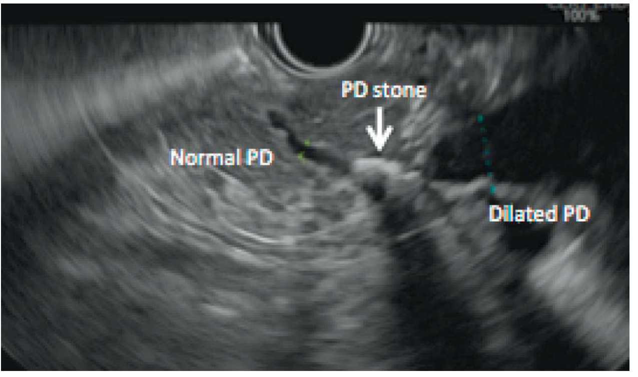
EUS showing a calcified intraductal stone in the pancreatic head (PD stone, arrow) with upstream normal duct and downstream dilation. - Clinical GI Endoscopy, Fig. 59.4
EUS in Subepithelial Lesions (SELs)
Key EUS features assessed for any intramural mass:
| Feature | What to Assess |
|---|---|
| Location | Organ + position |
| Size | 3 dimensions |
| Background echogenicity | Hypo/hyper/anechoic |
| Focal echogenicity | Foci present/absent |
| Shape/margins | Round, oval; smooth vs. irregular |
| Margin definition | Well- vs. poorly-defined |
| Wall layer of origin | Mucosa / submucosa / muscularis propria |
| Invasion | T-stage |
Characteristic appearances:
-
Lipomas: Brightly hyperechoic, uniform, well-demarcated, layer 3 (submucosal), easily deformed by transducer
-
Varices: Anechoic, vermiform, nearly always in groups
-
GISTs: Hypoechoic, layer 4 (muscularis propria) origin, malignant risk ↑ with size >3 cm, irregular margins, echogenic foci
-
EUS sensitivity/specificity for distinguishing lesion types: 64-92% / 80-100%
-
Clinical GI Endoscopy Expert Consult, 3e, p. [block5]
EUS vs. Other Modalities
| Modality | Advantage of EUS |
|---|---|
| CT scan | Better T-staging, detects small pancreatic masses, more sensitive for biliary sludge/stones |
| MRCP | Superior for biliary sludge/stones, luminal visualization, tissue acquisition; combined yield ~50% in IRAP (complementary) |
| Transcutaneous US | Far higher resolution due to intraluminal positioning and higher frequencies |
| ERCP | Non-invasive alternative for diagnosis (ERCP reserved for treatment) |
Training Requirements and Complications
EUS requires dedicated training in both scope manipulation and image interpretation. The wide, relatively inflexible echoendoscope typically requires sedation - many units use propofol-based anaesthesia for complex therapeutic procedures.
Complications:
-
Diagnostic EUS: Oversedation, esophageal perforation (rare)
-
Therapeutic EUS (FNA/drainage): Haemorrhage, perforation, pancreatitis (rare, <1% for pancreatic FNA), infection
-
All patients undergoing therapeutic EUS require a normal coagulation screen beforehand
-
Bailey and Love's Short Practice of Surgery, 28th ed., p. 182
Summary
EUS is a versatile tool occupying a unique niche - it provides high-resolution, real-time imaging of structures adjacent to the GI lumen combined with the ability to obtain tissue under direct ultrasound guidance. Its main clinical roles are:
- Cancer staging (GI, pancreatic, hepatobiliary)
- Pancreatic disease (masses, chronic pancreatitis, cysts)
- Tissue acquisition (FNA/FNB)
- Biliary stone detection (especially microlithiasis)
- Therapeutic drainage (pseudocysts, biliary)
- Pain palliation (coeliac plexus block)
This is a shared conversation. Sign in to Orris to start your own chat.