
CREATE A PPT ON SOT,CEOT,AOT FROM SHAFERS
adenomatoid odontogenic tumor AOT histology microscopy radiology
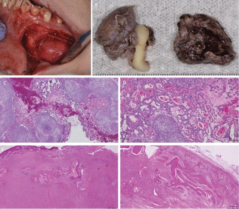
A composite image illustrating the intraoperative, macroscopic, and microscopic characteristics of an adenomatoid odontogenic tumor (AOT). Frame A is an intraoperative clinical photograph of the mandible showing a reddish, well-circumscribed mass exposed after a vestibular flap. Frame B shows the gross surgical specimen consisting of a brownish cystic capsule surrounding an impacted canine tooth and a separate thick-walled cystic fragment. Frames C-F are light microscopy images stained with Hematoxylin and Eosin (H&E). Micrographs C and D (20x magnification) demonstrate a biphasic odontogenic epithelial proliferation characterized by spindle-shaped cells arranged in solid nodules, whorled patterns, and characteristic duct-like structures with eosinophilic luminal material. Micrographs E and F show a different component of the lesion consisting of dense fibroblastic connective tissue proliferation containing irregular, mineralized bone-like or cementum-like trabeculae. The image illustrates the classic histopathological features of a hybrid odontogenic lesion, combining features of AOT with calcifying epithelial structures.
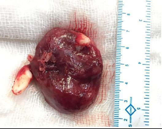
This clinical photograph shows a gross surgical specimen of an adenomatoid odontogenic tumor (AOT). The specimen is a thick-walled, spherical, and irregular cystic mass, approximately 5.5 cm in its longest dimension as indicated by the adjacent metric ruler. The tumor surface is smooth, slightly glistening, and exhibits a reddish-brown coloration suggestive of blood or fluid content. Pathognomonic for this type of odontogenic lesion, two unerupted teeth—specifically the left maxillary canine and the left upper lateral incisor—are seen firmly attached to the specimen. One tooth is embedded in the inferomedial portion, while the other is visible on the superior surface of the mass. The specimen's appearance reflects its origin from the maxillary sinus region, highlighting the follicular relationship between the tumor and impacted dental structures. This visual provides high-yield educational value for oral and maxillofacial pathology, illustrating the classic presentation of an intraosseous odontogenic neoplasm following enucleation.
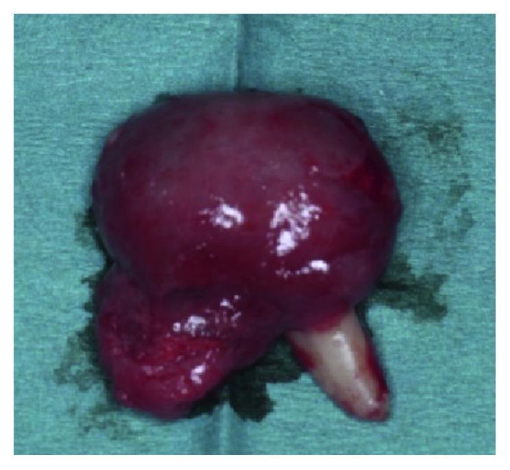
This intraoperative clinical photograph displays an enucleated specimen of an adenomatoid odontogenic tumor (AOT) associated with the maxillary left lateral incisor (tooth #22). The specimen consists of a large, well-encapsulated, bulbous soft tissue mass attached to the mid-root section of the tooth. The mass is approximately 2 cm in size, exhibiting a predominantly deep red to maroon color with a glistening, smooth, and highly vascularized surface. At its inferior aspect, a conical, creamy-white tooth root is visible, extending from the body of the tumor. The interface between the soft tissue and the tooth demonstrates a transition from a smooth capsule to a more irregular, lobulated texture near the point of attachment. This image illustrates the follicular type of AOT, which typically presents as a pericoronal radiolucency mimicking a dentigerous cyst, but is pathologically distinguished by its firm attachment to the tooth root and the characteristic histopathology of spindle-shaped epithelial cells forming whorled masses and duct-like structures.
calcifying epithelial odontogenic tumor Pindborg CEOT histology
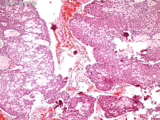
Calcifying epithelial odontogenic tumor (CEOT), also known as Pindborg tumor, is an odontogenic epithelial neoplasm typically arising in the jaw, most often the posterior mandible. This histopathology image depicts a micrograph of CEOT stained with hematoxylin and eosin. The tumor shows sheets, nests, and strands of polygonal epithelial cells embedded in a variably dense fibrous stroma. The cells possess abundant eosinophilic cytoplasm, distinct cell borders, and prominent intercellular bridges. Nuclear pleomorphism may be present in some fields, including giant nuclei, but mitotic activity remains low and is not indicative of malignancy. A hallmark feature is the extensive eosinophilic amyloid-like extracellular material that often fills the stromal spaces and may juxtapose calcified deposits. Calcifications develop in association with this amyloid, frequently forming concentric Liesegang rings. In some cases, epithelial component predominates with little amyloid or calcification; in others, sparse nests are separated by large amyloid-rich areas. Rarely, a clear cell variant with cystic growth has been described. The combination of epithelial cells, amyloid-like material, and characteristic calcifications is diagnostically distinctive and helps differentiate CEOT from ameloblastoma, calcifying odontogenic cyst, and other odontogenic tumors. Clinically, CEOT is generally benign but locally aggressive, requiring complete surgical excision, with potential for recurrence if incomplete.
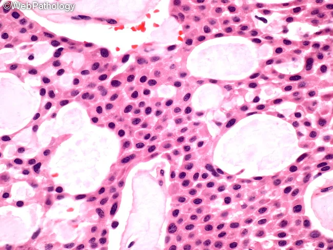
This 400x hematoxylin and eosin-stained histology image depicts a calcifying epithelial odontogenic tumor (CEOT), also known as Pindborg tumor, derived from odontogenic epithelium in the jaw. The tissue demonstrates cohesive sheets and nests of polygonal epithelial cells with abundant eosinophilic cytoplasm, distinct cell borders, and prominent intercellular bridges, most evident in the central portions. Nuclei are generally round to oval with mild pleomorphism and low mitotic activity. The tumor matrix is variably dense with scattered, round to oval extracellular deposits that resemble amyloid and are characteristic of CEOT. These amyloid-like deposits frequently calcify, forming irregular concentric rings or Liesegang-type calcifications that appear as basophilic or eosinophilic speckles disrupting the stroma and sometimes outlining cell clusters. The surrounding stroma is somewhat vascular and may contain dystrophic calcifications. Overall, the image shows a benign yet locally infiltrative neoplasm composed of epithelial cells in close association with mineralized material, with intact basal membrane in many areas. Diagnostic significance rests on the combination of epithelial sheets, amyloid-like material, and calcifications, a pattern that differentiates CEOT from other odontogenic tumors such as ameloblastoma. Clinically relevant features include potential jaw swelling and radiographic radiolucent lesions with calcifications, guiding surgical planning and prognosis. These features aid diagnosis and management.
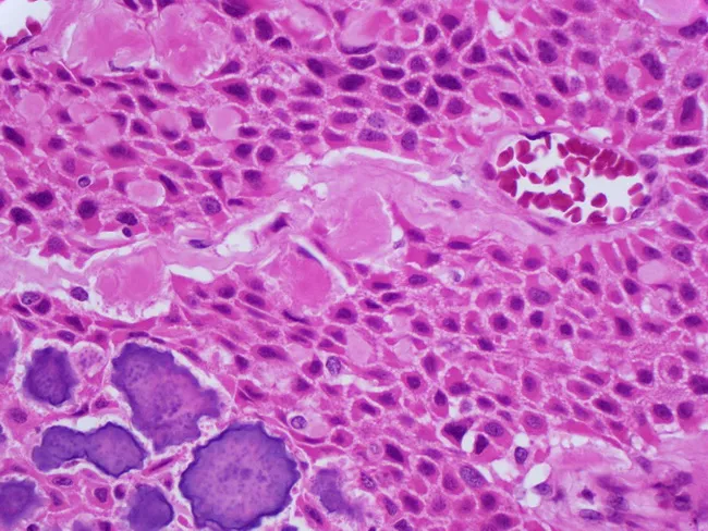
Imaging modality: Brightfield light microscopy of a hematoxylin and eosin stained histology section of jaw odontogenic tumor tissue. The specimen demonstrates classic calcifying epithelial odontogenic tumor (CEOT), also known as Pindborg tumor, arising in a tooth-bearing region of the mandible. Sheets, nests, and occasional strands of polygonal epithelial cells exhibit abundant eosinophilic cytoplasm with distinct cell borders and prominent intercellular bridges. The tumor nests are embedded in a fibrous stroma that ranges from moderately to densely collagenous. A characteristic feature is the presence of large extracellular amyloid-like material, sometimes forming concentric calcifications known as Liesegang rings. Calcifications are variably birefringent under polarized light and appear as rounded, ring-like deposits within the amyloid. The nuclei are generally small to moderately pleomorphic with mild mitotic activity, consistent with a benign yet locally invasive odontogenic neoplasm. Collectively, the combination of epithelial polygons, amyloid-like deposition, Liesegang rings, and calcifications strongly supports CEOT and helps distinguish it from ameloblastoma and other odontogenic lesions. Clinically, CEOT tends to present as a slow-growing jaw mass in adults; management is surgical excision with careful follow-up for recurrence. This image is valuable for educational purposes, differential diagnosis, and pathology reference. Correlation with radiographs and clinical behavior improves diagnostic confidence.
squamous odontogenic tumor SOT histology jaw
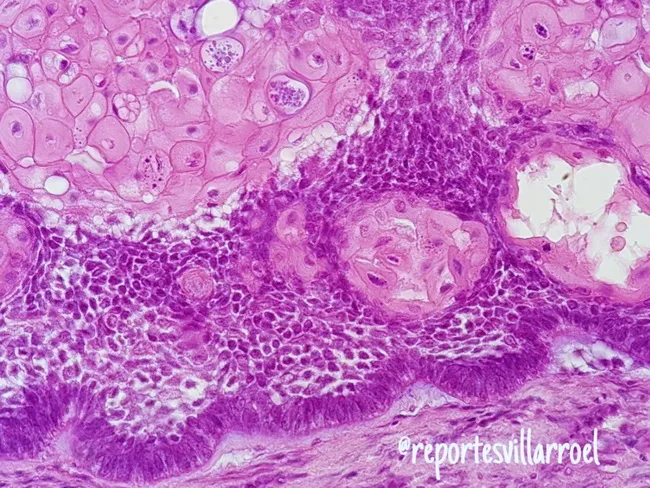
Imaging modality and technique: Light microscopy of hematoxylin and eosin (H&E) stained paraffin-embedded jaw lesion tissue, sectioned for routine histology. Anatomical localization: odontogenic tumor of the mandible, with epithelial islands embedded in a dense fibrous stroma consistent with ameloblastoma. The image demonstrates the acanthomatous variant, in which extensive squamous metaplasia occurs within the central portion of the epithelial islands, accompanied by keratin formation in the stellate reticulum. Peripheral ameloblast‑like cells show palisaded, polarized basilar nuclei; the central cells resemble the loosely arranged, eosinophilic stellate reticulum. The surrounding stroma is dense and vascular, with inflammatory cells in some fields. Notable architectural diversity is evident: nests and cords display follicular and plexiform arrangements, with focal desmoplastic areas and possible granular cell change in other regions. Such histology reflects intricate tumor biology while retaining characteristic features of ameloblastoma. Diagnostic relevance centers on distinguishing this entity from squamous odontogenic tumor and squamous cell carcinoma, as management and prognosis differ markedly. Key differentiators include peripheral palisading and reverse polarity, central stellate reticulum with metaplasia, and lack of overt malignant cytology. Clinically, recognizing acanthomatous ameloblastoma guides surgical planning and follow-up in jaw pathology and head‑neck oncology. This image serves as a reference for education and research.
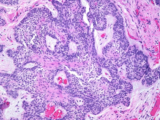
Imaging modality: light microscopy of Hematoxylin and Eosin stained, formalin-fixed, paraffin-embedded jaw tissue. The specimen shows ameloblastoma with prominent squamous metaplasia within tumor islands. Odontogenic epithelial nests exhibit peripheral ameloblast-like, tall-columnar cells with reverse polarity and a basaloid, dark-staining center resembling stellate reticulum. In many islands, there is extensive squamous differentiation with polygonal keratinizing cells and foci of keratinization, yet the cells retain minimal cytologic atypia and mitotic activity is low. The surrounding stroma is fibrous and vascular, with infiltrative margins indicating local aggressiveness characteristic of ameloblastoma. Absence of significant pleomorphism, hyperchromasia, or abnormal mitoses helps distinguish this process from squamous cell carcinoma, though extensive squamous metaplasia can mimic malignancy. The lesion is typical of an odontogenic tumor occurring in the jaw (mandible or maxilla). Clinically relevant differential diagnoses include squamous cell carcinoma arising in odontogenic keratocyst or de-differentiated ameloblastoma; correlation with radiology and clinical course is essential. The diagnostic significance lies in recognizing metaplastic squamous change to prevent misinterpretation as invasive carcinoma; complete surgical excision with careful follow-up is recommended due to potential recurrence. Clinicians should correlate with radiographic margins, molecular markers as available, and immunohistochemical profile showing p63 and cytokeratin expression patterns supporting odontogenic origin rather than carcinoma.
mkdir -p /tmp/workspace/odontogenic-tumors && ls /tmp/workspace/odontogenic-tumors/
~/odontogenic-tumors/create_ppt.py
import json, subprocess, base64, os
from io import BytesIO
from pptx import Presentation
from pptx.util import Inches, Pt, Emu
from pptx.dml.color import RGBColor
from pptx.enum.text import PP_ALIGN, MSO_ANCHOR
from pptx.util import Inches, Pt
from pptx.oxml.ns import qn
from lxml import etree

# ── Color palette ────────────────────────────────────────────────
DEEP_BLUE   = RGBColor(0x0D, 0x47, 0xA1)   # #0D47A1
LIGHT_BLUE  = RGBColor(0x42, 0xA5, 0xF5)   # #42A5F5
GOLD        = RGBColor(0xFF, 0xD6, 0x00)   # #FFD600
WHITE       = RGBColor(0xFF, 0xFF, 0xFF)
DARK_TEXT   = RGBColor(0x1A, 0x23, 0x7E)   # deep navy
LIGHT_BG    = RGBColor(0xE3, 0xF2, 0xFD)   # very light blue
ACCENT_RED  = RGBColor(0xD3, 0x2F, 0x2F)   # #D32F2F
ACCENT_GREEN= RGBColor(0x2E, 0x7D, 0x32)   # #2E7D32
SECTION_CEOT= RGBColor(0x4A, 0x14, 0x8C)   # purple for CEOT
SECTION_AOT = RGBColor(0x00, 0x69, 0x5C)   # teal for AOT
SECTION_SOT = RGBColor(0x1A, 0x23, 0x7E)   # deep blue for SOT
SLIDE_BG    = RGBColor(0xF5, 0xF5, 0xF5)

prs = Presentation()
prs.slide_width  = Inches(13.333)
prs.slide_height = Inches(7.5)
blank_layout = prs.slide_layouts[6]

# ── Helper: set solid fill on shape ──────────────────────────────
def fill_solid(shape, color):
    sp = shape._element
    spPr = sp.find(qn('p:spPr'))
    if spPr is None:
        spPr = etree.SubElement(sp, qn('p:spPr'))
    solidFill_parent = spPr
    # remove existing fill
    for tag in ['a:noFill','a:solidFill','a:gradFill','a:pattFill','a:blipFill','a:grpSpPr']:
        el = spPr.find(qn(tag) if ':' in tag else tag)
        if el is not None:
            spPr.remove(el)
    solidFill = etree.SubElement(spPr, qn('a:solidFill'))
    srgbClr   = etree.SubElement(solidFill, qn('a:srgbClr'))
    srgbClr.set('val', '{:02X}{:02X}{:02X}'.format(color.r, color.g, color.b))

def set_bg_color(slide, color):
    bg = slide.background
    fill = bg.fill
    fill.solid()
    fill.fore_color.rgb = color

def add_rect(slide, l, t, w, h, color):
    shape = slide.shapes.add_shape(1, Inches(l), Inches(t), Inches(w), Inches(h))
    fill_solid(shape, color)
    shape.line.fill.background()
    return shape

def add_tb(slide, text, l, t, w, h, size, bold=False, color=WHITE, align=PP_ALIGN.LEFT, italic=False, wrap=True):
    tb = slide.shapes.add_textbox(Inches(l), Inches(t), Inches(w), Inches(h))
    tf = tb.text_frame
    tf.word_wrap = wrap
    tf.margin_left = Pt(4)
    tf.margin_right = Pt(4)
    tf.margin_top = Pt(2)
    tf.margin_bottom = Pt(2)
    p = tf.paragraphs[0]
    p.alignment = align
    run = p.add_run()
    run.text = text
    run.font.size = Pt(size)
    run.font.bold = bold
    run.font.italic = italic
    run.font.color.rgb = color
    run.font.name = "Calibri"
    return tb

def add_bullet_slide(slide, title_text, bullets, accent_color, subtitle=None, image_url=None):
    """Generic bullet slide with a left accent bar and title"""
    set_bg_color(slide, SLIDE_BG)
    # Top header bar
    add_rect(slide, 0, 0, 13.333, 1.1, accent_color)
    add_tb(slide, title_text, 0.2, 0.05, 12.8, 1.0, 28, bold=True, color=WHITE, align=PP_ALIGN.LEFT)
    if subtitle:
        add_tb(slide, subtitle, 0.2, 0.65, 12.8, 0.5, 14, bold=False, color=GOLD, align=PP_ALIGN.LEFT)

    # Left accent strip
    add_rect(slide, 0, 1.1, 0.08, 6.4, GOLD)

    # Bullet text box
    tb = slide.shapes.add_textbox(Inches(0.3), Inches(1.2), Inches(12.8), Inches(6.0))
    tf = tb.text_frame
    tf.word_wrap = True
    tf.margin_left = Pt(6)
    tf.margin_right = Pt(6)
    tf.margin_top = Pt(4)
    tf.margin_bottom = Pt(4)

    first = True
    for item in bullets:
        if first:
            p = tf.paragraphs[0]
            first = False
        else:
            p = tf.add_paragraph()
        p.space_before = Pt(4)
        p.space_after  = Pt(2)

        if isinstance(item, dict):
            # heading bullet
            run = p.add_run()
            run.text = item['heading']
            run.font.bold = True
            run.font.size = Pt(item.get('size', 17))
            run.font.color.rgb = accent_color
            run.font.name = "Calibri"
            p.level = 0
        else:
            indent = 0
            text = item
            if item.startswith('   '):
                indent = 1
                text = item.lstrip()
            run = p.add_run()
            # bullet char
            if indent == 0:
                run.text = u'\u25CF  ' + text
                run.font.size = Pt(15)
                run.font.bold = False
                run.font.color.rgb = DARK_TEXT
            else:
                run.text = u'      \u25E6  ' + text
                run.font.size = Pt(13.5)
                run.font.bold = False
                run.font.color.rgb = RGBColor(0x37, 0x47, 0x4F)
            run.font.name = "Calibri"
            p.level = indent

# ─────────────────────────────────────────────────────────────────
# SLIDE 1 — Title Slide
# ─────────────────────────────────────────────────────────────────
slide = prs.slides.add_slide(blank_layout)
set_bg_color(slide, DEEP_BLUE)

# Diagonal accent shape (simulated via stacked rects)
add_rect(slide, 0, 0, 13.333, 7.5, DEEP_BLUE)
add_rect(slide, 0, 5.5, 13.333, 2.0, RGBColor(0x0A, 0x33, 0x7A))

# Gold banner
add_rect(slide, 0, 3.0, 13.333, 0.08, GOLD)
add_rect(slide, 0, 6.5, 13.333, 0.08, GOLD)

# Main title
add_tb(slide, "ODONTOGENIC TUMORS", 0.5, 0.5, 12.3, 1.2, 42, bold=True, color=WHITE, align=PP_ALIGN.CENTER)
add_tb(slide, "SOT  |  CEOT  |  AOT", 0.5, 1.6, 12.3, 1.0, 32, bold=True, color=GOLD, align=PP_ALIGN.CENTER)
add_tb(slide, "Squamous Odontogenic Tumor  •  Calcifying Epithelial Odontogenic Tumor  •  Adenomatoid Odontogenic Tumor",
       0.5, 2.5, 12.3, 0.6, 13, bold=False, color=LIGHT_BLUE, align=PP_ALIGN.CENTER)

add_tb(slide, "Based on Shafer's Textbook of Oral Pathology", 0.5, 3.6, 12.3, 0.6, 14, bold=False,
       color=RGBColor(0xB3, 0xD9, 0xFF), align=PP_ALIGN.CENTER)
add_tb(slide, "Oral & Maxillofacial Pathology", 0.5, 6.6, 12.3, 0.6, 13, bold=False,
       color=RGBColor(0xB3, 0xD9, 0xFF), align=PP_ALIGN.CENTER)

# ─────────────────────────────────────────────────────────────────
# SLIDE 2 — Overview / Classification
# ─────────────────────────────────────────────────────────────────
slide = prs.slides.add_slide(blank_layout)
set_bg_color(slide, SLIDE_BG)
add_rect(slide, 0, 0, 13.333, 1.1, DEEP_BLUE)
add_tb(slide, "Classification of Odontogenic Tumors", 0.2, 0.05, 12.8, 1.0, 26, bold=True, color=WHITE)
add_rect(slide, 0, 1.1, 0.08, 6.4, GOLD)

# Three columns
cols = [
    ("SOT", "Squamous Odontogenic Tumor", SECTION_SOT,
     ["Epithelial origin", "Extremely rare", "Benign, locally aggressive", "Resembles squamous cell carcinoma microscopically", "Arises from rests of Malassez"]),
    ("CEOT", "Calcifying Epithelial\nOdontogenic Tumor (Pindborg)", SECTION_CEOT,
     ["Also: Pindborg Tumor", "Epithelial origin", "Locally invasive benign neoplasm", "Liesegang ring calcifications", "Amyloid-like material"]),
    ("AOT", "Adenomatoid Odontogenic Tumor", SECTION_AOT,
     ["Also called '2/3 tumor'", "Epithelial origin", "Benign, non-invasive", "Associated with impacted canine", "Excellent prognosis"]),
]
x_starts = [0.3, 4.6, 8.9]
for (abbr, full, color, pts), x in zip(cols, x_starts):
    add_rect(slide, x, 1.2, 4.1, 0.7, color)
    add_tb(slide, abbr, x+0.05, 1.2, 4.0, 0.45, 22, bold=True, color=WHITE, align=PP_ALIGN.CENTER)
    add_tb(slide, full, x+0.05, 1.65, 4.0, 0.5, 10, bold=False, color=WHITE, align=PP_ALIGN.CENTER)
    add_rect(slide, x, 1.95, 4.1, 5.3, RGBColor(0xFF, 0xFF, 0xFF))
    # Add a subtle left border
    add_rect(slide, x, 1.95, 0.05, 5.3, color)
    tb2 = slide.shapes.add_textbox(Inches(x+0.1), Inches(2.05), Inches(3.95), Inches(5.1))
    tf2 = tb2.text_frame
    tf2.word_wrap = True
    first2 = True
    for pt in pts:
        if first2:
            p2 = tf2.paragraphs[0]
            first2 = False
        else:
            p2 = tf2.add_paragraph()
        p2.space_before = Pt(5)
        r2 = p2.add_run()
        r2.text = u'\u2714  ' + pt
        r2.font.size = Pt(13)
        r2.font.color.rgb = DARK_TEXT
        r2.font.name = "Calibri"

# WHO Classification note
add_tb(slide, "WHO Classification (2022): Benign Epithelial Odontogenic Tumors",
       0.3, 7.1, 12.8, 0.35, 11, bold=False, color=RGBColor(0x55, 0x55, 0x55), align=PP_ALIGN.CENTER)

# ─────────────────────────────────────────────────────────────────
# SLIDE 3 — SOT: Section Title
# ─────────────────────────────────────────────────────────────────
slide = prs.slides.add_slide(blank_layout)
set_bg_color(slide, SECTION_SOT)
add_rect(slide, 0, 0, 13.333, 7.5, SECTION_SOT)
add_rect(slide, 0, 3.5, 13.333, 0.08, GOLD)
add_tb(slide, "SECTION 1", 0.5, 1.0, 12.3, 0.8, 22, bold=False, color=LIGHT_BLUE, align=PP_ALIGN.CENTER)
add_tb(slide, "Squamous Odontogenic Tumor", 0.5, 1.7, 12.3, 1.2, 38, bold=True, color=WHITE, align=PP_ALIGN.CENTER)
add_tb(slide, "(SOT)", 0.5, 2.8, 12.3, 0.8, 28, bold=True, color=GOLD, align=PP_ALIGN.CENTER)
add_tb(slide, "First described by Pullon et al. (1975)", 0.5, 3.7, 12.3, 0.6, 15, bold=False,
       color=RGBColor(0xB3, 0xD9, 0xFF), align=PP_ALIGN.CENTER)
add_tb(slide, "Extremely rare benign odontogenic tumor • < 100 cases in literature",
       0.5, 4.4, 12.3, 0.6, 14, bold=False, color=RGBColor(0xCC, 0xCC, 0xFF), align=PP_ALIGN.CENTER)

# ─────────────────────────────────────────────────────────────────
# SLIDE 4 — SOT: Clinical Features
# ─────────────────────────────────────────────────────────────────
slide = prs.slides.add_slide(blank_layout)
add_bullet_slide(slide, "SOT — Clinical Features", [
    {"heading": "Incidence & Demographics", "size": 17},
    "Extremely rare — fewer than 100 cases reported in world literature",
    "No definite age predilection (variable age of occurrence)",
    "No significant sex predilection",
    "Familial and multifocal cases have been reported",
    {"heading": "Clinical Presentation", "size": 17},
    "Usually presents as 'inverted' triangular periodontal bone loss on radiograph",
    "   Base of triangle is toward tooth apex (opposite of periodontal disease)",
    "Gingival/peripheral tumors have also been described",
    "Generally small, slow-growing swelling",
    "May cause mild pain or tooth mobility",
    {"heading": "Location", "size": 17},
    "Any portion of the tooth-bearing area may be involved",
    "Both maxilla and mandible affected; no strong predilection",
], SECTION_SOT, subtitle="Demographics • Presentation • Location")

# ─────────────────────────────────────────────────────────────────
# SLIDE 5 — SOT: Radiographic & Histopathological Features
# ─────────────────────────────────────────────────────────────────
slide = prs.slides.add_slide(blank_layout)
add_bullet_slide(slide, "SOT — Radiographic & Histopathological Features", [
    {"heading": "Radiographic Features", "size": 17},
    "Radiolucent lesion (single)",
    "Poorly defined margins (unlike AOT and CEOT which are well-demarcated)",
    "'Inverted' triangular radiolucency adjacent to tooth root — CLASSIC sign",
    "May mimic periodontal disease radiographically",
    {"heading": "Histopathological Features (KEY)", "size": 17},
    "Islands of well-differentiated squamous epithelium in a mature fibrous stroma",
    "   Epithelial islands are completely benign cytologically",
    "No peripheral palisading of cells (unlike ameloblastoma)",
    "May be misinterpreted as invasion — can mimic squamous cell carcinoma",
    "   Also may be confused with acanthomatous ameloblastoma",
    "Calcifications may occasionally be present within epithelial nests",
    {"heading": "Histogenesis", "size": 17},
    "Arises from rests of Malassez in the periodontal ligament",
    "Also possibly from dental lamina rests or reduced enamel epithelium",
], SECTION_SOT, subtitle="Radiology • Microscopy • Histogenesis")

# ─────────────────────────────────────────────────────────────────
# SLIDE 6 — SOT: Treatment & Differentials
# ─────────────────────────────────────────────────────────────────
slide = prs.slides.add_slide(blank_layout)
add_bullet_slide(slide, "SOT — Treatment, Prognosis & Differentials", [
    {"heading": "Treatment", "size": 17},
    "Conservative local excision or curettage",
    "Enucleation and simple conservative curettage is sufficient",
    "No need for radical resection",
    "Multifocal cases may require more careful follow-up",
    {"heading": "Prognosis", "size": 17},
    "Excellent prognosis",
    "Recurrence is rare with adequate conservative treatment",
    {"heading": "Differential Diagnosis", "size": 17},
    "Squamous cell carcinoma (most important — cytologic features help)",
    "   SOT: completely benign squamous islands, no atypia or mitoses",
    "Acanthomatous ameloblastoma",
    "   Ameloblastoma: peripheral palisading, reverse nuclear polarity",
    "Periodontal disease (radiographically)",
    "Inflammatory jaw cysts with squamous metaplasia",
    {"heading": "Key Distinguishing Point", "size": 17},
    "Cytologically benign squamous islands in SOT — no malignant features",
], SECTION_SOT, subtitle="Management • Prognosis • Differential Diagnosis")

# ─────────────────────────────────────────────────────────────────
# SLIDE 7 — CEOT: Section Title
# ─────────────────────────────────────────────────────────────────
slide = prs.slides.add_slide(blank_layout)
set_bg_color(slide, SECTION_CEOT)
add_rect(slide, 0, 0, 13.333, 7.5, SECTION_CEOT)
add_rect(slide, 0, 3.5, 13.333, 0.08, GOLD)
add_tb(slide, "SECTION 2", 0.5, 1.0, 12.3, 0.8, 22, bold=False, color=LIGHT_BLUE, align=PP_ALIGN.CENTER)
add_tb(slide, "Calcifying Epithelial Odontogenic Tumor", 0.5, 1.7, 12.3, 1.2, 32, bold=True, color=WHITE, align=PP_ALIGN.CENTER)
add_tb(slide, "(CEOT  —  Pindborg Tumor)", 0.5, 2.8, 12.3, 0.8, 26, bold=True, color=GOLD, align=PP_ALIGN.CENTER)
add_tb(slide, "First described by Jens Pindborg (1955–1958)", 0.5, 3.7, 12.3, 0.6, 15, bold=False,
       color=RGBColor(0xD1, 0xB3, 0xFF), align=PP_ALIGN.CENTER)
add_tb(slide, "Locally invasive benign neoplasm • ~1% of all odontogenic tumors",
       0.5, 4.4, 12.3, 0.6, 14, bold=False, color=RGBColor(0xCC, 0xCC, 0xFF), align=PP_ALIGN.CENTER)

# ─────────────────────────────────────────────────────────────────
# SLIDE 8 — CEOT: Clinical Features
# ─────────────────────────────────────────────────────────────────
slide = prs.slides.add_slide(blank_layout)
add_bullet_slide(slide, "CEOT — Clinical Features", [
    {"heading": "Age & Sex", "size": 17},
    "Peak age: 30–50 years (most between 20–60 years)",
    "No significant sex predilection",
    {"heading": "Site", "size": 17},
    "Mandible > Maxilla (Mandibular:Maxillary ratio = 2:1)",
    "Posterior tooth-bearing areas most commonly affected",
    "Commonly associated with an unerupted/impacted tooth",
    "Peripheral (extraosseous) variant: ~6% of cases — presents in gingiva",
    {"heading": "Symptoms & Signs", "size": 17},
    "Painless, slowly progressive swelling — most common presentation",
    "Jaw expansion may be noted",
    "May cause displacement of adjacent teeth",
    "Rarely: paresthesia of inferior alveolar nerve (mandibular cases)",
    {"heading": "Clinical Significance", "size": 17},
    "Locally invasive — can cause significant bone destruction",
    "Recurrence rate ~14–20% after conservative treatment",
    "Malignant transformation rare but reported",
], SECTION_CEOT, subtitle="Demographics • Site • Symptoms")

# ─────────────────────────────────────────────────────────────────
# SLIDE 9 — CEOT: Radiographic Features
# ─────────────────────────────────────────────────────────────────
slide = prs.slides.add_slide(blank_layout)
add_bullet_slide(slide, "CEOT — Radiographic Features", [
    {"heading": "General Radiographic Pattern", "size": 17},
    "Radiolucent lesion — single, well-demarcated",
    "MULTILOCULAR (honeycomb or soap bubble pattern in larger lesions)",
    "   Unilocular pattern also seen in early lesions",
    "Mixed radiolucent-radiopaque lesion as calcifications increase",
    {"heading": "Classic Radiographic Appearance", "size": 17},
    "Radiolucency with 'driven snow' or 'snowstorm' calcification pattern",
    "   Small flocculent calcifications scattered throughout radiolucency",
    "Associated with crown of an impacted tooth (pericoronal location)",
    "Well-corticated border when unilocular",
    {"heading": "Advanced Lesion Features", "size": 17},
    "Root resorption of adjacent teeth",
    "Expansion and perforation of cortical plates",
    "Displacement of adjacent teeth",
    {"heading": "DDx on Radiograph", "size": 17},
    "Ameloblastoma (no calcifications)",
    "Calcifying odontogenic cyst (Gorlin cyst)",
    "Complex odontoma",
    "Ossifying fibroma",
], SECTION_CEOT, subtitle="Radiolucency • Calcification Pattern • 'Driven Snow' Appearance")

# ─────────────────────────────────────────────────────────────────
# SLIDE 10 — CEOT: Histopathological Features
# ─────────────────────────────────────────────────────────────────
slide = prs.slides.add_slide(blank_layout)
add_bullet_slide(slide, "CEOT — Histopathological Features (KEY)", [
    {"heading": "Epithelial Component", "size": 17},
    "Sheets, nests, and strands of polygonal epithelial cells",
    "Abundant eosinophilic cytoplasm with distinct cell borders",
    "Prominent intercellular bridges (desmosomal junctions)",
    "Nuclear pleomorphism may be present — but NOT indicative of malignancy",
    "   Mitotic activity is LOW despite nuclear atypia",
    {"heading": "Amyloid-Like Material (HALLMARK)", "size": 17},
    "Large areas of eosinophilic homogeneous amyloid-like extracellular material",
    "Stains positive with Congo red stain — shows apple-green birefringence",
    "Represents modified basement membrane/enamel proteins",
    {"heading": "Liesegang Ring Calcifications (HALLMARK)", "size": 17},
    "Calcifications form within the amyloid material",
    "Appear as concentric ring (lamellar) calcifications — 'Liesegang rings'",
    "   Pathognomonic feature of CEOT",
    "   Also called 'liesgang ring calcifications' or concentric rings",
    {"heading": "Clear Cell Variant", "size": 17},
    "Rare variant with cells showing clear cytoplasm due to glycogen",
    "More aggressive behavior reported",
], SECTION_CEOT, subtitle="Epithelial Cells • Amyloid • Liesegang Rings")

# ─────────────────────────────────────────────────────────────────
# SLIDE 11 — CEOT: Treatment & Special Notes
# ─────────────────────────────────────────────────────────────────
slide = prs.slides.add_slide(blank_layout)
add_bullet_slide(slide, "CEOT — Treatment, Prognosis & Differentials", [
    {"heading": "Treatment", "size": 17},
    "Resection with small margin of normal bone (preferred)",
    "   More aggressive than conservative enucleation due to local invasion",
    "Conservative enucleation + curettage for small, well-defined lesions",
    "Long-term follow-up mandatory (recurrence possible)",
    {"heading": "Prognosis", "size": 17},
    "Good prognosis overall",
    "Recurrence rate: ~14–20% with conservative surgery",
    "Malignant CEOT (malignant Pindborg tumor) — extremely rare",
    {"heading": "Differential Diagnosis", "size": 17},
    "Ameloblastoma (no amyloid, no Liesegang rings)",
    "Calcifying odontogenic cyst — Gorlin cyst",
    "Adenomatoid odontogenic tumor (AOT) — also has calcifications",
    "Odontoma",
    {"heading": "Mnemonic — CEOT Key Features", "size": 17},
    "C = Calcifications (Liesegang rings) + Congo red positive",
    "E = Eosinophilic amyloid-like material",
    "O = Odontogenic epithelium (polygonal cells)",
    "T = Tumor — Pindborg (30–50 yrs, mandible > maxilla)",
], SECTION_CEOT, subtitle="Management • Recurrence • Differentials")

# ─────────────────────────────────────────────────────────────────
# SLIDE 12 — AOT: Section Title
# ─────────────────────────────────────────────────────────────────
slide = prs.slides.add_slide(blank_layout)
set_bg_color(slide, SECTION_AOT)
add_rect(slide, 0, 0, 13.333, 7.5, SECTION_AOT)
add_rect(slide, 0, 3.5, 13.333, 0.08, GOLD)
add_tb(slide, "SECTION 3", 0.5, 1.0, 12.3, 0.8, 22, bold=False, color=LIGHT_BLUE, align=PP_ALIGN.CENTER)
add_tb(slide, "Adenomatoid Odontogenic Tumor", 0.5, 1.7, 12.3, 1.2, 36, bold=True, color=WHITE, align=PP_ALIGN.CENTER)
add_tb(slide, "(AOT)", 0.5, 2.8, 12.3, 0.8, 28, bold=True, color=GOLD, align=PP_ALIGN.CENTER)
add_tb(slide, "The 'Two-Thirds' Tumor  •  Most Innocuous Odontogenic Tumor", 0.5, 3.7, 12.3, 0.6, 15, bold=False,
       color=RGBColor(0xA7, 0xD9, 0xD0), align=PP_ALIGN.CENTER)
add_tb(slide, "Previously called: Adenoameloblastoma • Ameloblastic Adenomatoid Tumor (OBSOLETE)",
       0.5, 4.4, 12.3, 0.6, 13, bold=False, color=RGBColor(0xCC, 0xEE, 0xE8), align=PP_ALIGN.CENTER)

# ─────────────────────────────────────────────────────────────────
# SLIDE 13 — AOT: The "Two-Thirds" Rule
# ─────────────────────────────────────────────────────────────────
slide = prs.slides.add_slide(blank_layout)
set_bg_color(slide, SLIDE_BG)
add_rect(slide, 0, 0, 13.333, 1.1, SECTION_AOT)
add_tb(slide, "AOT — The 'Two-Thirds' Tumor (Classic Exam Fact)", 0.2, 0.05, 12.8, 1.0, 22, bold=True, color=WHITE)
add_rect(slide, 0, 1.1, 0.08, 6.4, GOLD)

# 4 boxes
boxes = [
    ("2/3 Female", "Female predominance\n(sex ratio 2:1)", SECTION_AOT),
    ("2/3 Maxilla", "Most in anterior maxilla\n(maxilla:mandible = 2:1)", RGBColor(0x00, 0x89, 0x7B)),
    ("2/3 Impacted Tooth", "Associated with unerupted teeth\nespecially the canine", RGBColor(0x00, 0x6B, 0x5C)),
    ("2/3 Teenagers", "Most common under 20\n(< 30 years in most cases)", RGBColor(0x00, 0x50, 0x48)),
]
bx = [0.3, 3.5, 6.7, 9.9]
for (title, desc, col), x in zip(boxes, bx):
    add_rect(slide, x, 1.3, 3.0, 1.5, col)
    add_tb(slide, title, x+0.1, 1.32, 2.8, 0.9, 17, bold=True, color=WHITE, align=PP_ALIGN.CENTER)
    add_tb(slide, desc, x+0.1, 2.05, 2.8, 0.8, 12, bold=False, color=RGBColor(0xDD,0xFF,0xFA), align=PP_ALIGN.CENTER)

add_tb(slide, "Additional Clinical Features:", 0.3, 3.2, 12.8, 0.5, 15, bold=True, color=DARK_TEXT)

tb = slide.shapes.add_textbox(Inches(0.3), Inches(3.6), Inches(12.8), Inches(3.6))
tf = tb.text_frame
tf.word_wrap = True
bullet_data = [
    "Usually discovered while investigating delayed eruption of a canine tooth",
    "Slow growing, relatively asymptomatic in most cases",
    "Occasionally arises peripherally within the gingiva (peripheral AOT)",
    "Unusual in patients over 30 years of age",
    "   Vague histologic similarity to ameloblastoma → previously misnamed 'adenoameloblastoma'",
    "   Unlike ameloblastoma: non-aggressive, does NOT recur, excellent prognosis",
]
first3 = True
for b in bullet_data:
    if first3:
        p3 = tf.paragraphs[0]
        first3 = False
    else:
        p3 = tf.add_paragraph()
    indent = 1 if b.startswith('   ') else 0
    r3 = p3.add_run()
    if indent == 0:
        r3.text = u'\u25CF  ' + b.strip()
        r3.font.size = Pt(13.5)
        r3.font.color.rgb = DARK_TEXT
    else:
        r3.text = u'      \u25E6  ' + b.strip()
        r3.font.size = Pt(12.5)
        r3.font.color.rgb = RGBColor(0x37, 0x47, 0x4F)
    r3.font.name = "Calibri"
    p3.space_before = Pt(4)

# ─────────────────────────────────────────────────────────────────
# SLIDE 14 — AOT: Radiographic Features
# ─────────────────────────────────────────────────────────────────
slide = prs.slides.add_slide(blank_layout)
add_bullet_slide(slide, "AOT — Radiographic Features", [
    {"heading": "General Pattern", "size": 17},
    "Unilocular radiolucency — CLASSIC",
    "Sharp, circumscribed border — well-corticated to sclerotic margins",
    "Single lesion",
    {"heading": "Relationship to Tooth", "size": 17},
    "Pericoronal location — associated with crown of unerupted tooth",
    "   CLASSIC: associated with impacted maxillary canine",
    "Radiolucency extends APICAL to the CEJ (cervico-enamel junction)",
    "   Dentigerous cyst: extends only to CEJ (coronal to CEJ)",
    "This extension DIFFERENTIATES AOT from dentigerous cyst",
    {"heading": "Calcifications (Distinguishing Feature)", "size": 17},
    "Small, flocculent calcifications within the radiolucency",
    "   Appear as 'snowflake' or 'driven snow' calcifications",
    "   These calcifications help distinguish AOT from dentigerous cyst",
    "May appear as mixed radiolucent-radiopaque lesion",
    {"heading": "Differential Diagnosis on X-ray", "size": 17},
    "Dentigerous (follicular) cyst — most common DDx",
    "CEOT with associated impacted tooth",
    "Lateral periodontal cyst",
], SECTION_AOT, subtitle="Unilocular • Pericoronal • Calcifications within Radiolucency")

# ─────────────────────────────────────────────────────────────────
# SLIDE 15 — AOT: Histopathological Features
# ─────────────────────────────────────────────────────────────────
slide = prs.slides.add_slide(blank_layout)
add_bullet_slide(slide, "AOT — Histopathological Features (KEY)", [
    {"heading": "Architecture", "size": 17},
    "Well-encapsulated tumor — thick fibrous capsule",
    "Cystic spaces are common within the tumor",
    {"heading": "Epithelial Component", "size": 17},
    "Spindle-shaped epithelial cells — forming whorled masses (rosettes)",
    "   Classic 'whorled' or 'rosetted' arrangement",
    "Duct-like structures (adenomatoid structures) — HALLMARK",
    "   Duct-like spaces lined by cuboidal or columnar epithelial cells",
    "   Eosinophilic material within the duct-like lumens",
    {"heading": "Mineralized Structures", "size": 17},
    "Foci of amyloid-like material (if present — in rosette/adenomatoid areas)",
    "Poorly organized calcifications — may resemble dentin, cementum",
    "   Aborted attempts at enamel production",
    "NOT well-organized Liesegang rings (unlike CEOT)",
    {"heading": "Stroma", "size": 17},
    "Scant fibrous stroma between epithelial whorls",
    "Variable cystic spaces — some true luminal surfaces adjacent to tooth crown",
    {"heading": "Histogenesis", "size": 17},
    "Derives from enamel organ epithelium or dental lamina rests",
], SECTION_AOT, subtitle="Well-Encapsulated • Whorled Epithelium • Duct-like Structures")

# ─────────────────────────────────────────────────────────────────
# SLIDE 16 — AOT: Treatment & Variants
# ─────────────────────────────────────────────────────────────────
slide = prs.slides.add_slide(blank_layout)
add_bullet_slide(slide, "AOT — Types, Treatment & Prognosis", [
    {"heading": "Types of AOT (3 Variants)", "size": 17},
    "1. Follicular type (most common ~70%) — pericoronal, enclosing impacted tooth",
    "2. Extrafollicular type (~25%) — not associated with impacted tooth (interradicular/periapical)",
    "3. Peripheral type (~5%) — extraosseous, within gingival soft tissue",
    {"heading": "Treatment", "size": 17},
    "Enucleation — sufficient due to well-encapsulated nature",
    "   Lesion separates easily from bone due to thick capsule",
    "Associated impacted tooth may be left in place for eruption",
    "   Or orthodontic therapy can be initiated after enucleation",
    "NO need for wide resection",
    {"heading": "Prognosis", "size": 17},
    "Excellent — NEVER recurs (practically)",
    "   'If never can be used in health science — odontomas and AOT never recur'",
    "No malignant transformation reported",
    "Most innocuous odontogenic tumor",
    {"heading": "Key Points vs. Ameloblastoma", "size": 17},
    "Superficial histologic similarity to ameloblastoma — only similarity",
    "AOT: well-encapsulated, no recurrence, benign behavior",
    "Ameloblastoma: aggressive, recurs, NOT well-encapsulated",
], SECTION_AOT, subtitle="Follicular • Extrafollicular • Peripheral Types")

# ─────────────────────────────────────────────────────────────────
# SLIDE 17 — Comparison Table
# ─────────────────────────────────────────────────────────────────
slide = prs.slides.add_slide(blank_layout)
set_bg_color(slide, SLIDE_BG)
add_rect(slide, 0, 0, 13.333, 1.0, DEEP_BLUE)
add_tb(slide, "Comparative Overview: SOT vs CEOT vs AOT", 0.2, 0.05, 12.8, 0.9, 24, bold=True, color=WHITE)
add_rect(slide, 0, 1.0, 0.08, 6.5, GOLD)

# Table as text boxes
headers = ["Feature", "SOT", "CEOT (Pindborg)", "AOT (2/3 Tumor)"]
col_colors = [DEEP_BLUE, SECTION_SOT, SECTION_CEOT, SECTION_AOT]
col_w = [2.4, 3.1, 4.0, 3.6]
col_x = [0.1]
for w in col_w[:-1]:
    col_x.append(col_x[-1] + w + 0.05)

# Header row
for hdr, col, w, x in zip(headers, col_colors, col_w, col_x):
    add_rect(slide, x, 1.05, w, 0.5, col)
    add_tb(slide, hdr, x+0.05, 1.07, w-0.1, 0.45, 13, bold=True, color=WHITE, align=PP_ALIGN.CENTER)

rows = [
    ["Age",            "Variable",        "30-50 years",        "< 20 years (teens)"],
    ["Sex",            "No predilection", "No predilection",    "Female 2:1"],
    ["Site",           "Any jaw; no\npredilection", "Mandible > Maxilla\n(2:1)", "Anterior Maxilla\n(2/3 cases)"],
    ["Radiology",      "Radiolucent,\npoorly defined", "Radiolucent,\nmultilocular,\n'driven snow'", "Unilocular,\nwell-defined,\npericoronal"],
    ["Histology",      "Sq. epithelial\nislands in\nfibrous stroma", "Amyloid material +\nLiesegang rings +\npolygonal cells", "Whorled spindle\ncells + duct-like\nstructures"],
    ["Key Stain",      "H&E (benign\nsquamous cells)", "Congo red (+)\namyloid", "H&E (rosettes,\nduct-like spaces)"],
    ["Treatment",      "Conservative\nexcision/curettage", "Resection with\nsmall margin", "Enucleation"],
    ["Prognosis",      "Excellent",       "Good\n(14-20% recur)", "Excellent\n(never recurs)"],
]
row_bg = [RGBColor(0xEE,0xF5,0xFF), RGBColor(0xFF,0xFF,0xFF)]
for ri, row in enumerate(rows):
    y = 1.6 + ri * 0.69
    for ci, (cell, w, x) in enumerate(zip(row, col_w, col_x)):
        bg = RGBColor(0xE3,0xF2,0xFD) if ci == 0 else row_bg[ri % 2]
        add_rect(slide, x, y, w, 0.68, bg)
        fcol = DEEP_BLUE if ci == 0 else DARK_TEXT
        add_tb(slide, cell, x+0.05, y+0.02, w-0.1, 0.64, 11, bold=(ci==0), color=fcol, align=PP_ALIGN.CENTER)

# ─────────────────────────────────────────────────────────────────
# SLIDE 18 — Key Mnemonics & Memory Aids
# ─────────────────────────────────────────────────────────────────
slide = prs.slides.add_slide(blank_layout)
set_bg_color(slide, SLIDE_BG)
add_rect(slide, 0, 0, 13.333, 1.0, RGBColor(0x1A, 0x23, 0x7E))
add_tb(slide, "Mnemonics & High-Yield Memory Aids", 0.2, 0.05, 12.8, 0.9, 24, bold=True, color=WHITE)
add_rect(slide, 0, 1.0, 0.08, 6.5, GOLD)

mnem_boxes = [
    ("AOT = '2/3 TUMOR'", SECTION_AOT, [
        "2/3 Female",
        "2/3 Maxilla (anterior)",
        "2/3 Impacted tooth (canine)",
        "2/3 Teenagers (< 20 yrs)",
        "NEVER recurs after enucleation",
        "Most innocuous odontogenic tumor",
    ]),
    ("CEOT = 'PINDBORG'", SECTION_CEOT, [
        "Polygonal epithelial cells",
        "Intercellular bridges prominent",
        "Nuclear pleomorphism (benign!)",
        "D = Driven snow calcifications",
        "B = Birefringent (Congo red +)",
        "O = Odontogenic, posterior jaw",
        "R = Resection needed (invasive)",
        "G = Good prognosis",
    ]),
    ("SOT = RARE MIMIC", SECTION_SOT, [
        "Rare — < 100 cases worldwide",
        "Mimics SCC microscopically",
        "Inverted triangular bone loss",
        "Cytologically BENIGN islands",
        "Conservative excision = cure",
        "Rests of Malassez = origin",
    ]),
]

bx2 = [0.2, 4.6, 9.0]
for (title, col, pts), x2 in zip(mnem_boxes, bx2):
    w2 = 4.2
    add_rect(slide, x2, 1.1, w2, 0.55, col)
    add_tb(slide, title, x2+0.1, 1.12, w2-0.2, 0.5, 14, bold=True, color=WHITE, align=PP_ALIGN.CENTER)
    tb3 = slide.shapes.add_textbox(Inches(x2+0.05), Inches(1.7), Inches(w2-0.1), Inches(5.4))
    tf3 = tb3.text_frame
    tf3.word_wrap = True
    add_rect(slide, x2, 1.68, w2, 5.55, RGBColor(0xFF,0xFF,0xFF))
    add_rect(slide, x2, 1.68, 0.04, 5.55, col)
    tf3.margin_left = Pt(8)
    first4 = True
    for pt2 in pts:
        if first4:
            p4 = tf3.paragraphs[0]
            first4 = False
        else:
            p4 = tf3.add_paragraph()
        r4 = p4.add_run()
        r4.text = u'\u2022  ' + pt2
        r4.font.size = Pt(13)
        r4.font.color.rgb = DARK_TEXT
        r4.font.name = "Calibri"
        p4.space_before = Pt(5)

# ─────────────────────────────────────────────────────────────────
# SLIDE 19 — References & Thank You
# ─────────────────────────────────────────────────────────────────
slide = prs.slides.add_slide(blank_layout)
set_bg_color(slide, DEEP_BLUE)
add_rect(slide, 0, 0, 13.333, 7.5, DEEP_BLUE)
add_rect(slide, 0, 4.5, 13.333, 0.06, GOLD)
add_rect(slide, 0, 6.7, 13.333, 0.06, GOLD)

add_tb(slide, "References", 0.5, 0.3, 12.3, 0.8, 26, bold=True, color=GOLD, align=PP_ALIGN.LEFT)
refs = [
    "1. Shafer WG, Hine MK, Levy BM. Shafer's Textbook of Oral Pathology. 7th ed. Elsevier; 2012.",
    "2. K J Lee's Essential Otolaryngology. 11th ed. McGraw-Hill Education.",
    "3. Cummings Otolaryngology — Head & Neck Surgery. 7th ed. Elsevier.",
    "4. WHO Classification of Head and Neck Tumours. 4th ed. IARC Press, 2017/2022.",
    "5. Philipsen HP, Reichart PA. Calcifying epithelial odontogenic tumour. Oral Oncol 2000;36:17-26.",
    "6. Pullon PA, et al. Squamous odontogenic tumor. Oral Surg 1975;40:616-30.",
]
tb_ref = slide.shapes.add_textbox(Inches(0.5), Inches(1.1), Inches(12.3), Inches(3.2))
tf_ref = tb_ref.text_frame
tf_ref.word_wrap = True
for i, ref in enumerate(refs):
    p_r = tf_ref.paragraphs[0] if i == 0 else tf_ref.add_paragraph()
    r_r = p_r.add_run()
    r_r.text = ref
    r_r.font.size = Pt(11.5)
    r_r.font.color.rgb = RGBColor(0xCC, 0xDD, 0xFF)
    r_r.font.name = "Calibri"
    p_r.space_before = Pt(5)

add_tb(slide, "Thank You", 0.5, 4.7, 12.3, 1.0, 36, bold=True, color=WHITE, align=PP_ALIGN.CENTER)
add_tb(slide, "SOT  •  CEOT  •  AOT — Epithelial Odontogenic Tumors", 0.5, 5.6, 12.3, 0.6, 15, bold=False,
       color=GOLD, align=PP_ALIGN.CENTER)
add_tb(slide, "Shafer's Textbook of Oral Pathology", 0.5, 6.1, 12.3, 0.5, 12, bold=False,
       color=LIGHT_BLUE, align=PP_ALIGN.CENTER)

# ─────────────────────────────────────────────────────────────────
# SAVE
# ─────────────────────────────────────────────────────────────────
out_path = "/tmp/workspace/odontogenic-tumors/SOT_CEOT_AOT_Shafers.pptx"
buf = BytesIO()
prs.save(buf)
with open(out_path, "wb") as f:
    f.write(buf.getvalue())
print(f"Saved: {out_path}")
print(f"Slides: {len(prs.slides)}")
python /tmp/workspace/odontogenic-tumors/create_ppt.py 2>&1
~/odontogenic-tumors/create_ppt.py
Successfully replaced 1 instance(s) of the string in '/tmp/workspace/odontogenic-tumors/create_ppt.py'
python /tmp/workspace/odontogenic-tumors/create_ppt.py 2>&1
SOT CEOT AOT Shafers
Presentation · PPTX
What's inside — 19 slides, fully content-rich:
| Slide | Content |
|---|---|
| 1 | Title Slide — Deep blue design with gold accents |
| 2 | Overview / Classification — 3-column comparison of SOT, CEOT, AOT |
| 3 | SOT Section Title |
| 4 | SOT — Clinical Features — Age, sex, presentation, location, "inverted" bone loss |
| 5 | SOT — Radiographic & Histopathological Features — Poorly defined margins, benign squamous islands, rests of Malassez |
| 6 | SOT — Treatment, Prognosis & DDx — Conservative curettage, vs SCC, vs ameloblastoma |
| 7 | CEOT Section Title — Pindborg Tumor |
| 8 | CEOT — Clinical Features — 30-50 yrs, mandible > maxilla, painless swelling |
| 9 | CEOT — Radiographic Features — "Driven snow" calcifications, multilocular |
| 10 | CEOT — Histopathological Features — Amyloid material, Liesegang rings, Congo red staining |
| 11 | CEOT — Treatment & DDx — Resection with margins, 14-20% recurrence |
| 12 | AOT Section Title — "Two-Thirds Tumor" |
| 13 | AOT — The 2/3 Rule — Visual 4-box layout (female, maxilla, impacted tooth, teenager) |
| 14 | AOT — Radiographic Features — Unilocular, pericoronal, extends beyond CEJ (DDx from dentigerous cyst) |
| 15 | AOT — Histopathological Features — Whorled spindle cells, duct-like structures (HALLMARK) |
| 16 | AOT — Types, Treatment & Prognosis — Follicular/Extrafollicular/Peripheral variants, enucleation, never recurs |
| 17 | Comparison Table — All three tumors side by side (age, sex, site, radiology, histology, treatment, prognosis) |
| 18 | Mnemonics & Memory Aids — High-yield exam tips for all three |
| 19 | References & Thank You |