
wilsom disease
Wilson disease Kayser-Fleischer ring copper
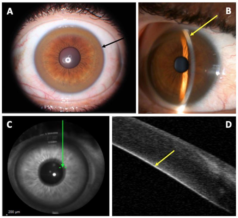
This composite educational graphic illustrates clinical and diagnostic findings of the Kayser–Fleischer ring (KFR), a pathognomonic sign of Wilson disease. Panel A is a clinical photograph from a slit-lamp examination showing a diffuse, circumferential golden-brown pigment ring (black arrow) in the corneal limbic area. Panel B utilizes a fine-slit lamp beam to demonstrate the precise localization of copper deposits at the posterior aspect of the cornea (yellow arrow). Panel C provides a localized infrared corneal B-scan view, while Panel D presents an Anterior Segment Optical Coherence Tomography (AS-OCT) cross-sectional image. The AS-OCT scan reveals a distinct, linear hyper-reflective layer at the level of the Descemet’s membrane (yellow arrow), corresponding exactly to the clinical site of copper deposition. This visual comparison highlights how diagnostic imaging can verify clinical observations of ocular copper accumulation in patients with copper metabolism disorders.
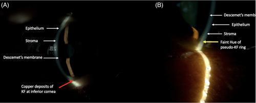
A side-by-side clinical slit-lamp photograph comparing two types of corneal rings. Image (A) shows a Kayser-Fleischer (KF) ring in a patient with Wilson’s Disease, characterized by distinct copper-colored deposits located at the level of the Descemet's membrane in the inferior cornea. Image (B) demonstrates a Pseudo-Kayser-Fleischer (PKF) ring associated with hyperbilirubinemia, presenting as a faint yellow hue within the posterior stroma rather than the Descemet's membrane. Both images include anatomical labels for the epithelium, stroma, and Descemet's membrane to provide depth context. The comparison highlights the different anatomical localization and coloration of copper versus bilirubin deposits, which is a critical diagnostic distinction in patients presenting with liver disease and ocular findings.
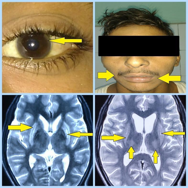
This composite figure illustrates the clinical and radiological hallmarks of Wilson's disease. The top-left clinical photograph shows a close-up of an eye with a yellow arrow pointing to a Kayser-Fleischer ring, characterized by a brownish-gold copper deposition at the corneal limbus. The top-right photograph depicts a patient with a 'mask-like facies' (hypomimia), noted by arrows pointing toward the mouth and facial muscles. The bottom panels consist of two axial MRI brain images (T2-weighted/FLAIR sequences). Yellow arrows indicate bilateral, symmetrical hyperintensities within the basal ganglia, specifically involving the lentiform nuclei (globus pallidus and putamen) and the thalami. These findings are highly characteristic of copper accumulation in the central nervous system and are critical for the diagnostic evaluation of hepatolenticular degeneration. The visual evidence correlates with neurological manifestations such as tremors and spasticity in pediatric or young adult patients.
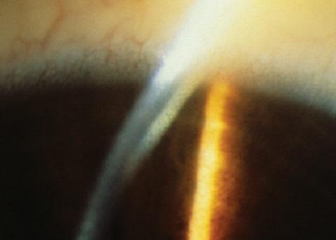
This clinical photograph shows a close-up slit-lamp examination of the eye, specifically focusing on the anterior segment and cornea. A prominent Kayser-Fleischer ring is visible as a golden-brown or greenish-yellow circumferential band located at the corneal periphery. This pigment deposition occurs within Descemet's membrane near the limbus. The slit-lamp beam provides cross-sectional illumination, highlighting the depth of the copper deposition and the relative transparency of the central corneal stroma. Subtle limbal vascularization is observed in the peripheral sclera. This finding is a pathognomonic sign of Wilson's disease (hepatolenticular degeneration), reflecting systemic copper overload and abnormal metabolism of ceruloplasmin. The image serves as a high-yield diagnostic reference for ophthalmology and internal medicine, illustrating the classic ocular manifestation of this metabolic disorder.
"Wilson disease"[MeSH Terms] AND treatment
Wilson Disease (Hepatolenticular Degeneration)
Genetics & Pathophysiology
- In the TGN: Incorporates copper into apoceruloplasmin to form ceruloplasmin, which is secreted into blood.
- In lysosomes: Transports non-ceruloplasmin-bound copper into bile canaliculi for excretion (the major route of copper elimination).
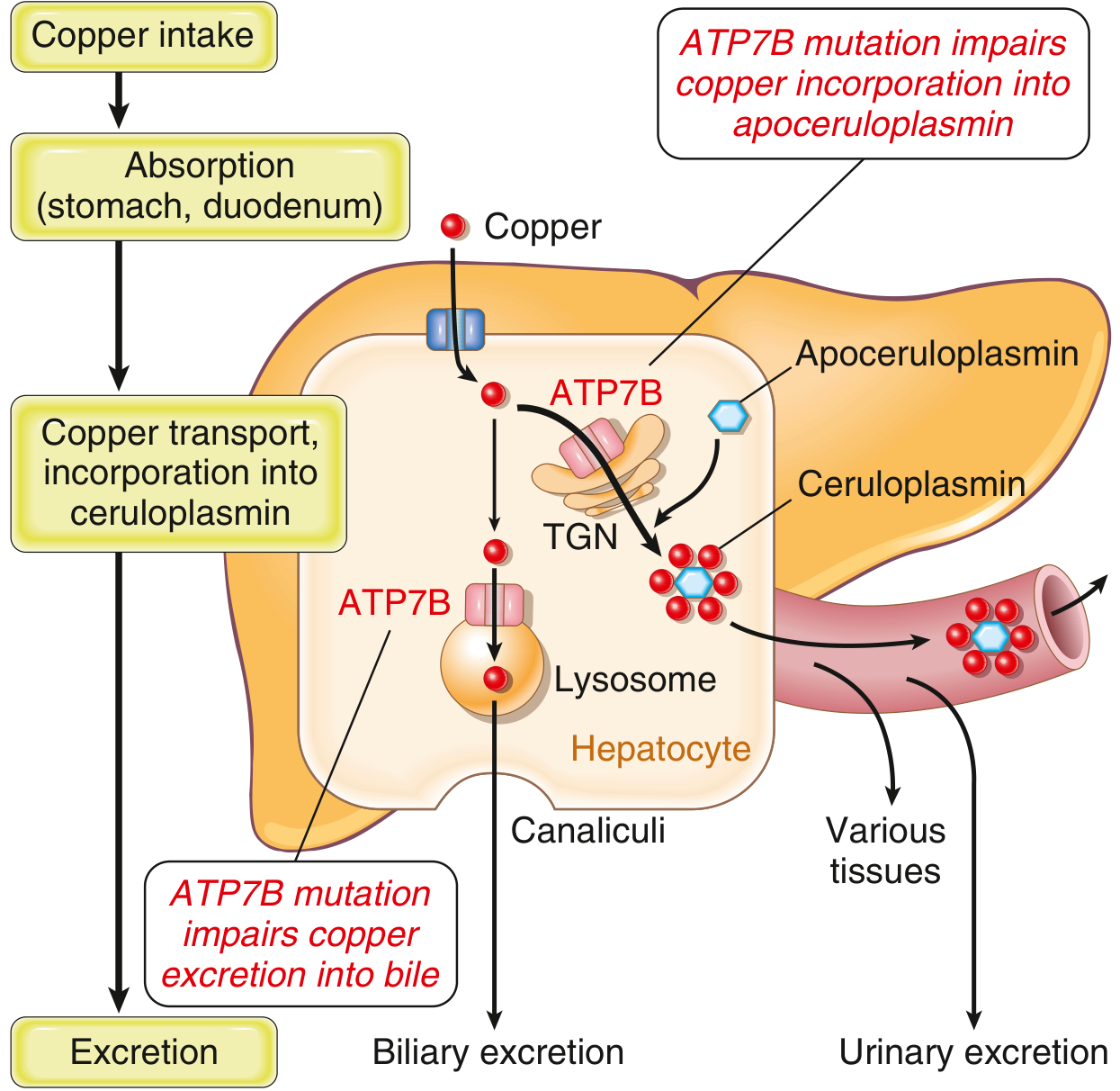
Clinical Features
1. Hepatic Disease (~1/3 of cases)
- Ranges from mild steatosis and focal necrosis to chronic hepatitis, cirrhosis, or acute fulminant liver failure (ALF)
- In ALF, key clues: ALP:bilirubin ratio <4 and AST:ALT ratio >2.2 (distinguishes Wilson ALF from other causes)
- Ascites is an early feature in the acute presentation, typically in the second decade
2. Neurological Disease (~1/3 of cases)
- Predominantly extrapyramidal: chorea, tremor (sometimes "wing-beating"), dystonia
- Also: dysphagia, dysarthria, ataxia, gait disturbance, a fixed sardonic smile
- MRI shows abnormal T2/FLAIR signal in the putamen, thalamus, midbrain, pons, cerebellum; atrophy is common
3. Neuropsychiatric Disease (~1/3 of cases)
- Personality and mood changes are the most common early features
- Depression in ~30%, bipolar spectrum ~20%, suicidal ideation in 5-15%
- Patients can be unusually sensitive to neuroleptics
- Despite long-term treatment, ~70% of patients develop psychiatric symptoms
Characteristic Sign: Kayser-Fleischer Rings
- Present in ~98% of patients with neurological disease
- Present in ~80% of all Wilson disease cases
- Best visualized with slit-lamp examination
- Must be distinguished from pseudo-KF rings (due to hyperbilirubinemia, which deposits in the stroma rather than Descemet membrane)
Morphology (Pathology)
| Organ | Findings |
|---|---|
| Liver | Steatosis, focal necrosis, chronic hepatitis, steatohepatitis (ballooning + Mallory hyaline), cirrhosis; copper stain (rhodanine/orcein) shows red-brown lysosomal deposits |
| Brain | Toxic injury to basal ganglia (putamen/lenticular nuclei), thalamus; T2 hyperintensities on MRI |
| Cornea | KF rings at Descemet membrane |
| Kidney | Renal tubular dysfunction (Fanconi syndrome) |
| Bones/Joints | Osteoporosis, arthropathy |
Diagnosis
| Test | Finding | Notes |
|---|---|---|
| Serum ceruloplasmin | Low | Hallmark, but doesn't cause pathology |
| 24-hr urinary copper | Elevated | Most specific test |
| Hepatic copper content | >250 µg/g dry weight | Most sensitive test (~80%) |
| Slit-lamp exam | KF rings | Essential in neurological cases |
| Liver biopsy | Steatosis, hepatitis, copper deposits | Sometimes necessary |
| MRI brain | T2 hyperintensity in basal ganglia | In neurological disease |
| Serum copper | Low, normal, or elevated | Not diagnostically useful |
Treatment
| Therapy | Mechanism | Use |
|---|---|---|
| D-penicillamine | Copper chelation | First-line; 20-50% of neurological patients worsen acutely |
| Trientine | Copper chelation | Lower risk of neurological worsening; preferred if penicillamine not tolerated |
| Tetrathiomolybdate | Copper chelation | May have the lowest neurological worsening risk |
| Zinc salts | Blocks intestinal copper absorption (induces metallothionein in enterocytes) | Presymptomatic patients, maintenance therapy, pregnancy |
| Liver transplantation | Curative (restores ATP7B function) | Advanced cirrhosis, fulminant hepatic failure |
Key Points for Exam
- Gene: ATP7B (chromosome 13) - autosomal recessive
- Inheritance: 1 in 30,000-40,000 live births
- KF rings are pathognomonic when combined with neurological disease (absent in ~20% of all cases)
- Low ceruloplasmin + elevated 24-hr urine copper + elevated hepatic copper = diagnosis
- ALF clue: ALP/bilirubin <4 and AST/ALT >2.2
- Penicillamine can cause paradoxical neurological worsening - trientine is preferred in such cases
- Liver transplantation is curative because it replaces the defective ATP7B-expressing hepatocytes