Peptic acid disease

Reading File
Finding Sources
Finding Sources
Searching PubMed

"peptic ulcer disease" AND management

Reading File
Reading File
Reading File
Reading File
Reading File
Reading File
Searching Images

peptic ulcer disease gastric duodenal endoscopy histology
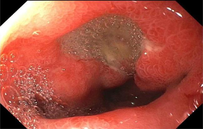
This endoscopic clinical photograph displays a 7 mm cratered ulcer located in the duodenal bulb, a common manifestation of peptic ulcer disease. The ulcer is characterized by a central, depressed crater filled with a brownish-gray fibrinous base and mucus. The surrounding duodenal mucosa is intensely erythematous and edematous, indicating active inflammation (duodenitis). Visible mucosal changes include a granular texture and prominent mucosal folds. Bubbles from gastric secretions are present in the foreground, indicating the fluid-rich environment of the upper gastrointestinal tract. This visual is representative of H. pylori-associated ulceration or NSAID-induced mucosal injury, providing a classic example for gastroenterology and internal medicine education regarding diagnostic upper endoscopy (esophagogastroduodenoscopy) and the morphological evaluation of duodenal lesions.
This endoscopic clinical photograph displays a 7 mm cratered ulcer located in the duodenal bulb, a common manifestation of peptic ulcer disease. The ulcer is characterized by a central, depressed crater filled with a brownish-gray fibrinous base and mucus. The surrounding duodenal mucosa is intensely erythematous and edematous, indicating active inflammation (duodenitis). Visible mucosal changes include a granular texture and prominent mucosal folds. Bubbles from gastric secretions are present in the foreground, indicating the fluid-rich environment of the upper gastrointestinal tract. This visual is representative of H. pylori-associated ulceration or NSAID-induced mucosal injury, providing a classic example for gastroenterology and internal medicine education regarding diagnostic upper endoscopy (esophagogastroduodenoscopy) and the morphological evaluation of duodenal lesions.

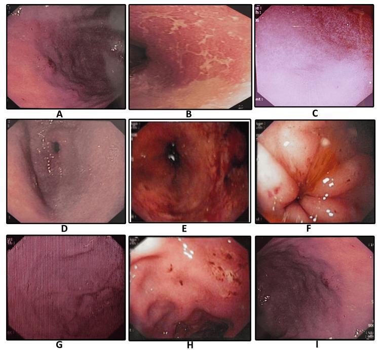
A composite of nine endoscopic images (labeled A-I) illustrating the diverse clinical manifestations of Helicobacter pylori-associated gastritis and peptic ulcer disease within the gastric and duodenal mucosa. The images provide a comparative overview of mucosal pathology: (A) demonstrates mucosal erosion with punctate dark lesions; (B) shows adherent sticky mucus appearing as whitish-yellow patches; (C) illustrates diffuse mucosal erythema; (D) depicts antral gastritis with generalized hyperemia; (E) highlights spotty redness across the mucosal surface; (F) reveals a duodenal ulcer with surrounding inflammatory changes; (G) displays classic nodular gastritis characterized by a uniform, 'cobblestone' or granular mucosal texture; (H) shows a gastric ulcer with evidence of active erosion; and (I) exhibits hypertrophic gastropathy with significantly enlarged rugal folds. This educational compilation is essential for gastroenterology training to differentiate specific morphological patterns such as nodularity, erosions, and fold thickening during diagnostic upper gastrointestinal endoscopy.
A composite of nine endoscopic images (labeled A-I) illustrating the diverse clinical manifestations of Helicobacter pylori-associated gastritis and peptic ulcer disease within the gastric and duodenal mucosa. The images provide a comparative overview of mucosal pathology: (A) demonstrates mucosal erosion with punctate dark lesions; (B) shows adherent sticky mucus appearing as whitish-yellow patches; (C) illustrates diffuse mucosal erythema; (D) depicts antral gastritis with generalized hyperemia; (E) highlights spotty redness across the mucosal surface; (F) reveals a duodenal ulcer with surrounding inflammatory changes; (G) displays classic nodular gastritis characterized by a uniform, 'cobblestone' or granular mucosal texture; (H) shows a gastric ulcer with evidence of active erosion; and (I) exhibits hypertrophic gastropathy with significantly enlarged rugal folds. This educational compilation is essential for gastroenterology training to differentiate specific morphological patterns such as nodularity, erosions, and fold thickening during diagnostic upper gastrointestinal endoscopy.

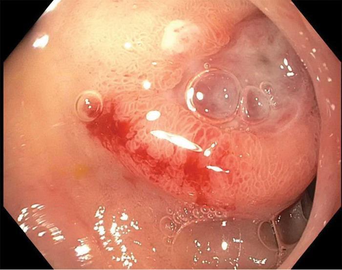
This endoscopic clinical photograph from an upper gastrointestinal endoscopy (EGD) displays the lumen of the distal duodenal bulb. Centered in the frame is a single, approximately 1-cm ulcerated lesion. The ulcer is characterized by a clean base and a slightly protuberant, elevated morphology. Distinctive features include irregular, 'heaped-up' mucosal edges surrounding the ulcer crater, and a cluster of translucent air bubbles typical of the insufflation process. Clinically significant signs of hemorrhage are visible, specifically active oozing of bright red blood (Forrest classification 1B) from the ulcer's margin. The surrounding mucosa shows slight erythema and texture changes indicative of gastric heterotopia or secondary inflammation. This image is an educational example of a peptic ulcer with features suspicious for an underlying mass lesion, requiring differential diagnosis between benign peptic ulcer disease and malignant or stromal tumors such as a Gastrointestinal Stromal Tumor (GIST).
This endoscopic clinical photograph from an upper gastrointestinal endoscopy (EGD) displays the lumen of the distal duodenal bulb. Centered in the frame is a single, approximately 1-cm ulcerated lesion. The ulcer is characterized by a clean base and a slightly protuberant, elevated morphology. Distinctive features include irregular, 'heaped-up' mucosal edges surrounding the ulcer crater, and a cluster of translucent air bubbles typical of the insufflation process. Clinically significant signs of hemorrhage are visible, specifically active oozing of bright red blood (Forrest classification 1B) from the ulcer's margin. The surrounding mucosa shows slight erythema and texture changes indicative of gastric heterotopia or secondary inflammation. This image is an educational example of a peptic ulcer with features suspicious for an underlying mass lesion, requiring differential diagnosis between benign peptic ulcer disease and malignant or stromal tumors such as a Gastrointestinal Stromal Tumor (GIST).

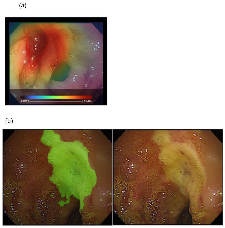
This composite image illustrates deep learning (DL) applications for identifying and segmenting gastrointestinal pathology during endoscopy, specifically for peptic ulcer disease (PUD). Panel (a) presents an endoscopic view of an active bleeder with a Grad-CAM heatmap overlay. The heatmap uses a pseudocolor scale where warmer tones (red, orange) signify areas of high clinical significance or high probability of active bleeding, concentrated around the lesion, while cooler tones (blue, green) represent lower-risk areas. Panel (b) shows a side-by-side comparison of a gastric or duodenal ulcer. The right frame displays the original endoscopic image showing a yellowish-white ulcer base with irregular borders and surrounding erythema. The left frame demonstrates computer-aided semantic segmentation, where the ulcer bed is highlighted with a green overlay, precisely demarcating the lesion's boundaries from healthy mucosa. These visualizations serve as educational examples of how artificial intelligence can assist in the endoscopic classification of bleeding risks, such as the Forrest classification, by providing objective lesion identification and risk assessment.

This composite image illustrates deep learning (DL) applications for identifying and segmenting gastrointestinal pathology during endoscopy, specifically for peptic ulcer disease (PUD). Panel (a) presents an endoscopic view of an active bleeder with a Grad-CAM heatmap overlay. The heatmap uses a pseudocolor scale where warmer tones (red, orange) signify areas of high clinical significance or high probability of active bleeding, concentrated around the lesion, while cooler tones (blue, green) represent lower-risk areas. Panel (b) shows a side-by-side comparison of a gastric or duodenal ulcer. The right frame displays the original endoscopic image showing a yellowish-white ulcer base with irregular borders and surrounding erythema. The left frame demonstrates computer-aided semantic segmentation, where the ulcer bed is highlighted with a green overlay, precisely demarcating the lesion's boundaries from healthy mucosa. These visualizations serve as educational examples of how artificial intelligence can assist in the endoscopic classification of bleeding risks, such as the Forrest classification, by providing objective lesion identification and risk assessment.

Reading File
Reading File
Loading Image
This is a shared conversation. Sign in to Orris to start your own chat.