
How to know a reflex is exaggerated or not
Reading File
A reflex is considered exaggerated (hyperreflexia) when it produces a stronger-than-normal response to a standard stimulus - typically indicating an upper motor neuron (UMN) lesion. Normal reflexes require an intact reflex arc plus intact descending inhibitory control from the brain/spinal cord.
Searching sources...
Finding Sources
Finding Sources
Finding Sources
Finding Sources
Reading File
Finding Sources
Reading File
Finding Sources
Finding Sources
Finding Sources
Reading File
Reading File
Finding Sources
Reading File
Now I have enough to give a thorough answer. Let me also fetch the reflex stick figure image to include in the response.
Loading Image
Here is a complete guide to determining whether a reflex is exaggerated.
How to Determine if a Reflex is Exaggerated
1. The Grading Scale (NIMS / Standard 0-4+ Scale)
Reflexes are graded on a 0 to 4+ scale:
| Grade | Description |
|---|---|
| 0 | Absent - no response even with reinforcement |
| 1+ | Hypoactive - diminished, barely present |
| 2+ | Normal |
| 3+ | Brisk - more lively than average, but may be normal |
| 4+ | Exaggerated / Hyperreflexic - associated with clonus; clearly abnormal |
- 3+ alone may be normal in some individuals (especially anxious patients or those with a naturally brisk baseline)
- 4+ is always abnormal
- A reflex is definitively considered exaggerated when it is 3+ with other upper motor neuron signs, or 4+ with or without clonus
2. Key Features That Tell You a Reflex is Exaggerated
A. Clonus
The most reliable sign of true hyperreflexia. Clonus is a series of rhythmic involuntary contractions (at 5-7 Hz) triggered by an abruptly applied and sustained stretch. Common sites: ankle (dorsiflex the foot suddenly and hold) and patella (push the patella sharply downward). Sustained clonus (>5 beats) is always pathological. - Adams and Victor's Principles of Neurology, 12th Ed.
B. Spread / Irradiation of Reflexes
Tapping one tendon produces contraction in a distant muscle group. For example, tapping the radius elicits not only the brachioradialis reflex but also the biceps, triceps, and finger flexors. This "radiation" strongly suggests an upper motor neuron (UMN) lesion. - Adams and Victor's Principles of Neurology, 12th Ed.
C. Hoffmann's Sign (upper limb)
With the patient's hand relaxed, flick the nail of the middle finger downward and release it. An abnormal (positive) response is thumb flexion and adduction. This is a pathological reflex equivalent to hyperreflexia in the upper limbs. - Neuroanatomy through Clinical Cases, 3rd Ed.
D. Babinski's Sign (lower limb)
Stroke the lateral sole from heel to ball, then arc medially. Normal: toes curl downward. Abnormal (Babinski positive): big toe extends upward, other toes fan out. In any adult, this is always pathological and indicates a UMN/corticospinal tract lesion. - Neuroanatomy through Clinical Cases, 3rd Ed.
E. Crossed Adductor Reflex
Tapping the patellar tendon causes not only the expected quadriceps jerk but also adduction of the opposite thigh. This is a sign of hyperreflexia.
F. Inverted Reflexes
At a specific spinal level, the expected reflex is absent but a reflex below it is exaggerated. For example, in C5/C6 myelopathy: tapping the biceps tendon fails to elicit the biceps reflex (LMN lesion at that level) but instead produces finger flexion or triceps jerk (hyperreflexia below the lesion). This "inverted reflex" is a hallmark of UMN involvement. - Adams and Victor's Principles of Neurology, 12th Ed.
3. Compare Both Sides
Always compare left vs. right for the same reflex. Asymmetry is often more meaningful than the absolute grade. A 3+ reflex on one side with 2+ on the other, combined with other signs, points to pathology on the brisk side.
4. Context Matters: UMN vs LMN
| Feature | UMN Lesion (exaggerated reflexes) | LMN Lesion (diminished reflexes) |
|---|---|---|
| Deep tendon reflexes | Increased / hyperreflexic | Decreased or absent |
| Tone | Spastic (velocity-dependent, clasp-knife) | Flaccid |
| Babinski | Present (extensor plantar) | Absent |
| Clonus | Present | Absent |
| Muscle wasting | Minimal/late | Early and prominent |
| Fasciculations | Absent | Present |
ROSEN's Emergency Medicine states: "Muscle paralysis associated with intact deep tendon reflexes indicates an UMN (spinal cord) lesion, whereas paralysis with absent reflexes indicates an LMN (nerve root or cauda equina) lesion."
5. Reinforcement Techniques (for borderline reflexes)
If a reflex seems absent or borderline, use the Jendrassik maneuver before calling it normal or exaggerated:
- Upper limb reinforcement: Ask the patient to clench teeth or squeeze their thighs together while you test an arm reflex.
- Lower limb reinforcement: Ask the patient to hook their fingers together and pull (Jendrassik maneuver) while you test the patellar or Achilles reflex.
If a reflex only appears with reinforcement, it is graded 1+ (hypoactive). If a reflex is already brisk without reinforcement and becomes more exaggerated with it, that supports hyperreflexia.
6. Reflex Stick Figure Documentation
Reflexes are commonly summarized using a stick figure, with each joint labeled with the grade:
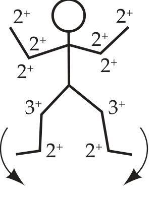
The arrows at the feet represent plantar responses (down = normal; up = Babinski positive). This patient has normal upper limb reflexes (2+) but brisk patellar reflexes (3+) - warranting further evaluation for UMN pathology. - Neuroanatomy through Clinical Cases, 3rd Ed.
7. Causes of Exaggerated Reflexes
- UMN lesions: stroke, brain tumors, MS, cervical/thoracic myelopathy, ALS (combined with LMN signs)
- Metabolic: hyperthyroidism (generalized hyperreflexia), hypocalcemia
- Anxiety/physiological: mild bilateral symmetrical briskness without other signs can be benign
- Pre-eclampsia: hyperreflexia with clonus is a warning sign of eclampsia
Bottom line: A reflex is exaggerated when it is grade 3+ in the presence of other UMN signs, or grade 4+ at any time, especially with clonus, Babinski sign, spread of reflexes, or Hoffmann's sign. Always compare sides, use reinforcement for borderline cases, and interpret the reflex in the full clinical context. - Adams and Victor's Principles of Neurology, 12th Ed.; Neuroanatomy through Clinical Cases, 3rd Ed.
This is a shared conversation. Sign in to Orris to start your own chat.