Pancreatitis severity score

Reading File
Finding Sources
Searching PubMed

"acute pancreatitis" AND "severity scoring"

Finding Sources
Searching PubMed

"acute pancreatitis" AND "severity"

Finding Sources
Finding Sources
Reading File
Reading File
Reading File
Reading File
Reading File
Reading File
Reading File
Searching Images

CT severity index acute pancreatitis Balthazar grading

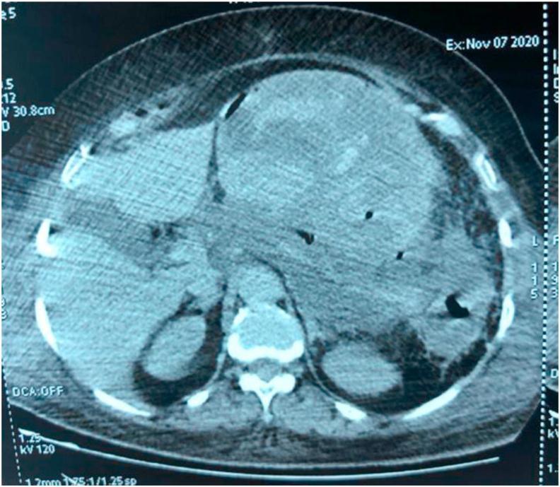
This diagnostic image is an axial contrast-enhanced CT scan of the abdomen demonstrating severe acute pancreatitis, specifically Balthazar Grade E. The pancreas appears markedly enlarged and edematous, with poor definition of its anatomical borders. The primary finding is the presence of extensive, heterogeneous peripancreatic fluid and necrotic collections with irregular outlines. These collections involve the anterior pararenal space and the lesser sac, characterized by varying densities including hypodense areas consistent with fluid and higher-density components suggestive of necrotic debris or hemorrhage. There is evidence of fat stranding and significant inflammatory changes in the surrounding retroperitoneal tissues. Small foci of gas (hypodense bubbles) within the collections are visible, which may indicate superinfection or emphysematous changes. The image serves as a clinical example of high-severity pancreatitis with significant local complications and peripancreatic necrosis, suitable for medical education regarding the Balthazar scoring system and CT severity index (CTSI).

This diagnostic image is an axial contrast-enhanced CT scan of the abdomen demonstrating severe acute pancreatitis, specifically Balthazar Grade E. The pancreas appears markedly enlarged and edematous, with poor definition of its anatomical borders. The primary finding is the presence of extensive, heterogeneous peripancreatic fluid and necrotic collections with irregular outlines. These collections involve the anterior pararenal space and the lesser sac, characterized by varying densities including hypodense areas consistent with fluid and higher-density components suggestive of necrotic debris or hemorrhage. There is evidence of fat stranding and significant inflammatory changes in the surrounding retroperitoneal tissues. Small foci of gas (hypodense bubbles) within the collections are visible, which may indicate superinfection or emphysematous changes. The image serves as a clinical example of high-severity pancreatitis with significant local complications and peripancreatic necrosis, suitable for medical education regarding the Balthazar scoring system and CT severity index (CTSI).

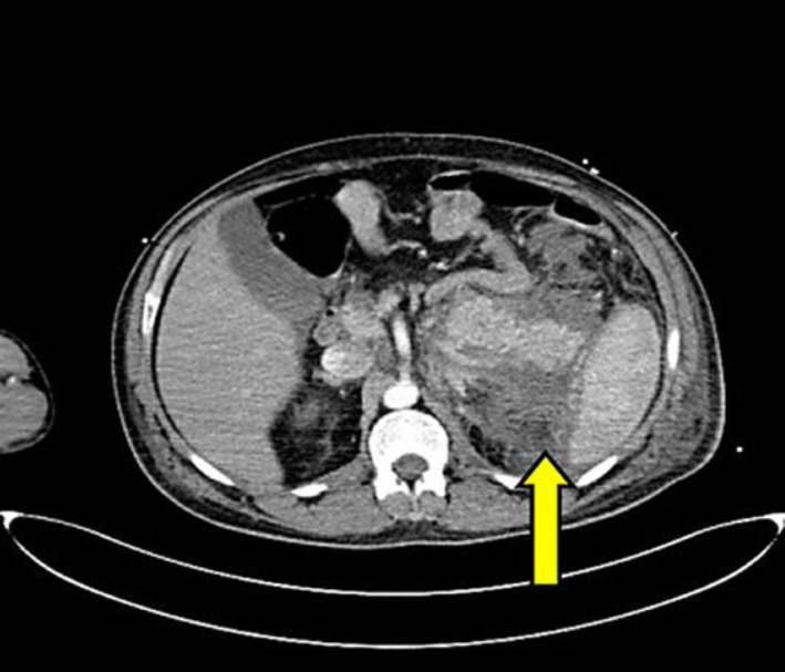
**Imaging Modality:** Contrast-enhanced computed tomography (CECT) of the abdomen.

**Anatomical Region:** Axial section at the level of the upper abdomen, showing the pancreas, liver, spleen, and kidneys.

**Observed Pathology:** The image demonstrates findings consistent with acute pancreatitis. There is significant inflammatory changes and edema involving the pancreatic parenchyma. A prominent, low-attenuation peripancreatic fluid collection is visible (indicated by the yellow arrow).

**Characteristic Visual Features:**
*   **Pancreas:** Diffuse enlargement with ill-defined borders and heterogeneous enhancement.
*   **Fluid Collection:** An acute peripancreatic fluid collection (APFC) is noted in the anterior pararenal space, extending laterally into the splenorenal space.
*   **Fat Stranding:** Increased density and "dirty" appearance of the peripancreatic fat, indicating active inflammation.
*   **Adjacent Structures:** The collection is bordered by the tail of the pancreas and the medial aspect of the spleen.

**Key Diagnostic Features:** The combination of pancreatic swelling, heterogeneous contrast enhancement, and organized peripancreatic fluid in the splenorenal space are characteristic radiologic markers for grading the severity of acute pancreatitis using the Balthazar or CT Severity Index (CTSI) criteria.

**Imaging Modality:** Contrast-enhanced computed tomography (CECT) of the abdomen. **Anatomical Region:** Axial section at the level of the upper abdomen, showing the pancreas, liver, spleen, and kidneys. **Observed Pathology:** The image demonstrates findings consistent with acute pancreatitis. There is significant inflammatory changes and edema involving the pancreatic parenchyma. A prominent, low-attenuation peripancreatic fluid collection is visible (indicated by the yellow arrow). **Characteristic Visual Features:** * **Pancreas:** Diffuse enlargement with ill-defined borders and heterogeneous enhancement. * **Fluid Collection:** An acute peripancreatic fluid collection (APFC) is noted in the anterior pararenal space, extending laterally into the splenorenal space. * **Fat Stranding:** Increased density and "dirty" appearance of the peripancreatic fat, indicating active inflammation. * **Adjacent Structures:** The collection is bordered by the tail of the pancreas and the medial aspect of the spleen. **Key Diagnostic Features:** The combination of pancreatic swelling, heterogeneous contrast enhancement, and organized peripancreatic fluid in the splenorenal space are characteristic radiologic markers for grading the severity of acute pancreatitis using the Balthazar or CT Severity Index (CTSI) criteria.

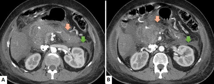
Contrast-enhanced abdominal CT images in axial planes (A and B) demonstrating findings characteristic of severe acute pancreatitis, graded as Balthazar Score E. The images show a swollen, edematous pancreas with ill-defined borders. An orange arrow indicates prominent peripancreatic fat stranding, visualized as increased attenuation and a hazy, reticular pattern in the normally hypodense adipose tissue surrounding the pancreatic head and body. A green arrow highlights a localized fluid collection associated with significant thickening and increased density of the left perinephric fascia (Gerota's fascia), indicating an inflammatory process extending into the retroperitoneum. These radiological features are consistent with necrotizing or severe interstitial edematous pancreatitis and are critical for assessing disease severity and guiding clinical management in cases of acute epigastric pain and elevated serum lipasemia.

Contrast-enhanced abdominal CT images in axial planes (A and B) demonstrating findings characteristic of severe acute pancreatitis, graded as Balthazar Score E. The images show a swollen, edematous pancreas with ill-defined borders. An orange arrow indicates prominent peripancreatic fat stranding, visualized as increased attenuation and a hazy, reticular pattern in the normally hypodense adipose tissue surrounding the pancreatic head and body. A green arrow highlights a localized fluid collection associated with significant thickening and increased density of the left perinephric fascia (Gerota's fascia), indicating an inflammatory process extending into the retroperitoneum. These radiological features are consistent with necrotizing or severe interstitial edematous pancreatitis and are critical for assessing disease severity and guiding clinical management in cases of acute epigastric pain and elevated serum lipasemia.

I now have comprehensive data from multiple authoritative textbooks. Let me compile the full answer.

Pancreatitis Severity Scoring Systems

Predicting the severity of acute pancreatitis (AP) at presentation guides triage (ward vs. step-down vs. ICU), fluid resuscitation strategy, nutritional planning, and timing of interventions. Multiple scoring systems exist; none is definitively superior to the others, and most have high false-positive rates.

1. Revised Atlanta Classification (2012)

The current standard framework - not a numeric score but a clinical classification:
SeverityCriteria
MildNo organ failure, no local/systemic complications
Moderately SevereTransient organ failure (<48 h) OR local/systemic complications
SeverePersistent organ failure (>48 h)
Organ failure is defined by a Modified Marshall score ≥2 in any of:
  • Respiratory: PaO2 <60 mmHg
  • Cardiovascular: Systolic BP <90 mmHg or HR >130 bpm
  • Renal: Serum creatinine >2.0 mg/dL
Limitation: Patients cannot be classified as severe until 48 hours after presentation - limits ED utility.
  • Rosen's Emergency Medicine, p. 1268

2. Ranson's Criteria (1974)

The oldest and most recognized score. Uses 11 parameters - 5 at admission and 6 at 48 hours.

Nongallstone Pancreatitis

At AdmissionAt 48 Hours
Age >55 yearsHematocrit fall >10%
WBC >16,000/mm³BUN rise >5 mg/dL
Blood glucose >200 mg/dLSerum calcium <8 mg/dL
Serum LDH >350 IU/LPaO2 <60 mmHg
Serum AST >250 IU/LBase deficit >4 mEq/L
Fluid sequestration >6 L

Gallstone Pancreatitis

At AdmissionAt 48 Hours
Age >70 yearsHematocrit fall >10%
WBC >18,000/mm³BUN rise >2 mg/dL
Blood glucose >220 mg/dLSerum calcium <8 mg/dL
Serum LDH >400 IU/LBase deficit >5 mEq/L
Serum AST >250 IU/LFluid sequestration >4 L
Interpretation:
  • <3 criteria - Mild, uncomplicated disease
  • ≥3 criteria - Severe pancreatitis (defines severe AP)
  • >6 criteria - ~50% mortality risk
Performance: PPV ~50%, NPV ~90% - mainly used to rule out severe disease, not rule it in.
  • Schwartz's Principles of Surgery, p. 1472; Sabiston Textbook of Surgery, p. 2000

3. BISAP Score (2008)

Bedside Index of Severity in Acute Pancreatitis - the most practical ED tool. Calculated within the first 24 hours.
VariableCriterionPoints
B - BUN>25 mg/dL1
I - Impaired mental statusGCS <151
S - SIRS≥2 of 4 SIRS criteria present1
A - Age>60 years1
P - Pleural effusionPresent on imaging1
Interpretation:
  • Score 0-1: <1% mortality
  • Score ≥3: Substantially increased in-hospital mortality, high risk for organ failure and pancreatic necrosis
  • Score 5: >20% mortality
Advantage: Simple, calculable at ED presentation, correlates well with organ failure and necrosis risk.
  • Harrison's Principles of Internal Medicine 22E, p. 681; Sabiston Textbook of Surgery

4. APACHE II Score

Originally designed for ICU use. Consists of 12 physiologic variables + age points + chronic health points. Not calculated quickly at bedside.
  • APACHE II ≥8 = severe acute pancreatitis (high risk)
  • The score can be recalculated daily to track disease progression
  • More useful for ongoing ICU monitoring than initial ED triage
  • Rosen's Emergency Medicine, p. 1268; Sabiston Textbook of Surgery

5. CT Severity Index (CTSI) - Balthazar Score

Imaging-based scoring using contrast-enhanced CT (ideally portal venous phase, 65-70 sec post-contrast).

Scoring (Total max = 10 points)

Pancreatic Inflammation (0-4 points):
CT FindingPoints
Normal pancreas0
Focal/diffuse enlargement only1
Peripancreatic fat inflammatory changes2
Single fluid collection or phlegmon3
Two or more fluid collections or gas in/adjacent to pancreas4
Pancreatic Necrosis (0-6 points):
NecrosisPoints
None0
≤30%2
30-50%4
>50%6
Outcome by CTSI:
ScoreMortalityMorbidity
0-33%8%
4-66%35%
7-1017%92%
  • CTSI ≥4 = high risk for severe disease
  • Sabiston Textbook of Surgery, p. 2001
CT example of Balthazar Grade E pancreatitis:
Balthazar Grade E - severe acute pancreatitis with extensive peripancreatic necrosis

6. Modified Glasgow (Imrie) Criteria

An 8-parameter score calculated at 48 hours. Used widely in the UK.
ParameterThreshold
Age>55 years
WBC>15,000/mm³
Blood glucose>180 mg/dL (no diabetic history)
Serum urea>45 mg/dL
PaO2<60 mmHg
Serum calcium<8 mg/dL
Serum albumin<3.2 g/dL
Serum LDH>600 IU/L
Score ≥3 at 48 hours = severe pancreatitis.

7. SIRS Criteria - Simplest Predictor

Persistent SIRS (present at admission AND at 48 hours) is as predictive of severity as the complex scores, and simpler to apply.
SIRS statusMortality
Never meets SIRS criteria~0%
Transient SIRS (resolves before 48 h)~8%
Persistent SIRS (>48 h)~25%
SIRS requires ≥2 of: Temp >38°C or <36°C; HR >90; RR >20 or PaCO2 <32; WBC >12,000 or <4,000 or >10% bands.
  • Sabiston Textbook of Surgery

8. Biochemical Markers

MarkerThresholdNotes
CRP>150 mg/LPeaks at 48-72 h; not useful at admission
BUN (admission)ElevatedRising BUN during hospitalization = higher mortality
HematocritElevated (>44%)Hemoconcentration = dehydration and necrosis risk
ProcalcitoninElevatedCorrelates with severity; not widely standardized

Summary - Severity Cutoffs for "Severe" AP

Scoring SystemCutoff for Severe
Atlanta 2012Persistent organ failure >48 h
Ranson≥3 criteria
BISAP≥3 points
APACHE II≥8
CTSI≥4
Glasgow/Imrie≥3 at 48 h
SIRSPersistent at 48 h

Clinical Triage Guidance (Harrison's 22E)

  • BISAP 0-1 + low BUN/hematocrit, good response to fluids - regular ward
  • Persistent SIRS at 24 h or comorbidities - step-down unit
  • High BISAP + high BUN/hematocrit + no response to fluids + organ failure - direct ICU admission
Harrison's Principles of Internal Medicine 22E; Rosen's Emergency Medicine; Sabiston Textbook of Surgery
This is a shared conversation. Sign in to Orris to start your own chat.