
All bursa in shoulder
Reading File
The shoulder has several bursae, the most clinically important being the subacromial (subdeltoid) bursa. Others include the subscapularis, subcoracoid, and coracobrachial bursae.
Searching sources...
Finding Sources
Finding Sources
Good results. Let me read the most relevant sections - the Tintinalli BURSAE section and the Imaging anatomy section, plus fetch the anatomical image.
Reading File
Reading File
Reading File
Excellent content. Now let me fetch the key anatomical diagram showing shoulder bursae and the THIEME atlas image.
Loading Image
Loading Image
Loading Image
I now have comprehensive information from multiple authoritative textbooks. Here is the complete answer:
Bursae of the Shoulder
There are 8 bursae in the shoulder complex. They facilitate frictionless motion between the shoulder's components. Only a few are clinically significant.
Anatomical Diagram (THIEME Atlas of Anatomy)
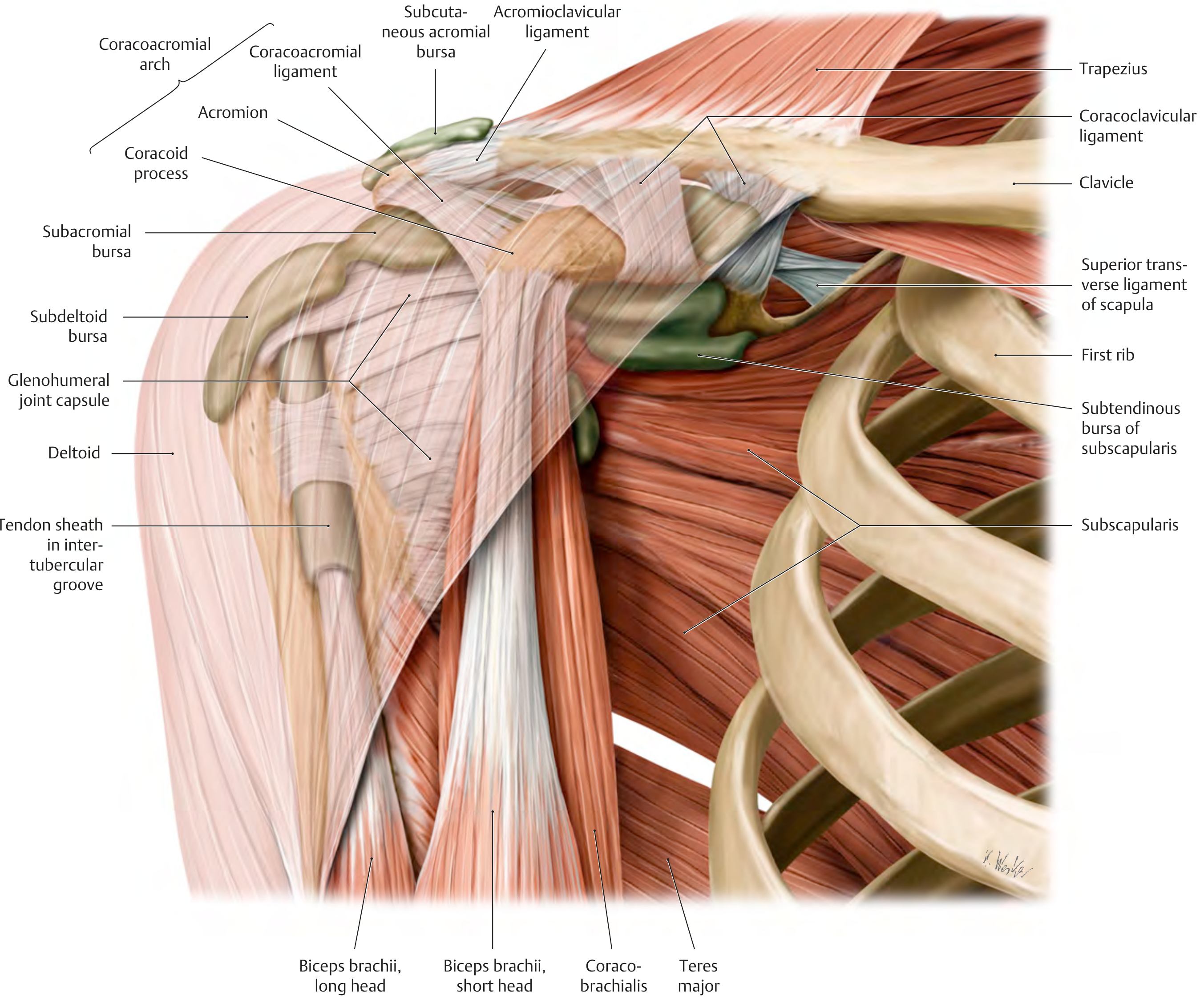
The diagram clearly labels the subacromial bursa, subdeltoid bursa, subcutaneous acromial bursa, and subtendinous bursa of the subscapularis.
The Major Bursae
1. Subacromial Bursa (Most Clinically Important)
- Located between the coracoacromial arch (above) and the rotator cuff / supraspinatus tendon (below)
- Roof: adherent to undersurface of deltoid, acromion, and coracoacromial ligament
- Floor: adherent to the rotator cuff (especially supraspinatus)
- Lubricated by synovial fluid; surrounded by a layer of peribursal fat
- Its role is to reduce friction on the supraspinatus tendon during abduction/elevation
- Does not normally communicate with the glenohumeral joint - but communication develops in full-thickness rotator cuff tears
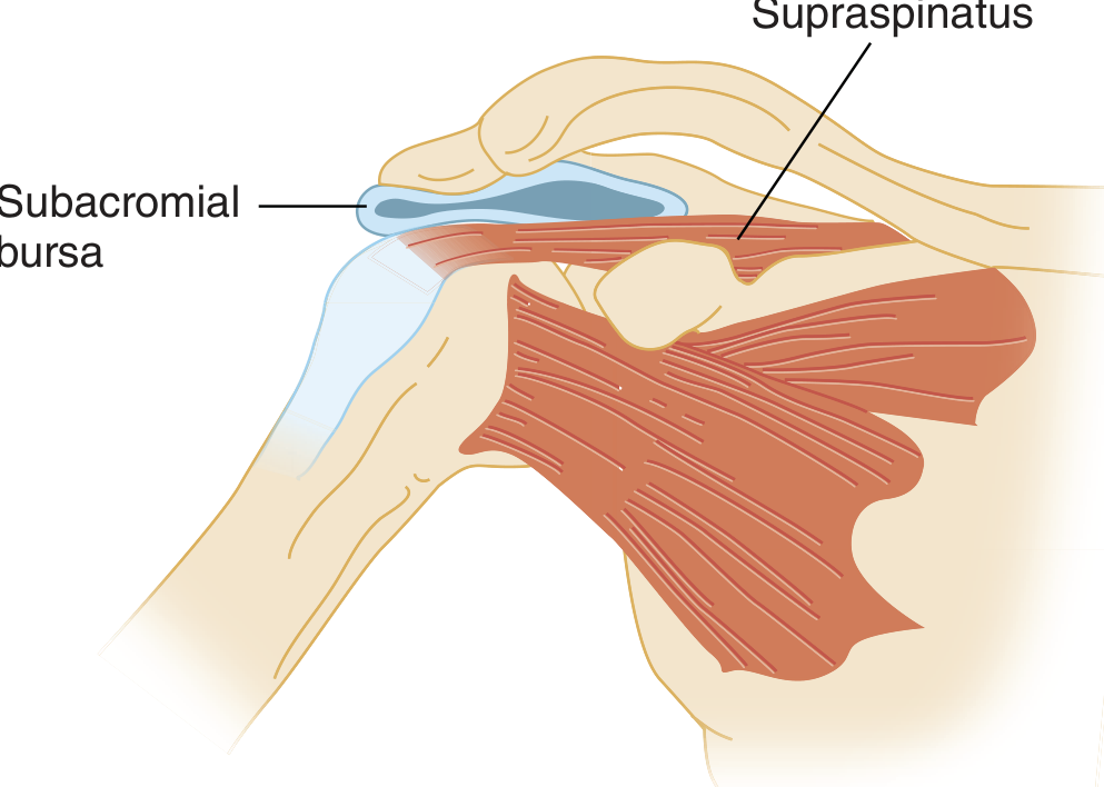
2. Subdeltoid Bursa
- Located between the deltoid muscle and the rotator cuff
- Consistently communicates with the subacromial bursa - together they are often referred to as the subacromial-subdeltoid (SASD) bursa, one large continuous structure
- Functions together with the subacromial bursa to allow frictionless gliding of the humeral head and rotator cuff tendons beneath the coracoacromial arch
3. Subcutaneous Acromial Bursa
- Located superficially over the acromion, just beneath the skin
- Smaller and less clinically relevant
4. Subcoracoid Bursa (Coracoid Bursa)
- Located between the subscapularis muscle and the coracoid process
- Does not normally communicate with the glenohumeral joint
- Separated from the subscapular recess by an identifiable fibrous septum
- May communicate with the subacromial-subdeltoid bursa in about 10% of patients
- Not normally visible on MRI unless distended by fluid
5. Subtendinous Bursa of Subscapularis (Subscapular / Superior Subscapular Recess)
- Located between the anterior surface of the scapula and the subscapularis muscle
- Also called the subscapular bursa or superior subscapular recess
- Frequently communicates with the glenohumeral joint (through the subscapularis recess, which is a normal variant)
6-8. Smaller Bursae (Minor, Not Routinely Seen on MRI)
These do not communicate with the glenohumeral joint and are rarely clinically significant:
- Infraspinatus bursa - near the infraspinatus tendon insertion
- Teres major bursa - near the teres major tendon
- Pectoralis major bursa - near the pectoralis major tendon
Summary Table
| Bursa | Location | Communicates with Joint | Clinical Significance |
|---|---|---|---|
| Subacromial | Between coracoacromial arch and rotator cuff | No (unless rotator cuff tear) | High - most common site of bursitis |
| Subdeltoid | Between deltoid and rotator cuff | No (communicates with subacromial) | High - part of SASD complex |
| Subcutaneous acromial | Over acromion, subcutaneous | No | Low |
| Subcoracoid | Between subscapularis and coracoid | No (10% communicates with SASD) | Moderate |
| Subscapular (subtendinous of subscapularis) | Between scapula and subscapularis | Yes (normal variant) | Moderate |
| Infraspinatus | Near infraspinatus tendon | No | Low |
| Teres major | Near teres major tendon | No | Low |
| Pectoralis major | Near pectoralis major tendon | No | Low |
Key Clinical Points
- The subacromial bursa is the target of steroid injections for subacromial impingement syndrome and bursitis
- In rotator cuff tears (especially full-thickness), fluid communicates between the glenohumeral joint and the subacromial-subdeltoid bursa - this is detectable on MRI/ultrasound
- Subacromial impingement syndrome encompasses subacromial bursitis, rotator cuff tendinitis, supraspinatus tendinitis, and painful arc syndrome
Sources: Tintinalli's Emergency Medicine, 9e | Imaging Anatomy: Bones, Joints, Vessels and Nerves | THIEME Atlas of Anatomy: General Anatomy & Musculoskeletal System
Mri ke andr mobile phone kyu nahi le ja skte hai
MRI room mein mobile phone isliye nahi le ja sakte kyunki MRI machine ek bahut powerful magnet hoti hai jo hamesha ON rehti hai.
3 Main Reasons:
1. 🧲 Powerful Magnetic Field (Projectile Effect)
- MRI machine 1.5 Tesla ya 3 Tesla ka magnetic field generate karti hai — ye Earth ke magnetic field se 30,000 - 60,000 guna zyada strong hota hai
- Mobile phone mein iron/steel/metal components hote hain (camera lens, battery casing, screws)
- Ye phone ko ek missile ki tarah machine ki taraf khich leta hai — isse patient ko serious injury ho sakti hai
- Isse "projectile effect" kehte hain
2. 📱 Electronic Interference
- MRI machine radio frequency (RF) waves use karti hai images banane ke liye
- Mobile phone ke electronic circuits in RF waves ko disturb kar dete hain
- Isse MRI image mein artifacts (distortions/errors) aa jaate hain — diagnosis galat ho sakti hai
- Phone khud bhi damage ho sakta hai permanently
3. 🔥 Heating Effect
- RF energy se phone ke metal parts aur circuits garam ho jaate hain
- Isse patient ko burns ho sakte hain agar phone paas mein ho
- Battery overheat ya explode bhi kar sakti hai
Kitna Powerful Hota Hai Magnetic Field?
| Object | Behavior |
|---|---|
| Mobile phone | Strongly attracted, can become projectile |
| Keys / coins | Fly toward machine at high speed |
| Oxygen cylinder | Extremely dangerous missile hazard |
| Credit cards | Data wiped instantly |
| Pacemaker | Can malfunction - life threatening |
Simple Samajh ke liye:
MRI machine ko "always-on magnet" samjho. Jaise ek industrial electromagnet crane hoti hai jo cars uthati hai - waise hi ye chhoti chhoti metal cheezein apni taraf khich leti hai, chahe current band ho ya on ho - kyunki MRI ka magnet superconducting hota hai jo kabhi band nahi hota.
Isliye MRI room ke bahar ek screening zone hoti hai jahan sabhi metal aur electronic items rakhne hote hain.
Sar me kya frk pdega
Aapka matlab hai - agar mobile phone MRI room mein le jaayein toh PATIENT ke sar (head) par kya effect padega?
Sar/Head par Direct Effects:
1. 🔥 RF Heating (Radio Frequency Heating)
- MRI ke dauran RF waves continuously emit hoti hain
- Ye waves brain tissue aur skull mein heat generate karti hain
- Agar phone paas mein ho toh localized heating badh jaati hai
- Result: Headache, tissue damage, burns possible hain
2. ⚡ Induced Current
- Strong magnetic field metal components mein electric current induce karta hai
- Agar phone sar ke paas ho toh ye induced current brain tissue tak transfer ho sakta hai
- Ye seizures ya nerve stimulation cause kar sakta hai (rare but possible)
3. 💥 Projectile Injury (Sabse Dangerous)
- Agar phone haath mein hai aur magnetic field khichti hai -
- Phone bullet ki speed se machine ki taraf jaata hai
- Beech mein jo bhi aaye - sar, face, aankhein - serious fracture, laceration, internal bleeding ho sakti hai
- Ye sabse bada danger hai
4. 📡 Image Artifact
- Sar ki MRI le rahe hain toh phone ki wajah se brain images distort ho jaayengi
- Tumor, bleed, ya koi bhi pathology miss ho sakti hai
- Diagnosis completely wrong ho sakti hai
Real Incidents jo ho chuke hain:
- 2001, New York: Ek bachche ki MRI ke dauran oxygen cylinder room mein ghus gayi - wo missile ban gayi, bachche ki death ho gayi
- Multiple cases mein phones, keys, scissors se patient ko serious injuries hui hain
Short Answer:
| Risk | Effect on Head/Brain |
|---|---|
| Projectile impact | Skull fracture, brain injury |
| RF heating | Burns, headache, tissue damage |
| Induced current | Nerve stimulation, seizure |
| Image distortion | Wrong diagnosis of brain condition |
Isliye MRI room mein phone le jaana - sirf inconvenient nahi - genuinely life-threatening hai. 🚨
SAR value
SAR ka full form hai Specific Absorption Rate.
SAR Kya Hota Hai?
SAR = Wo rate jis par human body electromagnetic radiation (RF energy) ko absorb karti hai
Unit:
Watts per Kilogram (W/kg)
SAR ke 2 Contexts Hain:
📱 1. Mobile Phone SAR
- Har mobile phone RF radiation emit karta hai
- SAR batata hai ki 1 kg body tissue kitni RF energy absorb karta hai per second
- Ye head aur body dono ke liye measure hota hai
Safe Limits (International Standards):
| Country/Region | SAR Limit |
|---|---|
| USA (FCC) | 1.6 W/kg (per 1g tissue) |
| Europe (ICNIRP) | 2.0 W/kg (per 10g tissue) |
| India (DoT) | 1.6 W/kg (FCC standard follow karta hai) |
✅ Jitna kam SAR, utna better - less radiation absorbed by body
Phone ka SAR kahan dekhein:
- Phone Settings > About Phone > SAR value
- Phone ke box par likha hota hai
- Ya
*#07#dial karo - India mein SAR value show hoti hai
🏥 2. MRI SAR (Sabse Important in Medical Context)
- MRI ke dauran machine RF pulses use karti hai
- Ye RF energy patient ke body mein heat generate karti hai
- SAR monitor karna mandatory hai MRI mein - patient safety ke liye
MRI SAR Limits (IEC 60601-2-33):
| Body Region | SAR Limit |
|---|---|
| Whole body | 2 W/kg |
| Head | 3.2 W/kg |
| Trunk | 10 W/kg |
| Extremities | 20 W/kg |
MRI mein SAR badhane ke factors:
- Higher field strength (3T > 1.5T - zyada SAR)
- More RF pulses (fast sequences)
- Patient ka weight (bada patient = zyada SAR)
- Implants ya metal paas mein hona
MRI mein high SAR ke effects:
- Body temperature increase hona
- Burns (especially implants ke paas)
- Tissue damage
Simple Comparison:
| Mobile SAR | MRI SAR | |
|---|---|---|
| Source | Phone antenna | MRI RF coil |
| Typical value | 0.5 - 1.6 W/kg | 1 - 4 W/kg |
| Duration | Continuous (calls) | Minutes (scan) |
| Risk | Long-term debate | Acute heating |
| Regulated by | FCC/ICNIRP/DoT | IEC/FDA |
Key Point:
MRI mein agar phone andar ho - toh phone ka khud ka SAR + MRI ka SAR dono combine ho jaate hain - double radiation absorption hoti hai head mein - isliye aur bhi dangerous hai! ☢️
This is a shared conversation. Sign in to Orris to start your own chat.