
Tear film instability dynamic clinical features diagnosis management and recent updates detail notes for MS ophthalmology examination with standard books with images
Reading File
Now let me search the medical library comprehensively for tear film instability content.
Finding Sources
Finding Sources
Finding Sources
Now let me read the main Kanski tear film section and Wills Eye Manual dry eye section in parallel:
Reading File
Reading File
Searching PubMed
"tear film instability" AND "dry eye disease"
Excellent content found. Let me now read more of the Kanski block and get the Wills Eye treatment section and fetch key images in parallel:
Reading File
Reading File
Reading File
Reading File
Now let me fetch the key tear film images and read the diagnostic section from Kanski:
Loading Image
Loading Image
Loading Image
Reading File
Excellent data collected. Let me get the DEWS treatment levels and a couple more images:
Reading File
Loading Image
Loading Image
Now I have comprehensive content. Let me compile the complete MS ophthalmology examination notes:
Tear Film Instability - Comprehensive MS Ophthalmology Examination Notes
Sources: Kanski's Clinical Ophthalmology (10th ed.), Wills Eye Manual (7th ed.), Goodman & Gilman's Pharmacological Basis of Therapeutics
1. DEFINITIONS
Dry Eye Disease (DED) is a multifactorial disease of the ocular surface and tear film, characterized by increased osmolarity of the tear film and inflammation of the ocular surface. The hallmark pathophysiologic feature is tear film instability.
| Term | Meaning |
|---|---|
| Keratoconjunctivitis sicca (KCS) | Any eye with some degree of dryness |
| Xerophthalmia | Dry eye from vitamin A deficiency |
| Xerosis | Extreme dryness and keratinization in severe conjunctival cicatrization |
| Sjögren syndrome | Autoimmune inflammatory disease of which dry eye is a feature |
"Dry eye disease is a multifactorial disease of the ocular surface and tear film accompanied by increased osmolarity of the tear film and inflammation of the ocular surface." - Kanski's Clinical Ophthalmology, 10th ed.
2. TEAR FILM PHYSIOLOGY
2.1 Three-Layer Structure
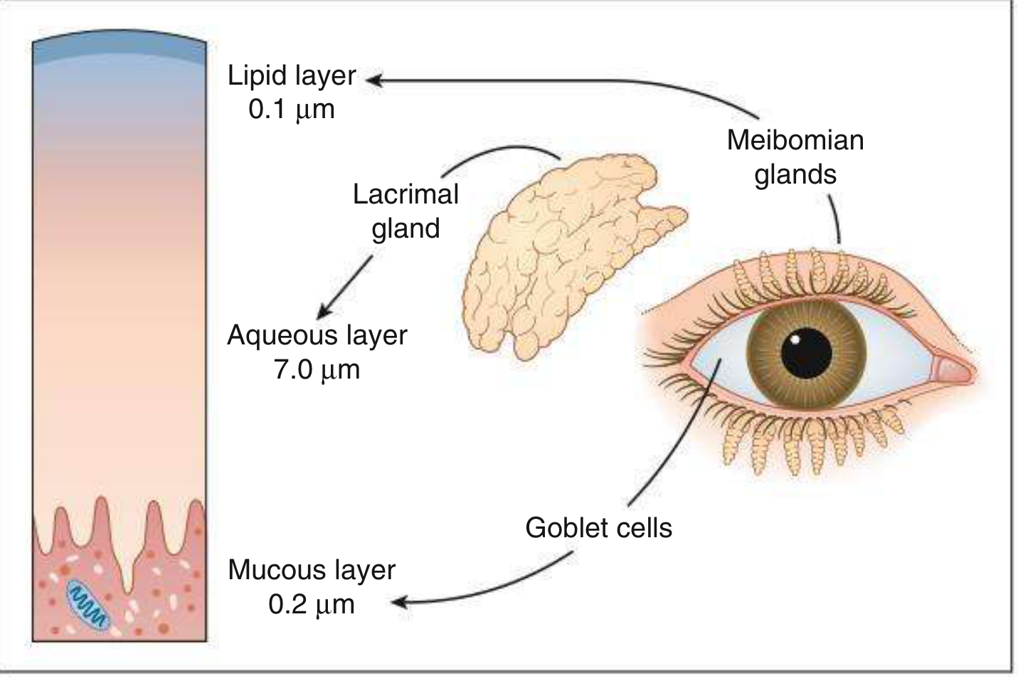
| Layer | Thickness | Source | Function |
|---|---|---|---|
| Lipid (outer) | 0.1 µm | Meibomian glands | Prevents evaporation; acts as surfactant; reduces surface tension |
| Aqueous (middle) | 7.0 µm | Lacrimal glands (main + accessory) | O2 supply; antibacterial (IgA, lysozyme, lactoferrin); washes debris |
| Mucous (inner) | 0.2 µm | Conjunctival goblet cells | Wetting by converting corneal epithelium from hydrophobic to hydrophilic; lubrication |
Key points on the lipid layer:
- Outer lipid: polar phase (phospholipids adjacent to aqueous-mucin) + non-polar phase (waxes, cholesterol esters, triglycerides)
- Polar lipids bound to lipocalins (small secretory proteins) within the aqueous layer
- Lid movement during blinking releases lipids from glands
- Deficiency → evaporative dry eye
Key points on mucous layer:
- Mucins are high-molecular-weight glycoproteins: transmembrane or secretory (gel-forming / soluble)
- Rose Bengal staining of diseased epithelium indicates absence of transmembrane and gel mucous layers
- Goblet cell loss occurs with cicatrizing conjunctivitis, vitamin A deficiency, chemical burns, medication toxicity
2.2 Tear Film Spread
Three factors required for effective resurfacing of the tear film:
- Normal blink reflex
- Contact between external ocular surface and eyelids
- Normal corneal epithelium
2.3 Hormonal Regulation
- Androgens: prime hormones for regulation of lipid production
- Oestrogens and progesterone receptors in conjunctiva and lacrimal glands
- Neural fibres adjacent to lacrimal glands and goblet cells stimulate aqueous and mucus secretion
3. PATHOPHYSIOLOGY / MECHANISM OF DISEASE
The four core inter-related mechanisms responsible for manifestations of dry eye:
Tear Instability
↓
Tear Hyperosmolarity ←→ Inflammation ←→ Ocular Surface Damage
↑_________________________________↑
- Inflammation in conjunctiva and accessory glands is present in 80% of KCS patients and may be both a cause AND consequence of dry eye (self-perpetuating cycle)
- Strong association between dry eye syndrome and reduced levels of systemic androsterone sulphate and epiandrosterone sulphate
- Tear instability → exposure of ocular surface epithelium → release of inflammatory mediators (IL-1, TNF-α) → T-cell mediated inflammation → goblet cell and epithelial damage → worsens instability
4. CLASSIFICATION
Based on the 2007 International Dry Eye Workshop (DEWS) - Updated by DEWS II (2017):
Primary Classification
DRY EYE DISEASE
├── AQUEOUS-DEFICIENT
│ ├── Sjögren syndrome dry eye (primary or secondary)
│ └── Non-Sjögren syndrome dry eye
│ ├── Lacrimal deficiency (primary or secondary)
│ ├── Lacrimal gland duct obstruction
│ └── Reflex hyposecretion
└── EVAPORATIVE
├── Intrinsic (meibomian gland dysfunction, blink-related)
└── Extrinsic (vitamin A deficiency, contact lens, medication)
4.1 Aqueous-Deficient Dry Eye
Sjögren Syndrome:
- Primary (sicca complex: dry eyes + dry mouth)
- Secondary (associated with RA, SLE, other connective tissue disease)
Non-Sjögren Syndrome:
- Primary: age-related dry eye, congenital alacrima, familial dysautonomia
- Secondary lacrimal deficiency: inflammatory/neoplastic lacrimal gland infiltration, AIDS, graft-versus-host disease, lacrimal gland/nerve ablation
- Lacrimal duct obstruction: trachoma, cicatricial pemphigoid, chemical injury, Stevens-Johnson syndrome
- Reflex hyposecretion - sensory block: contact lens wear, diabetes, refractive surgery, neurotrophic keratitis
- Reflex hyposecretion - motor block: CN VII damage, systemic drugs
4.2 Evaporative Dry Eye
Intrinsic:
- Meibomian gland dysfunction (MGD) - most common cause of evaporative dry eye
- Lid-related: low blink rate (prolonged computer use), incomplete blink, lid aperture disorders
- Drug-related (systemic retinoids reduce meibomian gland function)
Extrinsic:
- Vitamin A deficiency (goblet cell loss)
- Contact lens wear
- Ocular surface disease (allergy, preservative toxicity)
5. SYMPTOMS
Bilateral, chronic presentation. Discomfort often out of proportion to clinical signs.
Core Symptoms (DEWS II acronym: SPEED score / OSDI):
- Burning, stinging, dryness
- Foreign body / gritty sensation
- Itching
- Excess tearing (paradoxical reflex tearing)
- Mildly to moderately decreased / fluctuating vision
- Eye fatigue
Aggravating factors: smoke, wind, heat, low humidity, prolonged screen use (reduced blink rate), reading, television viewing
Pattern:
- Aqueous deficiency: symptoms worse later in the day
- Evaporative (MGD): symptoms worse in the morning
6. CLINICAL SIGNS (DYNAMIC FEATURES)
6.1 Tear Meniscus
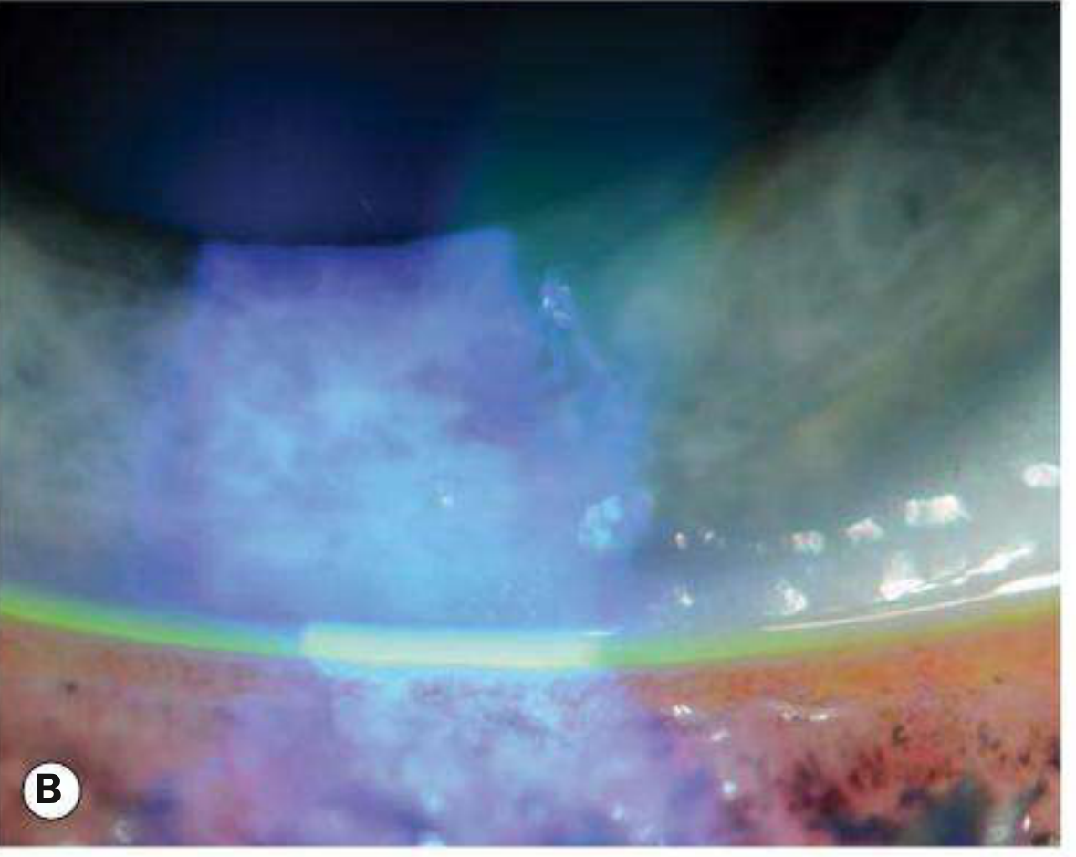
- Normal inferior tear meniscus: at least 0.5 mm in height with a convex shape
- Dry eye: scanty, concave, or absent tear meniscus
- Evaluate before instillation of any drops
6.2 Tear Film Break-up (Dynamic Assessment)
Assessed using fluorescein under cobalt blue light:
- Measure time from complete blink to first appearance of a dark spot (dry area) in the tear film
- Normal TBUT: >10 seconds
- Abnormal: <10 seconds (indicates tear film instability)
- Caution: Tear film defects must be randomly located - isolated early break-up at the same spot may indicate a focal corneal surface irregularity
6.3 Corneal and Conjunctival Staining
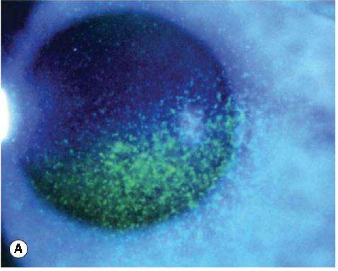
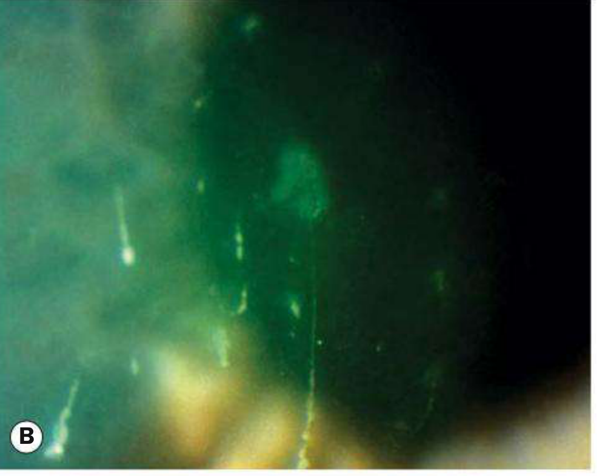
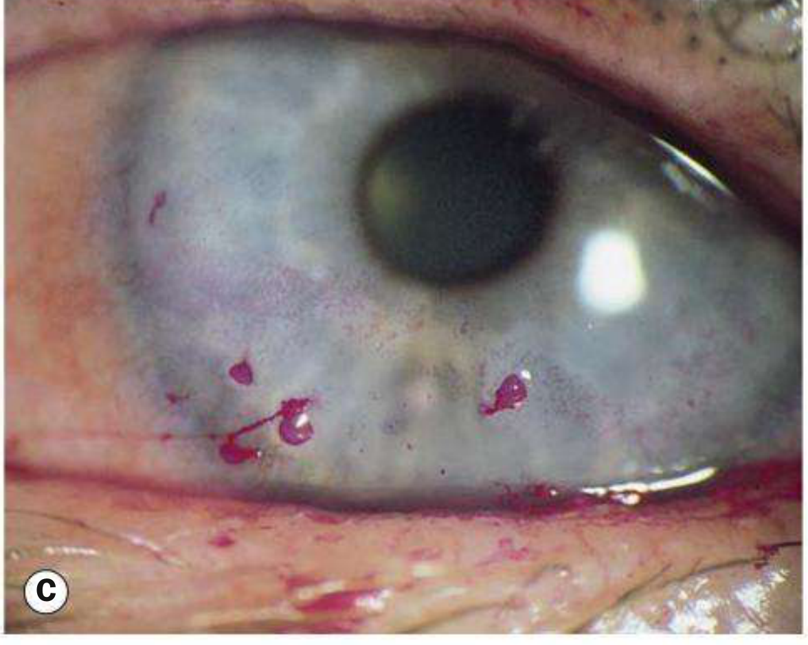
Staining agents and their patterns:
| Stain | What it detects | Pattern in dry eye |
|---|---|---|
| Fluorescein | Epithelial defects (intercellular gaps) | Punctate corneal erosions; interpalpebral |
| Rose Bengal | Devitalized/poorly mucin-coated cells | Interpalpebral conjunctiva + cornea |
| Lissamine green | Same as rose Bengal but less irritating - preferred | Same pattern |
Distribution of staining (diagnostic clue):
- Interpalpebral (cornea + conjunctiva): aqueous tear deficiency
- Superior conjunctival: superior limbic keratoconjunctivitis
- Inferior corneal + conjunctival: blepharitis or exposure keratopathy
6.4 Other Slit-Lamp Signs
- Mucous debris in tear film (Fig. 5.5A)
- Excess mucus threads or filaments on cornea (severe cases)
- Punctate corneal epithelial erosions (interpalpebral zone)
- Corneal filaments: degenerated epithelial cells + mucus core adherent to corneal surface (filamentary keratitis)
- Mucous plaque formation (severe)
6.5 Severe Corneal Complications
- Corneal melting (keratolysis)
- Corneal perforation with iris plugging
- Secondary bacterial infection
7. DIAGNOSIS
7.1 Diagnostic Tests (Order of Performance)
A. Tear Film Break-Up Time (TBUT) - Primary Dynamic Test
Technique:
- Instill fluorescein (strip or drop)
- Ask patient to blink 2-3 times, then hold gaze
- With cobalt blue illumination and wide slit beam, observe for first black spot / dry area
- Measure time in seconds (repeat 3x, take average)
Interpretation:
-
10 sec: Normal
- 5-10 sec: Borderline / mild instability
- <5 sec: Significant instability
"Less than 10 seconds indicates tear film instability." - Wills Eye Manual
B. Schirmer Tests
Technique: Whatman filter paper no. 41, 5mm × 35mm strip placed at junction of middle and lateral 1/3 of lower lid; eyes open with normal blinking for 5 minutes.
| Test | Anesthesia | Measures | Normal | Abnormal |
|---|---|---|---|---|
| Schirmer I (no anesthesia) | None | Basal + reflex secretion | ≥15 mm/5 min | <10 mm |
| Schirmer II (with anesthesia) | Proparacaine | Basal secretion only | - | ≤5 mm/5 min |
| Schirmer with nasal stimulation | With anesthesia | Reflex secretion | - | Low = reflex deficiency |
Wills Eye Manual: Anesthetized method preferred (less irritating); <10 mm may be borderline; ≤5 mm is abnormal.
C. Phenol Red Thread (PRT) Test
- Thread impregnated with pH-sensitive dye
- Placed over lower lid for 15 seconds
- Dye changes from yellow to red in tears
- Abnormal: ≤6 mm
- Comparable to Schirmer but faster; less reflex stimulation
D. Tear Osmolarity
- Emerging as accurate diagnostic measure
- Normal threshold: 308 mOsm/L (widely accepted)
- 316 mOsm/L distinguishes mild from moderate/severe dry eye
- Range of abnormality: 305-316 mOsm/L depending on degree of tear film instability
- Correlates with treatment response long-term
E. Vital Dye Staining (Rose Bengal / Lissamine Green / Fluorescein)
- van Bijsterveld score for rose Bengal staining (0-9 scale)
- Score >3.5: significant staining
F. Tear Constituent Measurement
- MMP-9 (matrix metalloproteinase-9): elevated in dry eye - tested with InflammaDry point-of-care device (positive if ≥40 ng/mL)
- Lactoferrin: decreased in aqueous-deficient dry eye
- Provides objective biomarker evidence
G. Other Tests (Rarely Clinical)
- Fluorescein clearance test / Tear function index: assesses delayed clearance in all dry eye states
- Tear meniscometry: quantifies volume of lower lid meniscus
- Impression cytology: determines goblet cell numbers
- Meibography: infrared imaging of meibomian glands - shows gland atrophy in MGD
7.2 DEWS II Diagnostic Algorithm (2017 Update)
STEP 1: Triaging questionnaire (OSDI/SPEED) + risk factors
↓
STEP 2: Homeostasis markers:
- TBUT
- Ocular surface staining (fluorescein/lissamine green)
- Symptom questionnaire
↓
STEP 3 (if positive): Etiologic subclassification
- Aqueous deficient? → Schirmer test
- Evaporative? → Meibomian gland assessment, LIPCOF, lipid layer thickness
↓
STEP 4: Contributing factors assessment
(lid margin, meibomian glands, conjunctival chalasis, nasolacrimal obstruction)
8. SEVERITY GRADING (DEWS)
| Grade | Discomfort | Visual symptoms | Conjunctival injection | Staining | TBUT (sec) | Schirmer (mm) |
|---|---|---|---|---|---|---|
| 1 | Mild, episodic | None or episodic | None to mild | None | ≥10 | ≥10 |
| 2 | Moderate, episodic or chronic | Episodic | None to mild | Variable | <10 | ≥10 |
| 3 | Severe, frequent | Constant, activity-limiting | Moderate-severe | Central corneal/conjunctival | <5 | <5 |
| 4 | Severe, disabling | Constant, disabling | Marked | Severe, filaments | Immediate | <2 |
9. MANAGEMENT
9.1 DEWS Treatment Levels (Step-Up Approach)
LEVEL 1 (Mild Dry Eye)
- Education and environmental/dietary modifications:
- Realistic expectations; compliance emphasis
- Blink exercises during screen use
- Screen below eye level (minimizes palpebral aperture)
- Increase humidity; avoid fans/drafts
- Omega-3 fatty acid supplementation (flaxseed oil, fish oil)
- Artificial tear substitutes (preserved, QID):
- Cellulose derivatives (hypromellose, methylcellulose): mild cases
- Carbomer gels: longer-lasting
- PVA (polyvinyl alcohol): mucin deficiency
- Sodium hyaluronate, povidone, glycerine, propylene glycol, polysorbate
- Diquafosol: topical secretagogue (newer agent)
- Eyelid therapy: warm compresses + lid hygiene for blepharitis/MGD
- Contact lens modification / discontinuation if symptomatic
LEVEL 2 (Moderate Dry Eye)
- Non-preserved tear substitutes (frequency >QID)
- Anti-inflammatory therapy (key for this level):
- Cyclosporine 0.05% or 0.09% b.i.d. (e.g., Restasis, Cequa): effective for chronic dry eye with decreased tears secondary to ocular inflammation; takes 1-3 months for significant improvement; burns on instillation initially; consider concurrent mild topical steroid for first month
- Lifitegrast 5% b.i.d. (Xiidra): LFA-1 antagonist; symptomatic improvement may occur within 2 weeks; causes burning, transient blurred vision, metallic taste
- Topical corticosteroids (short course): loteprednol 0.5%, fluorometholone 0.1% - used as bridge therapy
- Oral omega-3 fatty acids
- Tetracyclines (doxycycline 100 mg OD): for meibomianitis, rosacea (anti-inflammatory + antimicrobial)
- Punctal plugs (temporary collagen or permanent silicone):
- Lower puncta first; add upper puncta if needed
- Contraindicated in active lid/conjunctival inflammation
- Secretagogues: pilocarpine, cevimeline, rebamipide
- Moisture chamber spectacles / sidearm shields
LEVEL 3 (Severe Dry Eye)
- Autologous serum eye drops (20-100%): contains growth factors, vitamins, anti-inflammatory mediators; lacks preservatives; refrigerated storage
- Umbilical cord serum drops: similar to autologous, more available
- Bandage soft contact lens or scleral lens (fluid reservoir over cornea)
- Permanent punctal occlusion (thermal cautery)
- Filament removal with forceps + 10% N-acetylcysteine QID for filamentary keratitis
- Vitamin A topical drops
LEVEL 4 (Very Severe / Refractory)
- Systemic anti-inflammatory agents: hydroxychloroquine, immunosuppressants (for Sjögren syndrome)
- Surgery:
- Tarsorrhaphy (lateral, partial/complete): reduces palpebral aperture, decreases evaporation - permanent or temporary adhesive tape
- Salivary gland auto-transplantation
- Mucous membrane transplantation for severe conjunctival disease
- Amniotic membrane transplantation for corneal complications (perforation/melting)
- Moisture chamber goggles with humidification
9.2 Artificial Tear Formulations
| Type | Example | Notes |
|---|---|---|
| Cellulose-based | Hypromellose, methylcellulose | Mild-moderate |
| Carbomer gel | Viscotears | Longer-lasting; may blur |
| Polyvinyl alcohol | - | Mucin deficiency; increases tear persistence |
| Sodium hyaluronate | Hylo-Forte | Viscoelastic; long retention |
| Ointment (petrolatum) | Lacrilube | Bedtime only (blurs vision) |
| Eyelid spray (liposome) | - | Applied to closed eye; stabilizes lipid layer |
| Artificial tear insert | Lacrisert | Once/twice daily; extended duration |
Important: Use preservative-free formulations if dosing >QID to prevent preservative toxicity (benzalkonium chloride toxicity)
10. SPECIAL SITUATIONS
Post-LASIK / Post-Refractive Surgery Dry Eye
- Dry eye and tear film instability: commonest side effects of LASIK, occurring in up to 70% of patients
- Usually improve spontaneously with time
- Mechanism: disruption of corneal subbasal nerve plexus → reflex hyposecretion
- SMILE (small incision lenticule extraction) has lower incidence than LASIK due to smaller corneal nerve disruption
Sjögren Syndrome
- Increased incidence of lymphoma (50x baseline) - monitor
- Requires multidisciplinary follow-up: internal medicine, rheumatology, dental, gynecology
- Systemic immunomodulatory therapy (hydroxychloroquine) may help
- Increased risk with connective tissue diseases: RA (30%), SLE, primary biliary cirrhosis
Drug-Induced Dry Eye (Causes to Know for Examination)
Oral contraceptives, anticholinergics, antihistamines, antiarrhythmics, antipsychotics, antispasmodics, tricyclic antidepressants, beta-blockers, diuretics, retinoids, SSRIs, chemotherapy agents
11. RECENT UPDATES (2023-2026)
Based on recent peer-reviewed literature (PubMed):
11.1 Gut-Eye Axis in Dry Eye Disease
PMID 38898418 (2024) - Systematic Review: The gut microbiota influences dry eye via the gut-eye axis. Dysbiosis leads to increased intestinal permeability, systemic inflammation, and altered immune responses at the ocular surface. Probiotic supplementation is emerging as an adjunct therapy.
11.2 Botulinum Toxin A in Dry Eye
PMID 40204900 (2025) - Meta-Analysis (Eye, 2025): Botulinum toxin A (injected into lacrimal glands or used to treat blepharospasm-associated dry eye) shows effectiveness in specific subsets of DED.
11.3 NLRP3 Inflammasome as Therapeutic Target
PMID 40886875 (2025) - Systematic Review (Eur J Pharmacol, 2025): The NLRP3 inflammasome pathway is a key mediator of ocular surface inflammation in DED. NLRP3 inhibitors represent a promising new therapeutic direction.
11.4 Punctal Plugs - Updated Evidence
PMID 40393913 (2025) - Meta-Analysis (Contact Lens & Anterior Eye, 2025): Systematic review confirms punctal plugs are effective and safe for moderate DED, with significant improvement in TBUT, Schirmer scores, and symptom scores.
11.5 Exosome-Based Therapy (Emerging)
PMID 41808019 (2026) - Systematic Review (BMC Ophthalmol, 2026): Exosomes derived from mesenchymal stem cells have anti-inflammatory, regenerative, and immunomodulatory properties at the ocular surface. A potential future therapy for refractory DED.
11.6 DEWS II (2017) Key Updates (Still Current Standard)
- New definition emphasizes tear film homeostasis as central concept
- Added neurosensory abnormalities as a recognized component
- Introduced TFOS DEWS II Diagnostic Methodology Report with stepwise approach
- Validated OSDI and SPEED questionnaires as standard patient-reported outcome measures
- Recognized noninvasive TBUT (NITBUT) using Keratograph/topographers as gold standard to avoid fluorescein interference
12. HIGH-YIELD EXAMINATION POINTS
| Topic | Key Fact |
|---|---|
| Tear film layers from outside in | Lipid (0.1µm) → Aqueous (7µm) → Mucous (0.2µm) |
| TBUT normal | >10 seconds |
| TBUT significant instability | <5 seconds |
| Schirmer I (unanesthetized) normal | ≥15 mm/5 min |
| Schirmer II (anesthetized) abnormal | ≤5 mm/5 min |
| Phenol red thread abnormal | ≤6 mm/15 sec |
| Tear osmolarity threshold for dry eye | 308 mOsm/L (mild); 316 mOsm/L (moderate-severe) |
| MMP-9 threshold (InflammaDry) | ≥40 ng/mL = positive |
| Most common cause of evaporative dry eye | Meibomian gland dysfunction |
| LASIK dry eye incidence | Up to 70% |
| Aqueous deficiency symptoms pattern | Worse later in day |
| Evaporative (MGD) symptoms pattern | Worse in morning |
| Cyclosporine onset of action | 1-3 months |
| Lifitegrast onset | 2 weeks (symptoms) |
| Lissamine green advantage over rose Bengal | Less irritating, same staining pattern |
| Rose Bengal indicates | Absence of transmembrane + gel mucous layers |
| Inflammation present in DED | ~80% of KCS patients |
| 4 core mechanisms of DED | Tear instability, hyperosmolarity, inflammation, ocular surface damage |
13. DIFFERENTIAL DIAGNOSIS
| Condition | Distinguishing Features |
|---|---|
| Allergic conjunctivitis | Itching predominant, seasonal, papillary reaction |
| Superior limbic keratoconjunctivitis | Superior staining, redundant superior conjunctiva |
| Blepharitis | Lid margin disease, meibomian gland plugging |
| Exposure keratopathy | Inferior staining, lagophthalmos |
| Neurotrophic keratitis | Reduced corneal sensation, persistent epithelial defect |
| Conjunctivochalasis | Redundant bulbar conjunctiva along inferior lid margin |
| Mucous membrane pemphigoid | Cicatricial conjunctivitis, symblepharon |
References:
- Kanski's Clinical Ophthalmology: A Systematic Approach, 10th Edition (Chapter 5, pp. 173-182)
- Wills Eye Manual, 7th Edition (Section 4.3, pp. 171-176)
- Goodman & Gilman's Pharmacological Basis of Therapeutics, 14th Edition (Chapter on Treatment of Dry Eye)
- TFOS DEWS II Report 2017 (The Ocular Surface)
- PubMed: Recent meta-analyses and systematic reviews (2024-2026) as cited aboveThese are your complete, exam-ready MS Ophthalmology notes on tear film instability. Here is a brief summary of what's covered:
What these notes cover:
- Definitions - DED, KCS, xerophthalmia, xerosis, Sjögren syndrome
- Physiology - Three-layer tear film structure with textbook diagram (Kanski Fig 5.1), lipid/aqueous/mucous layer composition, spread mechanisms, hormonal regulation
- Pathophysiology - The four-core-mechanism cycle (tear instability → hyperosmolarity ↔ inflammation ↔ ocular surface damage)
- Classification - DEWS 2007 aqueous-deficient vs evaporative with full sub-classification tree
- Symptoms - Pattern differences between aqueous-deficient and evaporative subtypes
- Dynamic Clinical Features - Tear meniscus assessment, TBUT with technique and interpretation, vital dye staining patterns (fluorescein/rose Bengal/lissamine green), filamentary keratitis, severe complications - all with clinical photographs from Kanski
- Diagnosis - Complete technique for TBUT, Schirmer I/II, phenol red thread test, tear osmolarity, MMP-9 (InflammaDry), DEWS II diagnostic algorithm
- Severity Grading - DEWS grade 1-4 table with all parameters
- Management - DEWS four-level step-up approach with specific drugs, doses, and mechanisms
- Recent Updates (2024-2026) - Gut-eye axis, NLRP3 inflammasome, botulinum toxin, punctal plug meta-analyses, exosome therapy
- High-yield table for rapid examination revision
This is a shared conversation. Sign in to Orris to start your own chat.