
Write a short note on triceps surgery for Ms orthopedics theory examination gold medal level
triceps tendon repair surgery techniques orthopedics examination
distal triceps tendon rupture repair transosseous tunnel olecranon
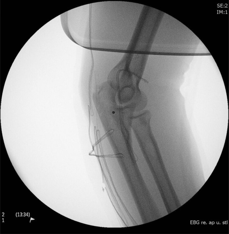
**Imaging Modality:** Intraoperative fluoroscopy (C-arm radiograph). **Anatomical Region:** Lateral view of the right elbow joint, including the distal humerus, proximal radius, and proximal ulna (olecranon). **Observed Pathology/Procedure:** Post-surgical repair of a triceps tendon rupture. The image demonstrates the implementation of a transosseous suture technique for tendon refixation to the olecranon process. **Characteristic Visual Features:** * **Hardware:** Multiple radiopaque, high-tensile sutures or wires are visible traversing the proximal ulna. * **Configuration:** The sutures follow a transosseous drill hole pattern, looping through the dorsal aspect of the olecranon to secure the distal triceps brachii tendon. * **Joint Alignment:** The ulnohumeral and radiocapitellar joints appear grossly aligned in a partially flexed position. * **Soft Tissue:** Subtle thickening of the posterior soft tissues is noted, consistent with the surgical site and tendon distal stump localization. **Key Diagnostic Features:** Identification of transosseous tunnels and suture material at the anatomical insertion point of the triceps tendon on the olecranon, confirming surgical stabilization of a previously ruptured extensor mechanism.
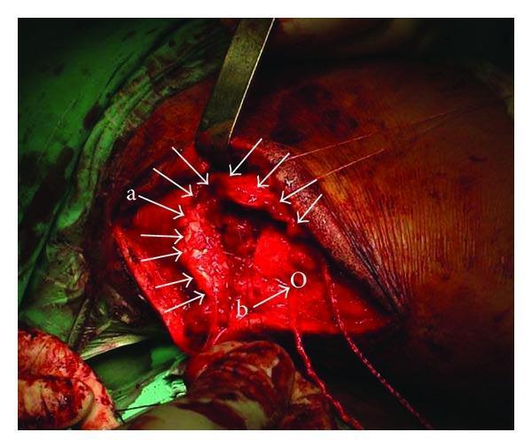
This intraoperative clinical photograph displays an orthopedic surgical procedure for the repair of a complete distal triceps tendon rupture in the posterior elbow region. The surgical field, accessed via a longitudinal approach, reveals the retracted and frayed distal triceps tendon (labeled 'a'), exhibiting irregular texture and discoloration indicative of high-grade injury. A synthetic tendon allograft (labeled 'b'), appearing as a structured, lighter-colored uniform band, is shown positioned through a transverse drill hole in the olecranon (labeled 'O'). Visible surgical instruments include a metal retractor holding back soft tissue to maintain exposure of the musculotendinous junction. Suture strands are present, integrated into the synthetic augmentation material to facilitate tensioning and reattachment of the triceps tendon to the bone. This image serves as an educational reference for complex tendon reconstruction techniques involving synthetic augmentation and allograft positioning.
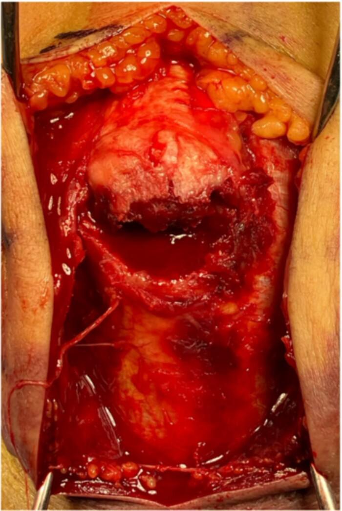
This clinical photograph displays an intraoperative view of an open orthopedic surgical field during the repair of a triceps tendon avulsion fracture of the right olecranon. The incision reveals a posterior midline approach to the elbow joint. Skin and subcutaneous adipose tissue are retracted laterally with surgical instruments, exposing the deep fascia and musculature. Central to the field is a displaced proximal ulnar bone fragment, characteristic of an olecranon avulsion. The fragment shows a clear irregular fracture line and remains attached to the triceps brachii tendon, which appears intact without longitudinal rupture. The surgical site contains significant serosanguinous fluid and highlights the anatomical relationship between the displaced bony component and the distal ulna. This visual serves to illustrate the surgical pathology of AO classification 2U1A1 fractures, emphasizing the pull of the triceps muscle on the proximal fragment and the necessity of anatomical reduction for restoring the elbow's extensor mechanism.
TRICEPS SURGERY
Applied Anatomy
- Long head - arises from the infraglenoid tubercle of the scapula
- Lateral head - arises from the posterior surface of the humerus (above the spiral groove)
- Medial head - arises from the posterior surface of the humerus (below the spiral groove, deepest component)
I. TRICEPS TENDON RUPTURE AND REPAIR
Epidemiology & Etiology
- Anabolic steroid use
- Multiple local corticosteroid injections (e.g., for olecranon bursitis)
- Chronic olecranon bursitis
- Systemic conditions: hyperparathyroidism, chronic renal failure, lupus, rheumatoid arthritis
- Sudden forceful flexion of an extended elbow (most common - eccentric overload)
- Direct blow to the posterior elbow
- Fall on an outstretched hand
- Olecranon insertion (most common - avulsion type)
- Musculotendinous junction
- Midsubstance (least common)
Clinical Features
- Pain and swelling over the posterior elbow
- Ecchymosis - may track distally
- Palpable gap/defect just proximal to the olecranon (may be masked by swelling acutely)
- Weakness of elbow extension; inability to actively extend against resistance in complete tears
- ~50% of injuries are initially missed (because the anconeus can still produce some extension)
Investigations
- X-ray: "Flake sign" - a small fleck of avulsed bone from the olecranon tip on lateral view (pathognomonic)
- MRI (gold standard): Sagittal T2 images most useful
- Partial rupture: small fluid-filled defect in the tendon (bright on T2)
- Complete rupture: large fluid-filled gap between the distal tendon and olecranon
- Ultrasound: Dynamic assessment of partial tears
Treatment
- All complete ruptures in active individuals
- Partial tears involving >50% of tendon width in active individuals
- Any complete tear where extension strength is significantly compromised
Surgical Techniques
1. Transosseous Suture Repair (Classic/Standard Technique)
- Mobilise subcutaneous tissues to expose the torn tendon
- Debride frayed edges of the ruptured tendon and curette the olecranon footprint to bleeding bone
- Drill two or three longitudinal tunnels through the olecranon (2.0 mm drill)
- Place heavy nonabsorbable sutures (No. 2 or No. 5, e.g., Ethibond) through the tendon using a Krackow (locking) stitch or Bunnell stitch
- Pass suture ends through the transosseous tunnels and tie over the olecranon with the elbow in full extension
- A tension-relief (cerclage) suture of No. 5 nonabsorbable material can be added around the repair construct to offload stress
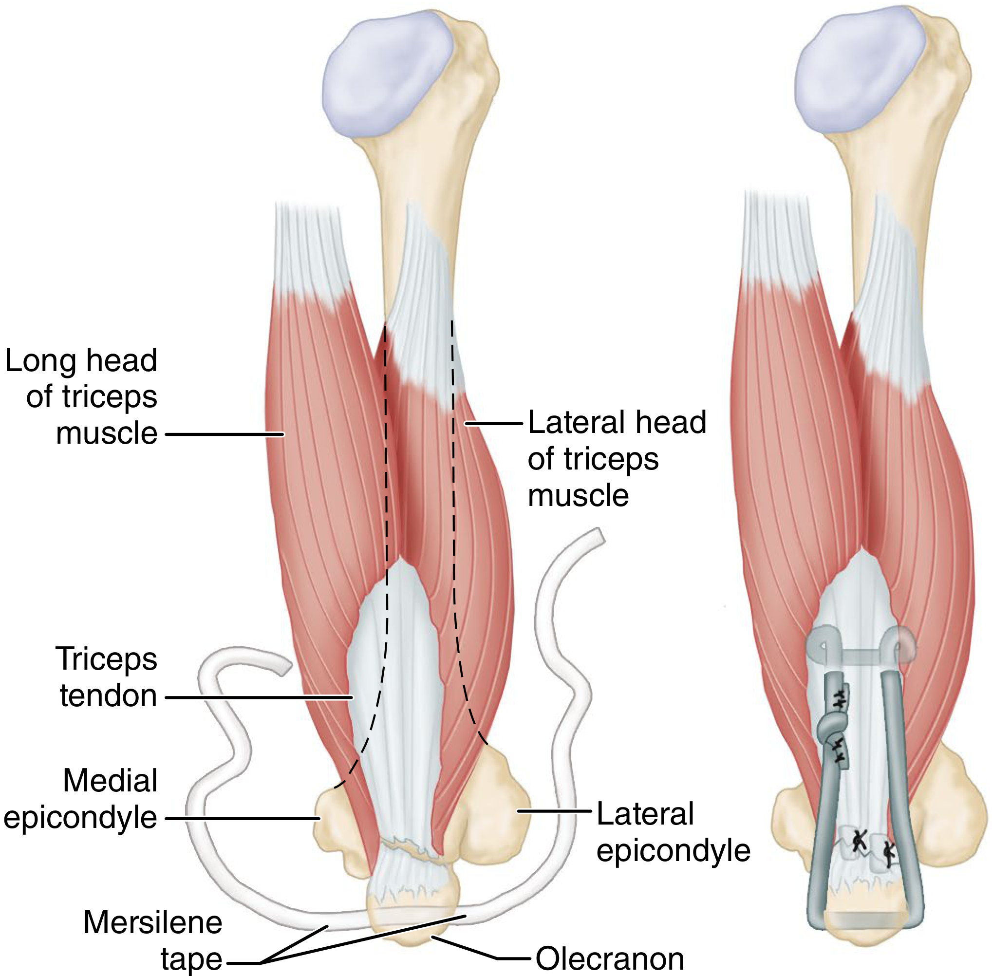
2. Double-Row Suture Anchor Repair (Azar Technique - Campbell's Operative Orthopaedics 2026)
- Patient supine, posterior midline incision 6-8 cm over olecranon
- Debride tendon edges; curette olecranon footprint to bleeding bone
- Place #2 FiberWire (Arthrex) through tendon using Krackow stitch
- Drill parallel transosseous tunnels (~2 cm and ~4 cm) using 2.0 mm drill
- Place 2-3 suture anchors just proximal to the insertion site for the proximal row (augmentation)
- Pass anchor sutures through tendon in horizontal mattress pattern - leave untied
- Pass FiberWire sutures into tunnels using a Hewson suture passer
- With elbow in full extension, reattach tendon to olecranon footprint
- Tie transosseous sutures first, then tie the bone anchor sutures
3. V-Y Plasty for Musculotendinous Junction Ruptures
4. Management of Chronic Ruptures (>6 weeks)
- Anconeus rotation flap
- Achilles tendon allograft or autograft
- Plantaris or hamstring tendon augmentation
- Fascia lata graft
- Ligament augmentation device (LAD)
Postoperative Protocol
- Splint in 0-30° flexion for 4 weeks
- Active ROM begins at 4-6 weeks, advancing elbow flexion by 10-15° per week
- Strengthening exercises begin at 8-10 weeks
- Full return to sport/heavy labour at 4-6 months
II. SURGICAL APPROACHES INVOLVING THE TRICEPS (Elbow Surgery)
| Approach | Description | Advantages | Disadvantages | Best Indication |
|---|---|---|---|---|
| Paratricipital (Triceps-On / Alonso-Llames) | Two windows alongside triceps, insertion preserved | No olecranon osteotomy; early ROM possible; preserves anconeus blood supply | Limited articular exposure; inadequate for type C3 | AO/OTA A2, A3, B1, B2, C1, C2 |
| Triceps-Splitting (Campbell) | Midline split of triceps tendon and insertion | Technically simple; easy conversion to TEA | Limited articular visibility; requires triceps repair; post-op protection needed | ORIF, TEA |
| TRAP (Triceps-Reflecting Anconeus Pedicle) | Entire triceps-anconeus flap reflected proximally (Kocher interval used distally) | Good posterior exposure; avoids olecranon osteotomy; preserves anconeus neurovascular pedicle | Risk of triceps dehiscence and extensor weakness | TEA, complex distal humerus |
| Van Gorder / Triceps Tongue | V-shaped transection at musculotendinous junction; allows V-Y lengthening | Can lengthen extensor mechanism if needed | Same weakness risk as TRAP; ~1/3 articular exposure only | TEA, associated triceps laceration |
| Olecranon Osteotomy | Chevron or transverse osteotomy of olecranon; triceps-olecranon unit reflected | Best articular visualization (~75%); gold standard for C3 fractures | Nonunion risk; symptomatic hardware; olecranon arthrosis | AO/OTA C2, C3 articular fractures |
III. TRICEPS TENDON TRANSFERS
A. Anterior Transfer of Triceps (Bunnell Technique)
- Posterolateral incision - divide triceps tendon at its insertion and dissect free from posterior distal humerus
- Transfer the tendon around the lateral aspect of the humerus
- Anterolateral incision - expose the radial tuberosity
- Prolong triceps tendon with a 4 cm fascia lata tube graft (as the tendon won't reach the tuberosity)
- Attach to roughened radial tuberosity with a pull-out steel suture through a drill hole; tie over a padded button
- Cast with elbow in acute flexion, forearm mid-rotation for 6 weeks; wire removed at 4 weeks
B. Posterior Deltoid-to-Triceps Transfer (Moberg Technique, Modified)
- Lateral decubitus - 10-13 cm incision along posterior deltoid border
- Elevate posterior 1/3 to 1/2 of deltoid with strip of periosteal insertion
- Split muscle fibers distally, preserving axillary nerve and posterior circumflex humeral vessels
- Separate posterior incision over distal triceps-olecranon; if adequate tendinous overlap, direct weave fixation; if not, interpose fascia lata graft (or great toe extensors, anterior tibial tendon)
- Tension adjusted so full passive elbow flexion is possible; stainless steel sutures through bone tunnel in olecranon
C. Distal Biceps-to-Triceps Transfer
IV. COMPLICATIONS OF TRICEPS SURGERY
| Complication | Notes |
|---|---|
| Rerupture | Higher with transosseous tunnels vs. suture anchors (Mirzayan et al.) |
| Loss of full extension | Most common functional deficit; ~10-15° extension lag is acceptable |
| Ulnar nerve injury | From direct injury or traction during medial exposure |
| Wound problems | Posterior elbow skin is thin and poorly vascularized |
| Olecranon bursitis | From prominent suture knots or hardware |
| Extensor weakness | After triceps-reflecting approaches (TRAP, Van Gorder) if repair fails |
| Triceps dehiscence | Most feared complication after triceps-splitting/reflecting approaches |
Key Examination Points (Gold Medal Level)
- "Rarest tendon rupture" - triceps is the least commonly ruptured of all major tendons
- "Flake sign" on lateral X-ray is pathognomonic of triceps avulsion
- ~50% missed acutely because anconeus preserves some elbow extension
- Suture anchors > transosseous tunnels - lower rerupture and reoperation rates (Mirzayan et al., 184 cases, Am J Sports Med 2018)
- Double-row technique restores the anatomic footprint and distributes load over wider area
- Paratricipital approach - preserves triceps insertion, allows early ROM, no osteotomy complications
- TRAP approach - preserves anconeus neurovascular supply (via Kocher interval), but risks triceps dehiscence
- Anterior triceps transfer (Bunnell) needs fascia lata graft to bridge to radial tuberosity
- Posterior deltoid-to-triceps (Moberg) - preferred for C7 tetraplegia restoration of elbow extension
- Repair tension: always set with elbow in full extension for distal repairs; full passive flexion for transfers