
Explain Whole Basic bacteriology/general bacteriology from warren levinson, brs, Lippincott book of bacteriology
gram positive gram negative bacterial cell wall structure comparison diagram
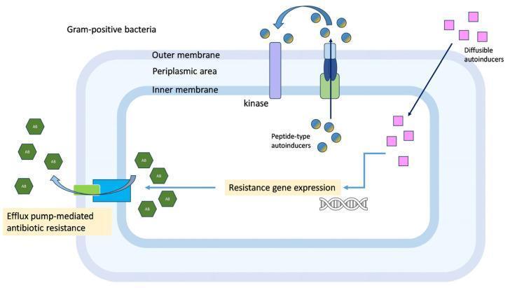
This pathophysiology diagram illustrates quorum-sensing-regulated resistance mechanisms in Gram-positive bacteria. The bacterial cell structure is depicted with three distinct layers: an inner membrane, a periplasmic area, and an outer membrane. The diagram highlights two primary signaling pathways involved in cell-to-cell communication. The first involves 'peptide-type autoinducers' (represented as blue/yellow spheres), which are processed and secreted via a transmembrane receptor and then detected by a membrane-spanning kinase. The second pathway involves 'diffusible autoinducers' (pink squares) that freely enter the cell. Both pathways converge to trigger 'resistance gene expression' at the DNA level. This genetic activation leads to physiological outcomes, specifically 'efflux pump-mediated antibiotic resistance,' where a transmembrane efflux pump actively transports antibiotic molecules (green hexagons) out of the intracellular space. This educational graphic demonstrates the interplay between bacterial signaling and multidrug resistance mechanisms.
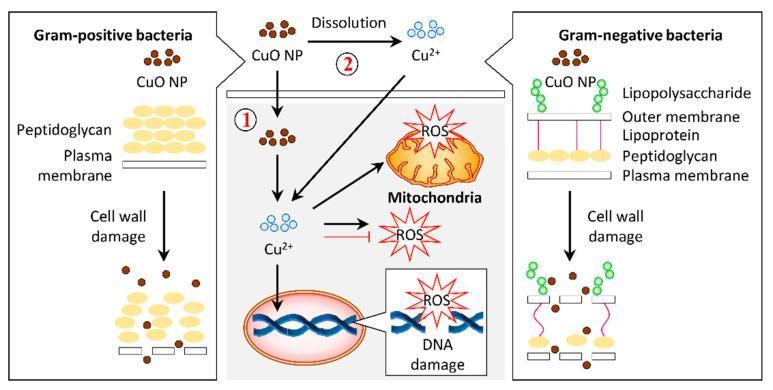
A pathophysiology diagram illustrating the antibacterial mechanisms of copper oxide nanoparticles (CuO NPs) on Gram-positive and Gram-negative bacteria. The left panel shows Gram-positive bacteria with a thick peptidoglycan layer above a plasma membrane. The right panel shows Gram-negative bacteria with a complex cell wall including lipopolysaccharides, an outer membrane, lipoproteins, and a thin peptidoglycan layer. Both panels demonstrate CuO NPs causing cell wall damage and lysis. The central panel details the intracellular bactericidal pathway labeled as mechanism 1 and 2. Mechanism 1 involves the internalization of CuO NPs into the cytosol, while mechanism 2 illustrates the dissolution of CuO NPs into copper ions (Cu2+) which then permeate the cell. Inside the cell, both Cu2+ ions and CuO NPs induce the production of reactive oxygen species (ROS). The resulting oxidative stress leads to mitochondrial disruption and DNA damage, ultimately resulting in cell death. This infographic highlights the role of nanoparticle morphology and ion release in targeting bacterial structural and metabolic integrity.
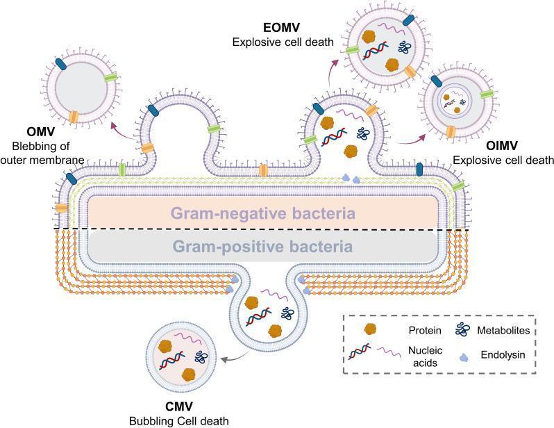
A medical microbiology diagram illustrating the biogenesis and classification of bacterial extracellular vesicles (BEVs) from Gram-negative and Gram-positive bacteria. The top half depicts a Gram-negative bacterium with its triple-layered cell envelope (outer membrane, periplasmic space, and cytoplasmic membrane). Three vesicle formation mechanisms are shown: blebbing of the outer membrane creating Outer Membrane Vesicles (OMV), and explosive cell death leading to Explosive Outer Membrane Vesicles (EOMV) and Outer-Inner Membrane Vesicles (OIMV). The bottom half represents a Gram-positive bacterium characterized by a thick peptidoglycan cell wall and a single cytoplasmic membrane. This section illustrates 'bubbling cell death' resulting in Cytoplasmic Membrane Vesicles (CMV). A legend identifies the internal cargo within these vesicles, including proteins (brown shapes), nucleic acids (wavy lines), metabolites (blue branched icons), and endolysin (light blue shapes). This pathophysiology diagram highlights the structural differences in bacterial envelopes and the various pathways—blebbing, explosive death, and bubbling—that contribute to BEV diversity and composition.
bacterial growth curve lag log stationary death phase
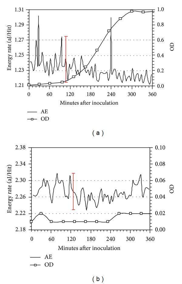
This figure presents two comparison charts, (a) and (b), depicting the growth kinetics and metabolic activity of Lactococcus lactis over 360 minutes. The primary metrics are Optical Density (OD), indicating bacterial concentration, and Energy Rate measured via Acoustic Emission (AE) in attojoules per hit (aJ/Hit). Graph (a) shows normal bacterial growth: the OD curve follows a characteristic sigmoidal pattern with lag, log, and stationary phases, while the AE data exhibits significant fluctuations and distinct peaks (e.g., around 108 and 240 minutes) representing metabolic energy release. Graph (b) demonstrates the effect of the metabolic inhibitor Sodium Azide (NaN3). In this panel, the OD remains near-baseline (indicating total inhibition of growth), and the AE signal shows only low-level background fluctuations without significant metabolic peaks. Red bars indicate ±3σ deviation standards for noise assessment. These graphs are utilized in microbiology and biomedical engineering to evaluate non-invasive methods for monitoring microbial activity and metabolic inhibition.
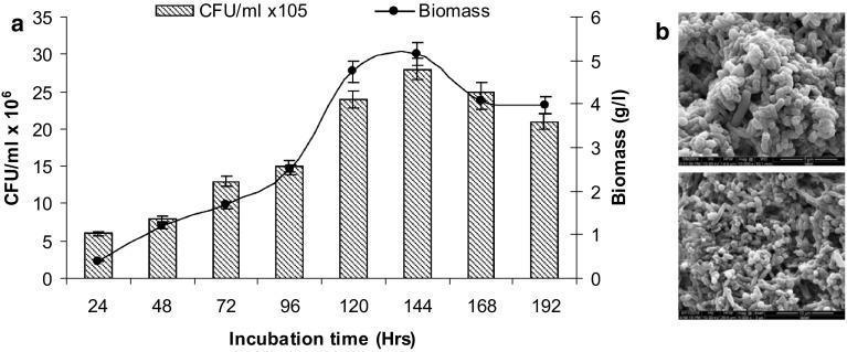
Educational microbiology graphic consisting of a growth curve plot (1a) and scanning electron micrographs (1b) documenting a bacterial consortium's development. Panel (a) presents a dual-axis line and bar graph showing the relationship between incubation time (24–192 hours), bacterial colony-forming units (CFU/mL x 10^6), and biomass concentration (g/L). The growth kinetics demonstrate a logarithmic increase in both parameters, peaking at 144 hours with a biomass of approximately 5.15 g/L and a CFU count of 28 x 10^6, followed by a slight decline indicating the stationary or death phase. Panel (b) features two scanning electron micrographs (SEM) at different magnifications, visualizing the bacterial morphology. The microbes are characterized as densely packed, rod-shaped bacilli forming a thick, interconnected biofilm matrix. The rough surface texture suggests the presence of an extracellular polymeric substance (EPS) coating. This figure illustrates bacterial growth dynamics and structural arrangement relevant to microbial degradation processes in biomedical or environmental health contexts.
bacterial spore structure endospore Bacillus Clostridium
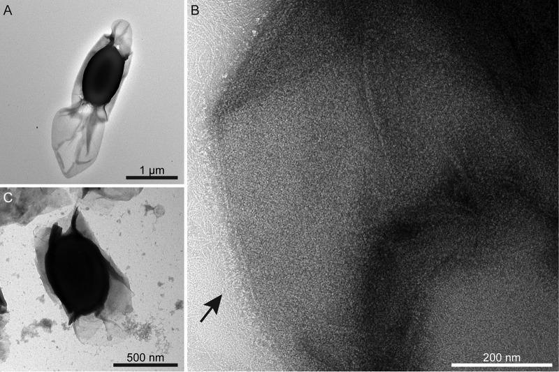
This composite image consists of three transmission electron micrographs (TEM) showcasing the ultrastructure of Clostridium sporogenes spores, emphasizing the exosporium. (A) Low-magnification TEM of a wild-type spore negatively stained with uranyl formate. It reveals a centrally located, electron-dense spore core enveloped by a translucent, thin exosporium layer that extends prominently at one pole. (B) High-magnification TEM providing a detailed view of the crystalline exosporium surface. An arrow indicates the 'hairy nap' fringe, consisting of fine filamentous appendages decorating the outer surface. (C) TEM of a csxA mutant spore, demonstrating a pathological disruption of the normal exosporium structure. The mutant spore core appears partially wrapped in irregular, broken sheets of material, with visible sloughed-off fragments. This comparison highlights the essential role of the CsxA protein as a core structural component of the crystalline basal layer in the exosporium. The image serves as an educational tool in microbiology and infectious diseases for understanding bacterial spore morphology and genetic determinants of protective surface layers.
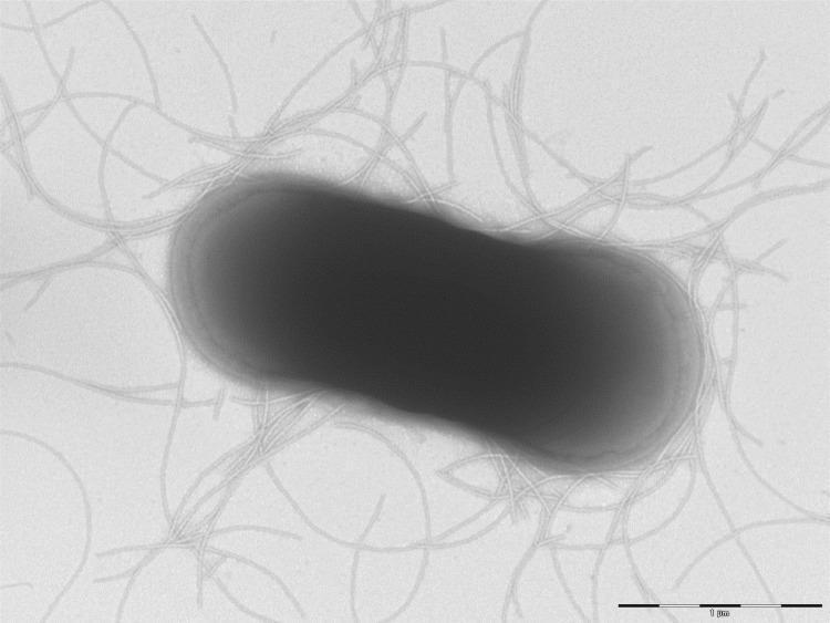
This Transmission Electron Microscopy (TEM) image displays the morphological characteristics of Clostridium dakarense (strain FF1T), a Gram-positive, anaerobic bacterium originally isolated from a human stool specimen. The image reveals a single, electron-dense, rod-shaped (bacillus) cell. Based on the 1 µm scale bar, the bacterium measures approximately 2.5 µm in length and 1.2 µm in width. A prominent feature is the presence of numerous peritrichous filamentous appendages extending from the bacterial surface, consistent with the motility observed in this species. These long, thread-like structures (likely flagella or fimbriae) are distributed relatively evenly around the cell perimeter. The cell boundary shows an undulating surface with a slightly translucent outer layer or halo, characteristic of the bacterial cell wall or capsule. The image demonstrates typical prokaryotic ultrastructure used in taxonomic classification and diagnostic microbiology to identify novel pathogenic or commensal species within the Clostridium genus.
gram stain procedure positive negative bacteria microscopy
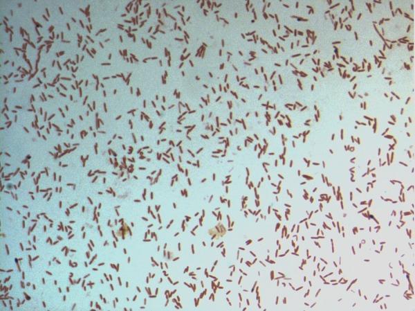
This diagnostic image shows a light microscopy view of a bacterial culture following Gram staining. The specimen displays numerous small, rod-shaped (bacilli) bacteria distributed across the field. The microorganisms exhibit a consistent pink-to-red hue, identifying them as Gram-negative. This staining characteristic indicates a cell wall structure with a thin peptidoglycan layer and an outer membrane that does not retain the crystal violet stain. The arrangement of the bacilli is predominantly individual or scattered, with occasional small clusters and pairs. Specifically identified as D. fastidiosa strain JC13T, this image serves as a primary microbiological reference for the morphological and staining properties of this novel genus within the family Erysipelotrichaceae. The visual demonstrates key diagnostic features including cell morphology, arrangement, and Gram reaction, which are essential for clinical microbiology classification and initial pathogen identification.
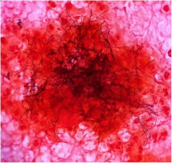
This light microscopy image demonstrates a Gram stain of an excised clinical sample, typically used in microbiology for diagnostic identification. The central focus shows a dense cluster of Gram-variable filamentous rods, characteristic of Actinomyces species. These organisms appear as dark, elongated, branching, thread-like structures. Some filaments exhibit a beaded appearance, staining more intensely (Gram-positive/purple) or less intensely (Gram-negative/pink) along their length. The background consists of host cellular debris and inflammatory cells, which are counterstained pink/red with safranin. These background elements include various eukaryotic cells and likely polymorphonuclear leukocytes. This visual is highly representative of Actinomycosis, demonstrating the classic 'sulfur granule' or 'tangled mass' morphology often seen in abscess aspirates or tissue samples. It serves as a critical educational tool for distinguishing filamentous bacteria from standard cocci or bacilli and highlights the importance of Gram stain variability in clinical diagnostics.
BASIC BACTERIOLOGY - Complete Notes
1. PROKARYOTES vs. EUKARYOTES
| Feature | Prokaryote (Bacteria) | Eukaryote (Human cell) |
|---|---|---|
| Nucleus | Absent (nucleoid) | Present (membrane-bound) |
| Chromosome | Single, circular, double-stranded DNA | Multiple, linear |
| Ribosome | 70S (50S + 30S subunits) | 80S (60S + 40S) |
| Cell wall | Peptidoglycan (most species) | Absent |
| Mitochondria | Absent | Present |
| Organelles | Absent | Present |
| Size | ~1 µm diameter | 7-100+ µm |
| DNA size (E. coli) | ~5 million bp, 1.3 mm length | ~2.9 × 10⁹ bp |
Key exam point: The 70S ribosome is the target of many antibiotics (aminoglycosides, macrolides, tetracyclines, chloramphenicol). This selectivity is what makes antibiotics useful against bacteria without harming human cells.
- Medical Microbiology 9e, p. 138
2. BACTERIAL CLASSIFICATION
2a. By Gram Stain (most important initial classification)
- Crystal violet (primary stain) - all cells turn purple
- Gram's iodine (mordant) - fixes crystal violet
- Acetone/alcohol (decolorizer) - removes crystal violet from gram-negative cells
- Safranin (counterstain) - gram-negative cells turn pink/red
- Gram-positive = Purple (thick peptidoglycan retains crystal violet)
- Gram-negative = Pink/Red (thin peptidoglycan, outer membrane, lost crystal violet)
2b. By Morphology
| Shape | Term | Examples |
|---|---|---|
| Spherical | Coccus | Staphylococcus, Streptococcus |
| Rod | Bacillus | E. coli, Klebsiella, Bacillus |
| Comma-shaped | Vibrio | Vibrio cholerae |
| Spiral | Spirochete | Treponema, Leptospira, Borrelia |
| Pleomorphic | Variable | Mycoplasma |
- Pairs = diplococci (Neisseria, Streptococcus pneumoniae)
- Chains = streptococci
- Clusters = staphylococci (grapes)
- Tetrads = Micrococcus
- Cuboidal packets of 8 = Sarcina
2c. By Oxygen Requirement
| Type | Definition | Example |
|---|---|---|
| Obligate aerobe | Requires O₂ | Mycobacterium tuberculosis, Pseudomonas |
| Obligate anaerobe | Killed by O₂ | Bacteroides, Clostridium |
| Facultative anaerobe | Grows with or without O₂ | E. coli, Staphylococcus |
| Microaerophile | Requires low O₂ | Helicobacter pylori, Campylobacter |
| Aerotolerant | Grows without O₂, not harmed by it | Streptococcus |
| Capnophile | Requires increased CO₂ | Neisseria, Brucella |
2d. By Staining Characteristics
| Stain | Purpose | Organisms |
|---|---|---|
| Gram stain | Routine | Most bacteria |
| Acid-fast (Ziehl-Neelsen) | Mycolic acid walls resist decolorization with acid-alcohol | Mycobacterium (TB, leprosy), Nocardia (partial) |
| Giemsa | Intracellular organisms | Chlamydia, Rickettsia, Borrelia |
| Silver stain | Spirochetes, Legionella, fungi | - |
| India ink | Capsule (negative stain) | Cryptococcus (fungus), Klebsiella |
| PAS | Whipple's disease | Tropheryma whipplei |
| Spore stain (Schaeffer-Fulton) | Endospores | Bacillus, Clostridium |
2e. By Temperature Preference
- Psychrophiles: 0-20°C (cold-loving)
- Mesophiles: 20-40°C (most human pathogens, optimal ~37°C)
- Thermophiles: 45-80°C
- Hyperthermophiles: >80°C (Archaea)
3. BACTERIAL CELL STRUCTURE
3a. Overview - Cell Envelope Layers
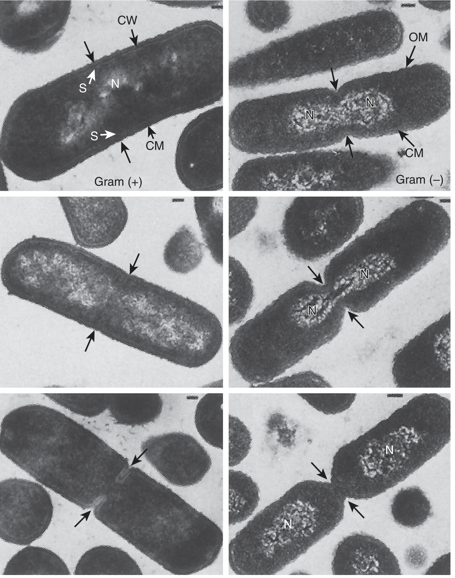
3b. Cytoplasmic (Plasma) Membrane
- Composed of phospholipids + proteins + enzymes
- No sterols (unlike eukaryotic membranes) - EXCEPT Mycoplasma, which incorporates host cholesterol
- Functions:
- Permeability barrier (selective)
- Site of electron transport chain and ATP synthesis (bacterial equivalent of mitochondria)
- Active transport of nutrients
- DNA replication initiation site
- Secretion systems (Type I-VI)
- Target of polymyxins (disrupts outer membrane in gram-negatives)
3c. Peptidoglycan (Murein/Mucopeptide) - THE KEY STRUCTURE
- A rigid mesh/lattice made of polysaccharide chains cross-linked by peptides
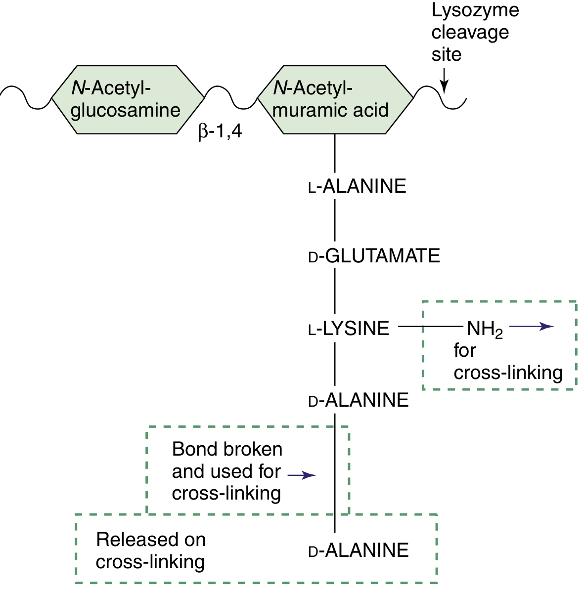
- Polysaccharide backbone: repeating disaccharides of N-acetylglucosamine (NAG/GlcNAc) and N-acetylmuramic acid (NAM/MurNAc) linked by β-1,4 glycosidic bonds
- A tetrapeptide hangs from each NAM unit, containing unusual D-amino acids (L-Ala → D-Glu → L-Lys or DAP → D-Ala → D-Ala)
- Cross-linking: The free amine of the diamino acid (3rd position) links to the D-Ala in the 4th position of an adjacent chain
- In gram-positive (e.g., S. aureus): via a pentaglycine bridge (Gly₅)
- In gram-negative (e.g., E. coli): direct cross-link via DAP (diaminopimelic acid)
-
Lysozyme (in tears, mucus, saliva) cleaves the β-1,4 bond between NAG and NAM → destroys bacteria
-
β-lactam antibiotics (penicillins, cephalosporins) block transpeptidases (PBPs - Penicillin-Binding Proteins) that form the cross-links → weak cell wall → lysis
-
Vancomycin binds the D-Ala-D-Ala terminus → blocks cross-linking
-
Gram-positive: thick (150-500 Å), multi-layered peptidoglycan
-
Gram-negative: thin (20-30 Å), single-layered
-
Mycoplasma: NO peptidoglycan → naturally resistant to β-lactams
-
Medical Microbiology 9e, p. 144-145
3d. Gram-Positive Cell Wall Components
| Component | Structure | Function/Significance |
|---|---|---|
| Thick peptidoglycan | 150-500 Å, multilayer | Structural rigidity; resists osmotic lysis |
| Teichoic acid | Polyribitol-phosphate or glycerol-phosphate, covalently bound to NAM | Strengthens wall; Ca²⁺ sequestration; activates complement |
| Lipoteichoic acid (LTA) | Lipid-anchored teichoic acid to cytoplasmic membrane | PAMP - triggers innate immunity (like LPS) |
| Surface proteins | Attached to peptidoglycan | Virulence - e.g., Protein A (S. aureus), M protein (Strep. pyogenes) |
3e. Gram-Negative Cell Wall Components
| Component | Structure | Function/Significance |
|---|---|---|
| Thin peptidoglycan | ~20-30 Å, single layer | Shape maintenance |
| Periplasmic space | Between inner and outer membrane | Contains transport proteins, β-lactamases, hydrolytic enzymes |
| Outer membrane (OM) | Lipid bilayer with proteins, LPS | Permeability barrier; held by Mg²⁺/Ca²⁺ links |
| LPS (Lipopolysaccharide/Endotoxin) | Three parts: Lipid A + Core polysaccharide + O antigen | Major PAMP; causes fever, shock; key virulence factor |
| Porins | β-barrel proteins in OM | Allow hydrophilic molecules to pass |
- Lipid A: Embedded in outer membrane; responsible for toxic effects - fever, hypotension, DIC, septic shock. Recognized by TLR-4 on macrophages
- Core polysaccharide: Links Lipid A to O antigen; contains unusual sugars (KDO, heptose)
- O antigen (O polysaccharide): Outermost repeating sugar units; species/strain specific; used for serotyping
Critical: Lipid A = endotoxin = responsible for gram-negative sepsis
3f. Outer Structures (External to Cell Wall)
CAPSULE
- Composition: Usually polysaccharide (exception: B. anthracis - poly-D-glutamate)
- Function: PRIMARY virulence factor - antiphagocytic (prevents opsonization)
- Capsulated bacteria: "Some Killers Have Pretty Nice Capsules" = Streptococcus pneumoniae, Klebsiella pneumoniae, Haemophilus influenzae, Pseudomonas aeruginosa, Neisseria meningitidis, Cryptococcus neoformans
- Quellung reaction: Capsule swells in the presence of specific antibody + India ink → used for serotyping Strep. pneumoniae
- Vaccines targeting capsular polysaccharides (e.g., pneumococcal, meningococcal, Hib vaccines)
FLAGELLA
- Structure: Made of flagellin protein (the H antigen in serotyping, e.g., E. coli O157:H7)
- Function: Motility; chemotaxis (move toward nutrients, away from toxins)
- Arrangements:
- Monotrichous: single polar flagellum (e.g., Vibrio cholerae)
- Lophotrichous: tuft at one pole
- Amphitrichous: flagella at both poles
- Peritrichous: flagella all around (e.g., E. coli, Salmonella)
- Note: Spirochetes have endoflagella (axial filaments) within the periplasm → corkscrew motility
PILI (Fimbriae)
| Type | Function |
|---|---|
| Common pili (Type I fimbriae) | Adhesion to host cells (attach to mannose residues) - most important for pathogenesis |
| Type IV pili | Twitching motility, adherence (Neisseria, Pseudomonas) |
| Sex pili (F pilus) | Conjugation - DNA transfer between bacteria |
Pili are a critical virulence factor - bacteria without pili cannot colonize mucosal surfaces.
3g. Internal Structures
NUCLEOID (Bacterial Chromosome)
- Single, circular, double-stranded DNA (covalently closed)
- Approximately 5 million base pairs in E. coli (~1.3 mm length!)
- Supercoiled and compacted by histone-like proteins (HU, H-NS)
- No nuclear membrane
- DNA replication: bidirectional from a single origin of replication (oriC)
PLASMIDS
- Extrachromosomal, small circular DNA molecules
- Replicate independently
- Carry genes for: antibiotic resistance, toxins, virulence, metabolic functions
- Can be transferred between bacteria by conjugation
- Types:
- F plasmid (fertility factor): Enables conjugation
- R plasmid (resistance plasmid): Antibiotic resistance genes
- Col plasmids: Bacteriocin production
- Virulence plasmids: Toxins (e.g., ST/LT toxin of ETEC)
RIBOSOMES
- 70S = 50S + 30S subunits
- Site of protein synthesis
- Antibiotic targets:
- 30S: aminoglycosides (block initiation), tetracyclines (block tRNA entry)
- 50S: macrolides/azithromycin (block translocation), chloramphenicol (blocks peptidyl transferase), linezolid (blocks initiation)
- "Buy AT 30, ECAM at 50" = Aminoglycosides + Tetracyclines at 30S; Erythromycin + Chloramphenicol + Aminoglycosides + clindaMycin at 50S
INCLUSIONS / GRANULES
- Metachromatic granules (volutin/Babes-Ernst granules): Polymetaphosphate storage; stain reddish-purple with methylene blue; key feature of Corynebacterium diphtheriae
- Sulfur granules: Energy storage in sulfur-oxidizing bacteria
- Lipid inclusions (PHB): Carbon/energy reserve
4. ENDOSPORES (SPORES)
- Only GRAM-POSITIVE bacteria (NEVER gram-negative)
- Genera: Bacillus (aerobic) and Clostridium (anaerobic)
- B. anthracis (anthrax), B. cereus, B. subtilis
- C. tetani, C. botulinum, C. perfringens, C. difficile
- Response to nutritional deprivation or harsh environmental conditions
- Sporulation is a survival mechanism - the spore is "suspended animation"
- Core (with chromosome, ribosomes, dipicolinic acid + Ca²⁺)
- Inner membrane
- Cortex (modified peptidoglycan)
- Spore coat (keratin-like protein)
- Exosporium (in some species)
- Contain calcium dipicolinate - responsible for heat resistance
- Dehydrated - metabolically dormant
- Refractile (bright/white) under light microscope
- Highly resistant to: heat, desiccation, UV light, many chemicals, disinfectants
| Location | Organism |
|---|---|
| Central | B. anthracis, C. perfringens |
| Subterminal | C. botulinum, B. cereus |
| Terminal (drumstick) | C. tetani |
| Terminal (tennis racket) | C. tertium |
- Must use autoclaving (121°C, 15 psi, 15 min) to kill spores - not regular heat or alcohol
- C. difficile spores survive on hospital surfaces for months
- B. anthracis spores used in bioterrorism
- C. botulinum spores in soil/honey - risk in canned food and infant botulism
5. BACTERIAL GROWTH AND METABOLISM
5a. Growth Curve
Log CFU
│ ___________
│ / \
│ / \ (Death)
│ / \
│____________/ \
│ Lag Log Stationary Death
└────────────────────────────────────→ Time
| Phase | Description | Features |
|---|---|---|
| Lag phase | Adaptation period | Bacteria adjust to new environment; synthesize enzymes; NO cell division; intense metabolic activity |
| Log (Exponential) phase | Exponential growth | Maximum growth rate; generation time is constant; most susceptible to antibiotics targeting cell wall synthesis; used for growth rate calculations |
| Stationary phase | Growth = death | Nutrient depletion and waste accumulation; toxin production peaks (many exotoxins made here); sporulation begins |
| Death (Decline) phase | Deaths > growth | Irreversible decline; accumulation of toxic waste products |
- E. coli: ~20 minutes (one of fastest)
- M. tuberculosis: ~20 hours (hence why TB treatment takes months)
- Treponema pallidum: ~30 hours
5b. Bacterial Metabolism
- Catabolism: Breakdown of substrates to generate energy (ATP)
- Anabolism: Synthesis of cellular components using that energy
- Central pathway: All substrates funnel through pyruvate
| Pathway | O₂ requirement | ATP yield | Examples |
|---|---|---|---|
| Aerobic respiration | Requires O₂ | ~38 ATP/glucose | Most aerobic bacteria |
| Anaerobic respiration | Alternate electron acceptors (NO₃⁻, SO₄²⁻) | Intermediate | Pseudomonas, Bacteroides |
| Fermentation | None | 2 ATP/glucose | Lactobacillus, Clostridium |
- Lactose fermentation: Differentiates E. coli (lactose fermenter, pink on MacConkey) from Salmonella/Shigella (non-fermenters, colorless on MacConkey)
- H₂S production: Salmonella, Proteus (black colonies on XLD agar)
- Urease: Helicobacter pylori, Proteus, Klebsiella (urea breath test for H. pylori)
6. BACTERIAL GENETICS
6a. Mutation
- Spontaneous mutation rate: ~10⁻⁶ to 10⁻⁹ per gene per generation
- Types: Point mutations, insertions, deletions, inversions
- Mutagens: UV light (thymine dimers), nitrous acid, alkylating agents, acridine dyes
- Bacteria use DNA repair mechanisms (SOS response, photoreactivation)
6b. Gene Transfer - THE BIG THREE
1. TRANSFORMATION
- Direct uptake of naked DNA from the environment
- Requires bacteria to be "competent" (capable of taking up DNA)
- Naturally competent: Streptococcus pneumoniae, Haemophilus, Neisseria
- Griffith experiment: Heat-killed smooth (S) + live rough (R) S. pneumoniae → live S bacteria (transforming principle = DNA; later shown by Avery, MacLeod, McCarty)
- Used in molecular biology to introduce plasmids into bacteria
2. CONJUGATION
- Direct cell-to-cell contact via sex pilus (F pilus)
- Requires F+ (donor) × F- (recipient) bacteria
- F plasmid encodes the F pilus and transfer machinery
- Types:
- F+ × F-: F plasmid transferred → F- becomes F+
- Hfr × F-: F plasmid integrated into chromosome → chromosomal genes transferred (rarely complete)
- F' (F prime): Hfr cell excises incorrectly → some chromosomal genes incorporated into F plasmid → sexduction
- Most important for transferring R plasmids (antibiotic resistance) between bacteria
- Works between different bacterial species - major driver of antibiotic resistance spread
3. TRANSDUCTION
- Transfer of DNA via bacteriophages (bacterial viruses)
| Type | Mechanism | Amount transferred |
|---|---|---|
| Generalized transduction | Phage accidentally packages random bacterial DNA | Any gene |
| Specialized transduction | Phage integrates (lysogeny), excises imprecisely → takes adjacent bacterial genes | Only specific genes near integration site |
- S. pyogenes + phage → erythrogenic toxin (scarlet fever)
- S. aureus + phage → Panton-Valentine leukocidin
- C. botulinum + phage → botulinum toxin
- C. diphtheriae + phage (β-phage) → diphtheria toxin
- V. cholerae + phage → cholera toxin
Remember: Many key bacterial toxins are encoded on phages or plasmids, not the main chromosome.
6c. Transposons (Jumping Genes)
- DNA sequences that can "jump" (transpose) from one location to another in the genome
- Carry genes including antibiotic resistance
- Can move between plasmids and chromosomes
- Important in spread of resistance genes
7. PATHOGENESIS OF BACTERIAL INFECTION
7a. Steps in Infection
- Entry - via respiratory, GI, genitourinary tract, skin, vector
- Adherence - pili, surface proteins bind to host receptors
- Colonization - establish in host tissue, evade normal flora competition
- Invasion - spread to deeper tissues (some pathogens are strictly extracellular)
- Evasion of host defenses
- Tissue damage - via toxins and/or immune response
- Transmission - to new host
7b. Virulence Factors
| Factor | Examples | Mechanism |
|---|---|---|
| Capsule | S. pneumoniae, K. pneumoniae, H. influenzae | Antiphagocytic; inhibits complement |
| Pili/fimbriae | N. gonorrhoeae, UPEC | Adhesion to mucosa |
| Cell wall components | LPS, LTA, M protein | Trigger inflammation; antiphagocytic |
| Enzymes | Coagulase (S. aureus), hyaluronidase, collagenase | Spread through tissue, evade defenses |
| IgA protease | N. gonorrhoeae, S. pneumoniae, H. influenzae | Cleave secretory IgA on mucosal surfaces |
| Protein A | S. aureus | Binds Fc region of IgG → blocks opsonization |
| M protein | S. pyogenes | Antiphagocytic; inhibits C3b deposition |
| Iron acquisition (siderophores) | Many gram-negative bacteria | Chelate iron from host transferrin |
7c. Exotoxins vs. Endotoxins - MASTER TABLE
| Feature | Exotoxin | Endotoxin |
|---|---|---|
| Source | Gram-positive AND gram-negative | Gram-negative ONLY |
| Chemical nature | Protein | Lipopolysaccharide (Lipid A) |
| Secreted? | Yes - secreted actively | No - part of outer membrane; released on lysis |
| Heat stability | Heat-labile (destroyed at 60-80°C) - exceptions: SE, TSST-1 | Heat-stable (withstands 250°C) |
| Antigenicity | Highly antigenic → toxoid possible | Weakly antigenic; no effective toxoid |
| Toxicity | High (nanogram quantities can kill) | Moderate; requires microgram quantities |
| Mechanism | Specific cell receptors and mechanisms | Binds TLR-4 → macrophages → TNF-α, IL-1, IL-6 |
| Clinical effects | Specific (tetanus = spasm; botulism = flaccid paralysis; cholera = watery diarrhea) | Fever, hypotension, DIC, septic shock |
| Limulus test | Not detected | Detected (Limulus amebocyte lysate test) |
| Examples | Tetanospasmin, botulinum toxin, cholera toxin, diphtheria toxin, TSST-1, anthrax toxin | Salmonella, E. coli, Neisseria LPS |
7d. Key Exotoxin Mechanisms
| Toxin | Organism | Mechanism | Effect |
|---|---|---|---|
| Cholera toxin | V. cholerae | ADP-ribosylates Gsα → permanently activates adenylyl cyclase → ↑↑cAMP | Profuse watery diarrhea |
| Pertussis toxin | B. pertussis | ADP-ribosylates Giα → permanently inactivates Giα → ↑cAMP | Whooping cough; lymphocytosis |
| Diphtheria toxin | C. diphtheriae (β-phage) | ADP-ribosylates EF-2 (elongation factor 2) → blocks protein synthesis | Pseudomembrane; myocarditis; neuropathy |
| Anthrax toxin | B. anthracis | Lethal factor (metalloprotease, kills macrophages) + Edema factor (adenylyl cyclase) + Protective antigen (receptor binding) | Cutaneous/pulmonary anthrax |
| Botulinum toxin | C. botulinum | Cleaves SNARE proteins → blocks Ach release at NMJ | Flaccid paralysis, descending |
| Tetanospasmin | C. tetani | Cleaves synaptobrevin → blocks glycine/GABA release at inhibitory synapses | Spastic paralysis (lockjaw) |
| TSST-1 | S. aureus | Superantigen → activates up to 20% of T cells → massive cytokine storm | Toxic shock syndrome |
| Shiga toxin | Shigella, EHEC | Cleaves 28S rRNA → inhibits protein synthesis + microangiopathy | Bloody diarrhea, HUS |
| Staphylococcal enterotoxins (A-E) | S. aureus | Superantigens; heat-stable | Food poisoning (preformed toxin) |
8. HOST DEFENSES AND BACTERIAL EVASION
8a. Normal Flora
- The human body is colonized by ~10¹³ bacteria (roughly equal to or exceeding human cell count)
- Normal flora: protect against pathogens by competition (colonization resistance)
- Disruption by antibiotics → overgrowth of pathogenic organisms (e.g., C. difficile colitis after broad-spectrum antibiotics)
- Transient flora: present temporarily, can be removed by handwashing
- Resident flora: permanent colonizers; harder to remove
| Site | Key organisms |
|---|---|
| Skin | S. epidermidis, S. aureus, Corynebacterium, Propionibacterium acnes |
| Oral | Viridans streptococci (S. mutans, S. sanguis), Bacteroides, Fusobacterium |
| Upper respiratory | S. pneumoniae, H. influenzae, N. meningitidis (carriers), Neisseria |
| Intestine | E. coli, Bacteroides (most abundant), Lactobacillus, Bifidobacterium |
| Vagina | Lactobacillus (maintains acidic pH) |
| Urethra | S. epidermidis, diphtheroids |
8b. Bacterial Evasion Strategies
- Capsule: Blocks phagocytosis, inhibits complement deposition
- Protein A (S. aureus): Binds Fc of IgG → prevents opsonization
- M protein (S. pyogenes): Blocks C3b deposition
- IgA protease: Destroys mucosal antibody
- Intracellular survival: Mycobacterium, Listeria, Salmonella survive inside macrophages
- Biofilm formation: Protects bacteria from antibiotics and immune cells; seen with S. epidermidis (implants), P. aeruginosa (CF lungs), H. pylori
- Antigenic variation: N. gonorrhoeae changes pili (pilin antigenic variation); Borrelia (relapsing fever)
- Phase variation: On/off switching of surface antigens
9. SPECIAL GROUPS
9a. Mycobacteria (Acid-Fast Bacteria)
- Cell wall: Peptidoglycan + arabinogalactan + mycolic acid (long-chain fatty acids, C60-C90)
- Mycolic acid makes them acid-fast - retain carbol fuchsin after acid-alcohol decolorization (Ziehl-Neelsen or Kinyoun stain = red/pink on blue background)
- Grow very slowly (M. tuberculosis: 20-hour generation time)
- Resistant to: desiccation, many disinfectants, detergents
- Also acid-fast: Nocardia (partially), Cryptosporidium, Isospora (oocysts)
9b. Mycoplasma
- Smallest free-living bacteria (~0.1-0.3 µm)
- NO cell wall (no peptidoglycan)
- Incorporate cholesterol from host into their membrane
- Not visible on Gram stain (no cell wall to stain)
- Resistant to β-lactams (no target)
- Treatment: Macrolides, tetracyclines, fluoroquinolones
- Examples: M. pneumoniae (atypical pneumonia), M. genitalium, Ureaplasma
9c. L-Forms
- Bacteria that have lost their cell wall (spontaneously or induced by antibiotics/lysozyme) but can still grow
- "Stable L-forms" have permanently lost cell wall
- Important in chronic/recurrent infections; hard to culture on standard media
9d. Chlamydia
- Obligate intracellular bacteria
- Two forms:
- Elementary body (EB): Small, infectious, metabolically inactive, can survive outside cells
- Reticulate body (RB): Larger, intracellular, metabolically active, replicates
- No peptidoglycan in their cell wall (anomalous gram-negative)
- Cannot synthesize their own ATP ("energy parasites")
9e. Rickettsia
- Obligate intracellular
- Gram-negative but poorly visualized by Gram stain
- Stained with Giemsa or Gimenez stain
- Transmitted by arthropod vectors
10. STERILIZATION AND DISINFECTION
10a. Key Definitions
| Term | Definition |
|---|---|
| Sterilization | Complete destruction/removal of ALL living organisms, including spores |
| Disinfection | Destruction of pathogenic organisms (not necessarily all spores) |
| Antisepsis | Use of disinfecting agents on living tissue (skin, mucous membranes) |
| Sanitization | Reduction of microbial load to safe levels (food/household contexts) |
| Asepsis | Practices to prevent microbial contamination of a protected environment |
| Pasteurization | Use of heat sufficient to kill pathogens (NOT sterilization); e.g., 74°C × 3-5 sec or 62°C × 30 min |
| Bactericidal | Kills bacteria |
| Bacteriostatic | Inhibits bacterial growth (bacteria can regrow if agent removed) |
10b. Physical Methods of Sterilization
| Method | Conditions | Kills Spores | Use |
|---|---|---|---|
| Autoclave (moist heat) | 121°C, 15 psi, 15-20 min | YES | Gold standard; surgical instruments, media |
| Dry heat oven | 160°C × 2h or 170°C × 1h | YES | Glassware, oils, powders |
| Incineration | Direct flame/burning | YES | Inoculation loops, biohazard waste |
| Boiling (100°C) | 10 minutes | NO (not reliable for spores) | Disinfection only |
| Pasteurization | 74°C × 3-5 sec | NO | Milk, beverages |
| Filtration | 0.22 µm membrane filter | YES (physical removal) | Heat-sensitive solutions, serum |
| UV radiation | ~260 nm (DNA absorption peak) | No (limited penetration) | Surface/air disinfection; induces thymine dimers |
| Ionizing radiation (gamma) | High-energy | YES | Medical devices, food |
| Tyndallization | Steam 100°C × 30 min on 3 consecutive days | YES (sporulating bacteria) | Historical method; intermittent sterilization |
10c. Chemical Methods
| Agent | Mechanism | Use |
|---|---|---|
| Glutaraldehyde (2%) | Cross-links proteins | High-level disinfection of endoscopes |
| Ethylene oxide gas | Alkylates DNA | Cold sterilization of heat-sensitive equipment |
| Formaldehyde | Cross-links proteins | Vaccines (inactivation), embalming |
| Chlorine/Hypochlorite (bleach) | Oxidation; protein denaturation | Water treatment, surface disinfection |
| Iodine/Iodophors (Betadine) | Oxidation | Antiseptic skin prep |
| Alcohols (70% ethanol/isopropanol) | Protein denaturation, membrane dissolution | Hand sanitizers, skin antisepsis (NOT effective vs. spores) |
| Quaternary ammonium compounds (quats) | Disrupt membranes | Surface disinfectant; LOW-level |
| Phenols | Denature proteins, disrupt membranes | Wound disinfectant (carbolic acid - Lister) |
| Heavy metals (Ag, Hg) | Bind sulfhydryl groups; inhibit enzymes | Silver sulfadiazine (burns), mercurochrome |
10d. Relative Resistance (from MOST to LEAST resistant)
Prions > Spores > Mycobacteria > Non-enveloped viruses > Fungi > Vegetative bacteria > Enveloped viruses
Remember: Enveloped viruses (HIV, Hepatitis B, influenza) are the EASIEST to kill. Non-enveloped viruses (poliovirus, norovirus) are harder. Spores are the hardest bacterial forms to kill.
11. ANTIBIOTIC MECHANISMS - OVERVIEW
| Antibiotic Class | Target | Bactericidal/Static |
|---|---|---|
| β-lactams (penicillins, cephalosporins, carbapenems) | Peptidoglycan synthesis (PBPs/transpeptidases) | Cidal |
| Vancomycin | D-Ala-D-Ala terminus of peptidoglycan precursor | Cidal |
| Aminoglycosides | 30S ribosome - cause misreading | Cidal |
| Tetracyclines | 30S ribosome - block aminoacyl-tRNA binding | Static |
| Chloramphenicol | 50S ribosome - block peptidyl transferase | Static (cidal for some) |
| Macrolides | 50S ribosome - block translocation | Static (usually) |
| Fluoroquinolones | DNA gyrase (topoisomerase II/IV) | Cidal |
| Rifampin | RNA polymerase | Cidal |
| Sulfonamides | Dihydropteroate synthase (folate synthesis) | Static |
| Trimethoprim | Dihydrofolate reductase (folate synthesis) | Static |
| Polymyxins | Cytoplasmic membrane (detergent effect) | Cidal |
12. KEY MNEMONICS - EXAM SHORTCUTS
13. SUMMARY COMPARISON TABLE - Gram-Positive vs. Gram-Negative
| Feature | Gram-Positive | Gram-Negative |
|---|---|---|
| Gram stain color | Purple | Pink/Red |
| Peptidoglycan | Thick (150-500 Å) | Thin (20-30 Å) |
| Outer membrane | Absent | Present |
| LPS/Endotoxin | Absent | Present (Lipid A) |
| Teichoic acid | Present | Absent |
| Periplasmic space | Thin/absent | Wide |
| Sporulation | Some (Bacillus, Clostridium) | None |
| Exotoxin production | Common | Some |
| Endotoxin | Absent | Present |
| Penicillin sensitivity | More sensitive | More resistant (OM barrier) |
| Lysozyme sensitivity | Sensitive | Resistant (OM protects) |
| Examples | S. aureus, Strep. pyogenes, Bacillus, Clostridium, Listeria | E. coli, Klebsiella, Pseudomonas, Neisseria, Salmonella, H. influenzae |
Explain Whole Basic bacteriology/general bacteriology from warren levinson
bacterial genetics conjugation transformation transduction horizontal gene transfer
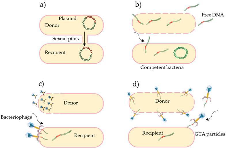
A multi-panel microbiological diagram illustrating the four primary mechanisms of horizontal gene transfer (HGT) in bacteria, which facilitate the spread of antibiotic resistance. (a) Conjugation: Shows a donor and recipient cell physically connected by a sexual pilus, mediating the direct transfer of a circular plasmid. (b) Transformation: Depicts a competent bacterial cell actively taking up naked, 'free DNA' fragments from the extracellular environment across its membrane. (c) Transduction: Illustrates viral-mediated transfer where a bacteriophage infects a donor, carrying genetic material to a recipient cell. (d) Gene Transfer Agents (GTAs): Shows the release of phage-like GTA particles from a donor cell (indicated by a disrupted membrane) to a recipient cell. The diagram effectively contrasts these pathways by highlighting their unique vectors: direct contact (pilus), environmental uptake, viral infection (bacteriophage), and specialized particles (GTAs). This educational resource is designed for microbiology and infectious disease curricula, emphasizing the mechanisms of genetic recombination and bacterial evolution.
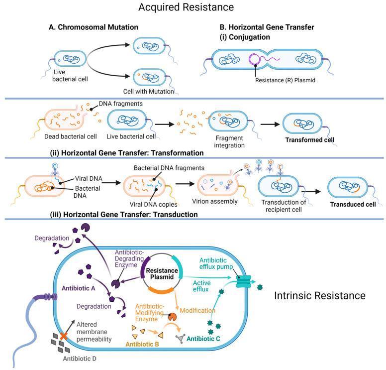
A multi-panel medical illustration detailing the mechanisms of bacterial antibiotic resistance, categorized into 'Acquired Resistance' and 'Intrinsic Resistance'. Acquired Resistance (upper panels) depicts four pathways: 1. Chromosomal Mutation: Shows a live bacterial cell undergoing spontaneous genetic changes. 2. Horizontal Gene Transfer (HGT) via Conjugation: Illustrates the direct transfer of a Resistance (R) Plasmid between two bacteria through a pilus. 3. HGT via Transformation: Shows a live cell uptaking DNA fragments released from a dead bacterial cell, leading to genetic integration. 4. HGT via Transduction: Demonstrates viral DNA (bacteriophage) incorporating bacterial DNA fragments and assembling into virions to infect a recipient cell. Intrinsic Resistance (lower panel) highlights structural and enzymatic mechanisms within a single bacterium: - Antibiotic Degradation: An enzyme chemically breaks down 'Antibiotic A'. - Antibiotic Modification: An enzyme alters 'Antibiotic B' to neutralize it. - Active Efflux: A membrane efflux pump actively transports 'Antibiotic C' out of the cytoplasm. - Altered Membrane Permeability: Structural changes block 'Antibiotic D' from entering the cell. This educational diagram is designed for microbiology and pharmacology studies to explain how pathogens evade antimicrobial therapy.
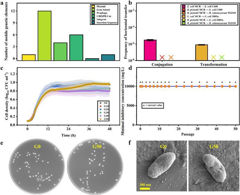
This composite educational graphic illustrates the horizontal and vertical transfer risk analysis of colistin resistance in the bacterium Ralstonia pickettii MCR. Panel a: Bar graph quantifying mobile genetic elements (MGEs), showing Gene Islands as the most abundant (12), followed by CRISPR-Cas (6), prophages (4), and single instances of plasmids and insertion sequences. Panel b: Bar graph of horizontal transfer frequency via conjugation and transformation experiments, indicating low or absent transfer to various recipients like E. coli and R. solanacearum. Panel c: Bacterial growth curves over 48 hours for different generations (G0 to G50), demonstrating stable cell density (log10 CFU/ml) across generations. Panel d: Plot showing stable colistin minimal inhibitory concentration (MIC) at >10,000 mg/L through 50 passages, confirming stable vertical transmission of resistance. Panel e: Comparative clinical photographs of bacterial colonies in Petri dishes at G0 and G50, showing identical circular, white phenotype morphology. Panel f: Scanning electron microscopy (SEM) images at 300 nm scale, showing consistent rod-shaped bacterial cell morphology between the initial (G0) and 50th (G50) generations.
exotoxin endotoxin comparison bacterial toxins mechanism
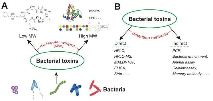
This educational infographic describes the classification and detection of bacterial toxins. Part A is a pathophysiology diagram categorizing toxins by molecular weight (MW). Low MW toxins are represented by a complex organic chemical structure (e.g., small molecules like microcystins), while High MW toxins include complex protein tertiary structures, polysaccharides (indicated by a chain of colored hexagons), and lipopolysaccharides (LPS). Part B is a flowchart detailing diagnostic pathways for toxin identification. Direct detection methods listed include High-Performance Liquid Chromatography (HPLC), HPLC-MS, MALDI-TOF, ELISA, and Strip tests, which focus on identifying the toxin molecule itself. Indirect detection methods include Polymerase Chain Reaction (PCR), bacterial enrichment, animal and cellular assays, and memory antibody testing, which identify the producing bacteria, their genes, or the physiological effects of the toxins. The diagram illustrates key concepts in clinical microbiology, immunology, and forensic toxicology for healthcare professionals and students.
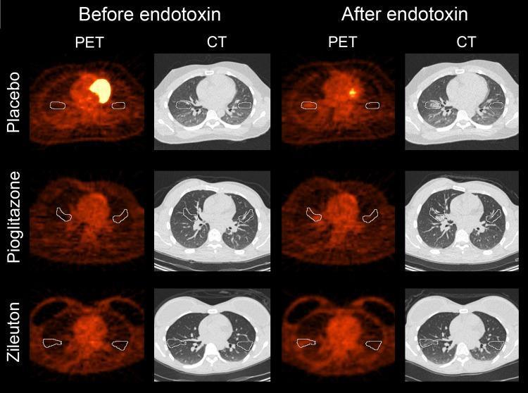
This diagnostic comparison display features axial PET and CT chest scans from three treatment cohorts (Placebo, Pioglitazone, and Zileuton) before and after experimental endotoxin instillation in the right middle lobe. The CT images show the anatomical structure of the lungs and heart, with white outlines indicating volumes of interest (VOIs) for analysis. In the 'After endotoxin' CT columns, subtle localized increases in parenchymal opacity (ground-glass opacities) are visible in the right lung field across all cohorts, representing acute airspace inflammation. The PET scans demonstrate radioactive tracer uptake, highlighting metabolic activity. Notably, the 'After endotoxin' PET scans show an increase in focal uptake within the targeted right middle lobe VOI compared to the baseline 'Before endotoxin' state. This increase in metabolic signal correlates with the inflammatory response triggered by the endotoxin. The comparison demonstrates that neither Pioglitazone nor Zileuton prevented the acute inflammatory metabolic changes or radiological opacities compared to the placebo group.
GENERAL BACTERIOLOGY - Warren Levinson Style
(Based on Jawetz, Melnick & Adelberg's Medical Microbiology 28e + Medical Microbiology 9e)
Note: Warren Levinson's Review of Medical Microbiology and Immunology is published by McGraw-Hill and shares the same framework, chapter organization, and USMLE-orientation as Jawetz. The content below is drawn directly from these library sources and follows Levinson's logical structure exactly.
CHAPTER 1: THE SCIENCE OF MICROBIOLOGY - INTRODUCTION
- There are 5 × 10³⁰ microbial cells on Earth; excluding cellulose, they constitute ~90% of the biomass of the biosphere
- 50-60% of the cells in our bodies are microbes
- The bacteria in the average human gut weigh ~1 kg
- Gut flora genes outnumber human genome genes by 150-fold
- 8% of human DNA is derived from remnants of viral genomes
Microorganisms and Their Classification
| Organism | Cell Type | Nucleus | Cell Wall |
|---|---|---|---|
| Bacteria | Prokaryote | Absent | Peptidoglycan |
| Fungi | Eukaryote | Present | Chitin |
| Protozoa | Eukaryote | Present | None |
| Algae | Eukaryote/Prokaryote | Varies | Cellulose/silica |
| Viruses | Not a cell | None | Protein coat only |
| Prions | Not a cell | None | None (protein only) |
| Viroids | Not a cell | None | None (RNA only) |
CHAPTER 2: BACTERIAL CELL STRUCTURE
PROKARYOTE vs. EUKARYOTE - The Fundamental Distinction
| Feature | Prokaryote | Eukaryote |
|---|---|---|
| Nucleus | Absent (nucleoid only) | Present, membrane-bound |
| Chromosome | Single, circular, double-stranded DNA | Multiple, linear |
| Ribosome | 70S (50S + 30S) | 80S (60S + 40S) |
| Cell wall | Peptidoglycan (most) | None in animal cells |
| Mitochondria | Absent (electron transport in plasma membrane) | Present |
| Sterols in membrane | No (exception: Mycoplasma) | Yes |
| Mitotic apparatus | Absent | Present |
| Size | ~1 µm (0.1-10 µm range) | 7-100 µm |
Levinson exam key: The 70S ribosome is uniquely prokaryotic - this is why antibiotics like aminoglycosides, tetracyclines, macrolides, and chloramphenicol can selectively kill bacteria without harming human (80S) ribosomes.
THE CELL ENVELOPE - Layers from Inside Out
A. THE CYTOPLASMIC (PLASMA) MEMBRANE
- Composition: Phospholipid bilayer + proteins + enzymes
- No sterols - EXCEPT Mycoplasma, which incorporates cholesterol from the host
- Functions (Levinson loves these - the plasma membrane is the "mitochondria" of bacteria):
- Selective permeability barrier
- Site of electron transport chain and ATP synthesis
- Active transport of nutrients into the cell
- DNA replication initiation
- Secretion of proteins (Types I-VI secretion systems)
Antibiotic target: Polymyxins (colistin) act as detergents, disrupting the outer membrane of gram-negative bacteria by competing with Mg²⁺ and Ca²⁺ that hold the LPS together.
B. THE CELL WALL - PEPTIDOGLYCAN (MOST HIGH-YIELD STRUCTURE)
- Polysaccharide backbone: alternating disaccharides of N-acetylglucosamine (NAG) and N-acetylmuramic acid (NAM), linked by β-1,4 glycosidic bonds
- A tetrapeptide side chain hangs from each NAM unit
- The tetrapeptide contains unusual D-amino acids (L-Ala - D-Glu - L-Lys or DAP - D-Ala - D-Ala)
- Cross-linking: The D-Ala at position 4 links to the diamino acid (position 3) of an adjacent chain via transpeptidase (PBP)
| Agent | Target | Mechanism |
|---|---|---|
| Lysozyme (tears, saliva) | β-1,4 bond between NAG-NAM | Cleaves backbone → osmotic lysis |
| β-lactams (penicillins, cephalosporins, carbapenems) | Transpeptidase (PBPs) | Blocks cross-linking → weak wall |
| Vancomycin | D-Ala-D-Ala terminus | Blocks transpeptidation |
| Bacitracin | Bactoprenol (lipid carrier) | Blocks recycling of peptidoglycan precursors |
C. GRAM-POSITIVE CELL WALL
- Thick peptidoglycan (150-500 Å, multiple layers)
- Teichoic acid: polyribitol-phosphate or glycerol-phosphate, covalently attached to NAM → strengthens wall; activates complement
- Lipoteichoic acid (LTA): anchored to cytoplasmic membrane through the peptidoglycan → major PAMP in gram-positive infections; triggers innate immunity
- Surface proteins: Protein A (S. aureus), M protein (S. pyogenes) - attached to peptidoglycan
D. GRAM-NEGATIVE CELL WALL
- Thin peptidoglycan (~20-30 Å, single layer) - sandwiched in the periplasmic space
- Periplasmic space: Contains hydrolytic enzymes (proteases, nucleases), β-lactamases (antibiotic resistance!), and transport proteins
- Outer membrane (OM): A second lipid bilayer with:
- Porins (OmpC, OmpF): β-barrel proteins that form pores for small hydrophilic molecules
- LPS (Lipopolysaccharide) on outer leaflet
- Lipoproteins: Connect OM to peptidoglycan
E. LPS (ENDOTOXIN) - THREE PARTS (HIGH YIELD)
[Lipid A] — [Core polysaccharide] — [O antigen/O polysaccharide]
| Part | Location | Clinical Significance |
|---|---|---|
| Lipid A | Inner, embedded in OM | THE TOXIC MOIETY → fever, hypotension, DIC, septic shock. Binds TLR-4 on macrophages → TNF-α, IL-1, IL-6 |
| Core polysaccharide | Middle | Contains KDO (ketodeoxyoctonate) and heptose sugars; links Lipid A to O antigen |
| O antigen | Outermost, hydrophilic | Repeating sugar units; species-specific; used for serotyping (e.g., E. coli O157:H7) |
Levinson point: Lipid A = endotoxin. It is released when gram-negative bacteria lyse (e.g., from antibiotics or complement). This is why gram-negative sepsis can paradoxically worsen with bactericidal antibiotics (Jarisch-Herxheimer-like reaction).
| Feature | Gram-Positive | Gram-Negative |
|---|---|---|
| Gram stain | Purple | Pink/Red |
| Peptidoglycan | Thick (150-500 Å) | Thin (20-30 Å) |
| Outer membrane | Absent | Present |
| LPS/Endotoxin | Absent | Present |
| Teichoic acid | Present | Absent |
| Periplasmic space | Narrow | Wide |
| Spores | Some | None |
| Penicillin sensitivity | More susceptible | More resistant (OM barrier) |
| Lysozyme | Sensitive | Resistant |
EXTERNAL STRUCTURES
F. CAPSULE
- Composition: Usually polysaccharide (exception: Bacillus anthracis - poly-D-glutamic acid)
- Primary virulence factor - anti-phagocytic
- Inhibits opsonization and phagocytosis
- Quellung reaction: Capsule swells visibly when exposed to type-specific antibody + India ink → used to serotype Streptococcus pneumoniae
- Streptococcus pneumoniae
- Haemophilus influenzae type b
- Neisseria meningitidis
- E. coli (K1 capsule - neonatal meningitis)
- Salmonella typhi (Vi capsule)
- Klebsiella pneumoniae
- invisible (Cryptococcus neoformans - fungus with polysaccharide capsule)
Capsular polysaccharide vaccines (pneumococcal, meningococcal, Hib) work by generating anti-capsule antibodies that opsonize the bacteria.
G. FLAGELLA - MOTILITY ORGANELLE
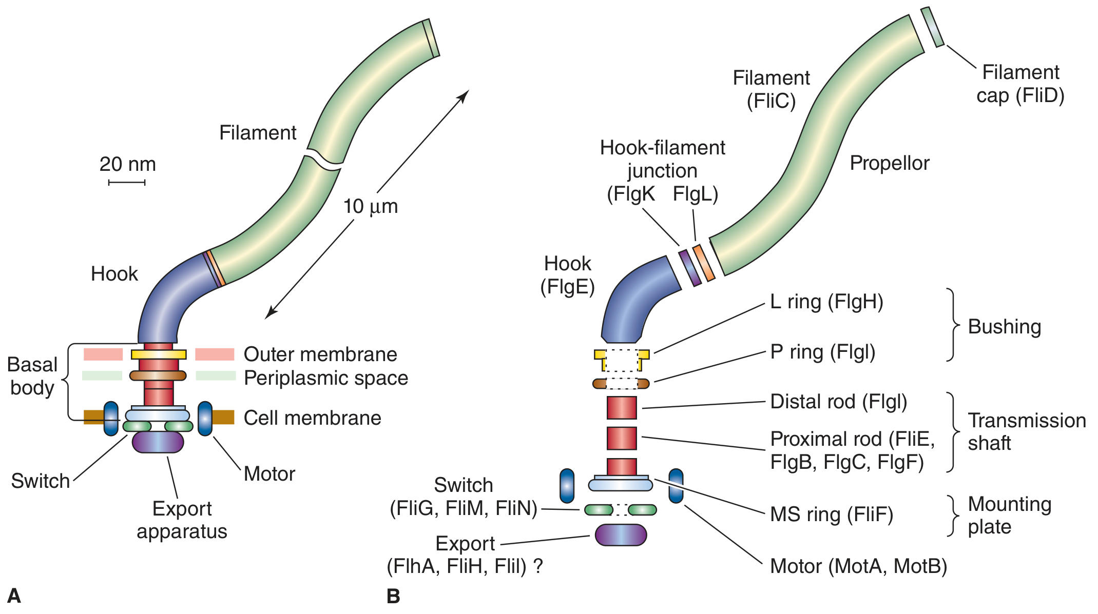
- Filament (FliC/flagellin) - the long external propeller
- Hook (FlgE) - curved connector
- Basal body - the molecular motor, embedded in cell wall; powered by proton motive force (H⁺ gradient)
| Type | Description | Example |
|---|---|---|
| Monotrichous | Single polar flagellum | Vibrio cholerae, Pseudomonas |
| Lophotrichous | Tuft at one pole | Spirillum |
| Amphitrichous | Flagella at both poles | Campylobacter |
| Peritrichous | All around the cell | E. coli, Salmonella, Proteus |
- Flagellin = the protein subunit = H antigen in serotyping (e.g., E. coli O157:H7 - O = O antigen, H = flagellin)
- Spirochetes have axial filaments (periplasmic flagella) between inner and outer membrane → corkscrew motility
- Flagella enable chemotaxis - bacteria move toward attractants (sugars, amino acids) and away from repellents
H. PILI (FIMBRIAE)
| Type | Function | Example |
|---|---|---|
| Common pili (Type I) | Adhesion to host epithelial cells - mannosylated glycoproteins | Uropathogenic E. coli (UPEC), N. gonorrhoeae |
| Type IV pili | Twitching motility, adherence, biofilm | Pseudomonas aeruginosa, Neisseria, V. cholerae |
| Sex pilus (F pilus) | Conjugation - connects donor to recipient | E. coli F+ strains |
Pili are the first step in infection - without adhesion to mucosa, pathogens cannot establish infection.
INTERNAL STRUCTURES
I. THE NUCLEOID (BACTERIAL CHROMOSOME)
- Single, circular, double-stranded DNA (Levinson: "haploid, circular, ds DNA")
- E. coli: ~5 million bp, ~1.3 mm length (1000× the cell diameter!)
- Exceptions to circularity: Borrelia burgdorferi and Streptomyces have linear chromosomes
- Exceptions to single chromosome: Vibrio cholerae and Brucella have two dissimilar chromosomes
- Supercoiled and condensed by DNA gyrase (topoisomerase II) → target of fluoroquinolones
- Rapidly growing bacteria have more copies of the nucleoid per cell than slowly growing ones
J. PLASMIDS
- Small, extrachromosomal, circular DNA (usually)
- Replicate independently of chromosome
- Carry accessory genes: antibiotic resistance, toxins, virulence, metabolic functions
- Transferred between bacteria by conjugation
| Plasmid Type | Genes Carried | Example |
|---|---|---|
| F plasmid (fertility factor) | Conjugation machinery, sex pilus | E. coli F+ |
| R plasmid (resistance plasmid) | Antibiotic resistance (β-lactamase, etc.) | Widespread in gram-negatives |
| Col plasmids | Colicins (bacteriocins) | E. coli |
| Virulence plasmids | Toxins, adhesins | ETEC (LT/ST toxins), S. aureus (TSST-1) |
K. RIBOSOMES
- 70S = 50S + 30S
- Ribosome assembly requires Mg²⁺; removed by chelation → ribosomes dissociate
- Antibiotic targets at 30S: Aminoglycosides, Tetracyclines
- Antibiotic targets at 50S: Macrolides, Chloramphenicol, Clindamycin, Linezolid
- 30S: Aminoglycosides, Tetracyclines
- 50S: Erythromycin (macrolides), Chloramphenicol, clindAmycin, lineMezolid (linezolid)
L. INCLUSION BODIES / GRANULES
- Metachromatic granules (Babes-Ernst / volutin granules): Stored polymetaphosphate; stain reddish-purple with methylene blue or toluidine blue when background is blue → diagnostic for Corynebacterium diphtheriae ("Chinese letter" or "V/L" arrangement + metachromatic granules)
- Poly-β-hydroxybutyrate (PHB): Lipid-like energy reserve in gram-negative bacteria
- Sulfur granules: Energy storage in sulfur bacteria
CHAPTER 3: ENDOSPORES
Who Makes Spores?
- Bacillus (aerobic spore formers)
- Clostridium (anaerobic spore formers)
Memory trick: B.C. - Bacillus and Clostridium - ancient and indestructible.
Why and When?
Spore Structure (inside out):
Core → Inner membrane → Cortex (modified PG) → Spore coat (keratin) → Exosporium
- Core: Contains complete chromosome, minimal enzymes, ribosomes, and high concentrations of calcium dipicolinate (chelated with Ca²⁺)
- Calcium dipicolinate: Responsible for the heat resistance of spores
- Dehydrated state: Also contributes to heat resistance
- Refractile (bright) under phase contrast microscopy
Properties of Spores:
- Highly resistant to: heat, desiccation, UV, most chemicals, disinfectants, alcohol
- Killed only by: autoclaving (121°C, 15 psi, 15-20 min), dry heat (160°C × 2h), ethylene oxide gas, glutaraldehyde, chlorine dioxide
Spore Location - Diagnostic:
| Spore Position | Shape | Organism |
|---|---|---|
| Central | Does NOT swell cell | B. anthracis, C. perfringens |
| Subterminal | Oval | C. botulinum, B. cereus |
| Terminal | "Drumstick" or "tennis racket" shape | C. tetani (drumstick) |
Germination:
Clinical importance:
- C. difficile: Spores persist on hospital surfaces for months; alcohol hand sanitizers do NOT kill them (need soap and water, chlorine bleach)
- B. anthracis: Spores used in bioterrorism (2001 anthrax letters); inhalation of spores → pulmonary anthrax
- C. botulinum: Spores in soil and honey; germinate under anaerobic conditions; honey should not be given to infants < 1 year (infant botulism)
- C. tetani: Spores in soil; penetrate wounds
CHAPTER 4: BACTERIAL GROWTH AND NUTRITION
Growth Curve - The 4 Phases
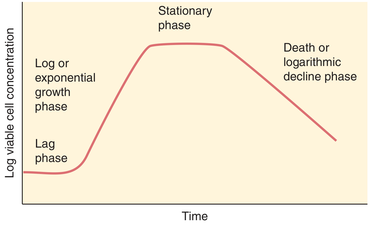
| Phase | Growth Rate | What Happens | Clinical Relevance |
|---|---|---|---|
| Lag | Zero | Cells adapt to new environment; synthesize enzymes and metabolites; NO division | Explains why infections take time to manifest |
| Log (Exponential) | Constant maximum | New material is catalytic → exponential increase; cell mass doubles each generation | Most susceptible to cell wall synthesis inhibitors (penicillins); used to measure generation time |
| Stationary | Zero (growth = death) | Nutrient depletion + toxic waste accumulation; sporulation begins | Toxin production peaks (many exotoxins like diphtheria, Shiga toxin produced here) |
| Death (Decline) | Negative | Deaths exceed growth; irreversible | Seen with antibiotic killing |
Generation Time (Doubling Time)
| Organism | Generation Time |
|---|---|
| E. coli | ~20 minutes (fastest common pathogen) |
| Staphylococcus | ~30 minutes |
| M. tuberculosis | ~20 hours (explains why TB therapy = 6 months) |
| Treponema pallidum | ~30 hours (explains why penicillin course is prolonged) |
| M. leprae | ~12 days (explains years of treatment) |
Oxygen Requirements (Levinson's Table)
| Category | O₂ Requirement | Mechanism | Examples |
|---|---|---|---|
| Obligate aerobe | Requires O₂ | Final electron acceptor is O₂ | M. tuberculosis (likes high O₂ → upper lobe TB), Pseudomonas, Nocardia |
| Obligate anaerobe | Cannot tolerate O₂ (killed) | Lack superoxide dismutase and catalase | Bacteroides fragilis, Clostridium, Fusobacterium, Actinomyces |
| Facultative anaerobe | Grows with or without O₂ | Uses O₂ when available; ferments when not | E. coli, Staphylococcus, Listeria, Enterobacteriaceae |
| Microaerophile | Requires low O₂ (2-10%) | High O₂ concentrations are toxic | Helicobacter pylori, Campylobacter |
| Aerotolerant | Grows without O₂; not harmed by it | Fermenters with some protective enzymes | Streptococcus (no cytochrome system) |
| Capnophile | Requires increased CO₂ (5-10%) | - | Neisseria, Brucella, Haemophilus |
- Superoxide dismutase: 2 O₂⁻ + 2H⁺ → O₂ + H₂O₂
- Catalase: 2 H₂O₂ → 2 H₂O + O₂
Temperature Requirements
| Category | Optimal Temperature | Example |
|---|---|---|
| Psychrophile | 0-20°C | Food spoilage organisms |
| Mesophile | 35-37°C | All human pathogens |
| Thermophile | 45-80°C | Thermus aquaticus (source of Taq polymerase for PCR) |
| Hyperthermophile | >80°C | Archaea |
Nutritional Requirements
- Autotrophs (lithotrophs): Can use inorganic carbon (CO₂) as carbon source → non-pathogens
- Heterotrophs (organotrophs): Require organic carbon sources → all human pathogens
- Fastidious organisms: Have complex nutritional requirements; need special media:
- N. gonorrhoeae: Thayer-Martin medium (chocolate agar + antibiotics)
- H. influenzae: Chocolate agar (factors X and V)
- Legionella: Buffered charcoal yeast extract (BCYE) agar with L-cysteine and iron
CHAPTER 5: BACTERIAL METABOLISM
Energy Production
| Pathway | O₂ | ATP yield | Key organisms |
|---|---|---|---|
| Aerobic respiration | Required | ~38 ATP/glucose | Aerobes and facultative anaerobes |
| Anaerobic respiration | Not O₂ (uses NO₃⁻, SO₄²⁻) | Intermediate | Pseudomonas, Bacteroides |
| Fermentation | None | 2 ATP/glucose | Lactobacillus, Clostridium, Streptococcus |
Metabolically Important End-Products (Diagnostic Use)
| Test | Principle | Organisms |
|---|---|---|
| Catalase test | H₂O₂ → H₂O + O₂ (bubbles) | Staphylococcus (+) vs. Streptococcus (-) |
| Oxidase test | Cytochrome c oxidase | Neisseria, Pseudomonas, Campylobacter (+) |
| Urease | Urea → NH₃ + CO₂ | H. pylori, Proteus, Klebsiella, Cryptococcus |
| Indole | Tryptophan → indole | E. coli (+), Klebsiella (-) |
| H₂S production | Thiosulfate reduction | Salmonella, Proteus |
| Coagulase | Fibrinogen clotting | S. aureus (+) vs. S. epidermidis (-) |
| CAMP test | β-hemolysin + streptolysin S → enhanced hemolysis | Group B Streptococcus (+) |
| Bile solubility | Autolysis in bile | S. pneumoniae (+) |
CHAPTER 6: BACTERIAL CLASSIFICATION AND STAINING
The Gram Stain - Step by Step
- Crystal violet (primary stain) → all cells purple
- Gram's iodine (mordant) → fixes crystal violet-iodine complex in cells
- Acetone-alcohol (decolorizer) → removes complex from thin-walled gram-negatives (outer membrane dissolves)
- Safranin (counterstain) → gram-negative cells turn pink/red
- Gram-positive = PURPLE (thick peptidoglycan traps the crystal violet-iodine complex)
- Gram-negative = PINK (thin peptidoglycan + outer membrane → decolorizer removes crystal violet)
| Organism | Reason | Better Stain |
|---|---|---|
| Mycobacterium | Mycolic acid wall resists stain | Acid-fast (Ziehl-Neelsen) |
| Mycoplasma | No cell wall | Phase contrast/electron microscopy |
| Treponema pallidum | Too thin (<0.2 µm) | Dark-field microscopy |
| Rickettsia, Chlamydia | Intracellular | Giemsa or Gimenez |
| Legionella | Stains poorly | Silver (Dieterle) stain |
Special Stains - High Yield
| Stain | What It Detects | Organism |
|---|---|---|
| Acid-fast (ZN/Kinyoun) | Mycolic acid (resists acid-alcohol decolorization) = red/pink bacilli | M. tuberculosis, M. leprae, Nocardia (partial) |
| India ink | Capsule (clear halo around dark background) | Cryptococcus neoformans, Klebsiella |
| Giemsa | Intracellular organisms, parasites | Chlamydia, Rickettsia, Borrelia, Plasmodium |
| Silver stain | Spirochetes, Legionella, fungi (PAS also) | Treponema, Pneumocystis jirovecii |
| PAS | Periodic acid-Schiff → glycogen, polysaccharides | Tropheryma whipplei (Whipple's disease) |
| Spore stain (Schaeffer-Fulton) | Spores stain green; vegetative cells red | Bacillus, Clostridium |
| Methylene blue | Metachromatic granules → reddish-purple | Corynebacterium diphtheriae |
| Fluorescent (auramine-rhodamine) | Mycobacterial cell wall | M. tuberculosis (faster than ZN) |
CHAPTER 7: BACTERIAL GENETICS
7A. The Bacterial Chromosome
- Haploid (single copy of each gene) - but rapidly growing bacteria may have 2-4 chromosome copies
- Single origin of replication (oriC) → bidirectional replication
- DNA gyrase (topoisomerase II): Relieves positive supercoiling ahead of replication fork → target of fluoroquinolones (ciprofloxacin, levofloxacin)
7B. Mutation
- Spontaneous mutation rate: ~10⁻⁶ to 10⁻⁹ per gene per cell division
- Types: Point mutation (missense, nonsense, frameshift), deletion, insertion, inversion
- Physical mutagens: UV light → thymine dimers → DNA repair required; ionizing radiation → double-strand breaks
- Chemical mutagens: Nitrous acid (deaminates bases), alkylating agents, base analogs, acridines (intercalating agents)
- SOS response: Emergency DNA repair system activated by extensive DNA damage; can introduce errors (mutagenic repair)
- Ames test: Uses histidine-requiring Salmonella mutants; a substance is mutagenic if it increases reversion to His⁺; mutagenic = potentially carcinogenic
7C. Three Mechanisms of Gene Transfer (HORIZONTAL GENE TRANSFER)
TRANSFORMATION - "DNA uptake from environment"
- Definition: Uptake of naked, free DNA from the environment by a competent bacterium
- Competence: Natural ability to take up DNA; requires specific surface proteins
- Naturally competent bacteria: Strep. pneumoniae, H. influenzae, Neisseria, Bacillus
- Artificially induced: Heat shock + CaCl₂ (laboratory technique)
- Smooth (S) Strep. pneumoniae (encapsulated, virulent) + heat-killed S → LIVE mice die
- Rough (R) Strep. pneumoniae (no capsule, avirulent) alone → mice survive
- S-strain "transforming principle" → R becomes S
- Later (Avery, MacLeod, McCarty 1944): Transforming principle = DNA → first proof that DNA is the genetic material
CONJUGATION - "Bacterial sex"
- Definition: Direct cell-to-cell transfer of DNA through a sex pilus (F pilus)
- Requires physical contact between donor and recipient
- The F (fertility) plasmid encodes the F pilus and all conjugation machinery
| Cross | What's transferred | Result |
|---|---|---|
| F⁺ × F⁻ | F plasmid only | F⁻ becomes F⁺; chromosomal genes rarely transferred |
| Hfr × F⁻ | F integrated into chromosome; chromosomal genes transferred | High frequency of recombination; rarely complete transfer; F⁻ stays F⁻ |
| F' (F prime) × F⁻ | F' plasmid (F + adjacent chromosomal genes) | Sexduction (F-duction); specific chromosomal genes transferred reliably |
Clinical relevance: Conjugation is the PRIMARY mechanism for spreading R plasmids (antibiotic resistance) between bacteria - even across different species.
TRANSDUCTION - "Bacteriophage-mediated gene transfer"
- Definition: Transfer of bacterial DNA from one bacterium to another by a bacteriophage (bacterial virus)
| Type | Mechanism | Genes Transferred |
|---|---|---|
| Generalized transduction | Phage accidentally packages a random fragment of bacterial DNA instead of its own; any gene can be transferred | Any gene |
| Specialized transduction | Temperate phage integrates (prophage), then excises imprecisely taking adjacent chromosomal genes with it; only genes near the integration site | Only specific adjacent genes |
| Bacterium + Phage → | New Toxin/Property |
|---|---|
| C. diphtheriae + β-phage | Diphtheria toxin (DT) - only toxigenic if the phage is present |
| S. pyogenes + phage | Erythrogenic toxin (scarlet fever) |
| V. cholerae + CTXφ phage | Cholera toxin |
| C. botulinum + phage | Botulinum toxin (types C and D) |
| S. aureus + phage | Panton-Valentine leukocidin (PVL), phage-encoded toxins |
Levinson exam trap: If you remove the phage from C. diphtheriae, the bacteria become non-toxigenic and harmless. The toxin gene is on the phage, NOT the bacterial chromosome.
7D. Transposons ("Jumping Genes")
- DNA sequences that can "transpose" (jump) from one location in the genome to another
- Carry their own transposase enzyme
- Can move between chromosomes and plasmids
- Often carry antibiotic resistance genes
- Enable rapid spread of resistance within and between bacterial strains
CHAPTER 8: PATHOGENESIS OF BACTERIAL INFECTIONS
Steps in Pathogenesis
Inoculation → Adherence → Colonization → Invasion → Evasion of host defenses → Tissue damage → Transmission
Virulence Factors
1. Adhesins
- Pili/fimbriae: Attach to specific host cell receptors (mannose, fibronectin, etc.)
- Lipoteichoic acid: Gram-positive adhesin; binds fibronectin on epithelial surfaces (S. pyogenes uses LTA + M protein to adhere to nasopharynx)
2. Invasins
- IgA protease: Cleaves secretory IgA on mucosal surfaces → removes antibody barrier; produced by N. gonorrhoeae, S. pneumoniae, H. influenzae, N. meningitidis
- Hyaluronidase: Breaks down hyaluronic acid in connective tissue → "spreading factor"; Strep. pyogenes, Clostridium
- Collagenase: Breaks down collagen → Clostridium
- Coagulase (S. aureus): Converts fibrinogen → fibrin → forms clot around bacteria → protects from phagocytosis
- Streptokinase/Fibrinolysin (S. pyogenes): Dissolves clots → spreads bacteria
3. Anti-phagocytic factors
- Capsule: Prevents opsonization (all encapsulated bacteria)
- Protein A (S. aureus): Binds Fc region of IgG → prevents opsonin recognition by Fc receptors on phagocytes
- M protein (S. pyogenes): Antiphagocytic; inhibits C3b deposition
- Intracellular survival: Mycobacterium (inhibit phagolysosome fusion), Listeria (escape from phagosome), Salmonella (survive within macrophage)
4. Iron Acquisition - Siderophores
- Iron is essential for bacterial growth; serum iron is normally kept low (bound to transferrin/lactoferrin)
- Bacteria produce siderophores (e.g., enterobactin, aerobactin) that chelate iron from host proteins
- Critical virulence mechanism for: E. coli, Klebsiella, Pseudomonas
CHAPTER 9: TOXINS
EXOTOXINS vs. ENDOTOXINS - Levinson's Master Comparison Table
| Feature | Exotoxin | Endotoxin (LPS) |
|---|---|---|
| Produced by | Gram-positive AND gram-negative | Gram-negative ONLY |
| Chemical nature | Protein | Lipopolysaccharide (Lipid A) |
| Location/Release | Actively secreted during growth | Part of outer membrane; released on bacterial death/lysis |
| Heat stability | Heat-labile (destroyed 60-80°C) - EXCEPTIONS: S. aureus enterotoxin, TSST-1 | Heat-stable (withstands 250°C) |
| Antigenicity | Highly antigenic | Weakly antigenic |
| Toxoid possible? | YES - formaldehyde converts to toxoid (DT vaccine, tetanus toxoid) | NO effective toxoid |
| Potency | Extremely potent (nanogram quantities lethal) | Moderate potency |
| Mechanism | Specific receptors and cellular targets | TLR-4 → macrophage activation → TNF-α, IL-1, IL-6 → fever, hypotension, DIC |
| Clinical syndrome | Specific (tetanus = spasm; cholera = watery diarrhea) | Generalized: fever, hypotension, septic shock, DIC |
| Detection | Specific bioassays, ELISA | Limulus amebocyte lysate (LAL) test - pyrogen testing of IV fluids |
Key Exotoxin Mechanisms - Levinson's Favourite Table
| Toxin | Organism | Mechanism | Effect |
|---|---|---|---|
| Cholera toxin | V. cholerae | ADP-ribosylates Gsα → permanently activates adenylyl cyclase → ↑↑↑ cAMP | Profuse rice-water diarrhea (loss of Cl⁻ and water) |
| Pertussis toxin | B. pertussis | ADP-ribosylates Giα → permanently inactivates Giα → ↑ cAMP | Whooping cough; lymphocytosis (blocks lymphocyte egress from nodes) |
| Diphtheria toxin | C. diphtheriae (from β-phage) | ADP-ribosylates EF-2 (elongation factor 2) → blocks protein synthesis | Pseudomembrane in pharynx; myocarditis; demyelinating neuropathy |
| Anthrax toxin | B. anthracis | Lethal factor (Zn²⁺ metalloprotease, cleaves MAP kinases) + Edema factor (adenylyl cyclase → ↑ cAMP) + Protective antigen (receptor binding/pore) | Cutaneous anthrax, pulmonary anthrax, septicemia |
| Botulinum toxin | C. botulinum | Cleaves SNARE proteins (VAMP/synaptobrevin) → blocks ACh release at neuromuscular junction | Flaccid descending paralysis; diplopia, dysarthria, dysphagia; no fever |
| Tetanospasmin | C. tetani | Cleaves synaptobrevin → blocks glycine and GABA release at inhibitory interneurons (Renshaw cells) | Spastic (rigid) ascending paralysis; lockjaw (trismus), risus sardonicus, opisthotonus |
| TSST-1 | S. aureus | Superantigen → cross-links MHC II + T-cell receptor → activates up to 20% of all T cells → cytokine storm | Toxic shock syndrome (fever, rash, hypotension, multi-organ failure) |
| Shiga toxin | Shigella, EHEC (O157:H7) | N-glycosidase cleaves 28S rRNA of 60S ribosome → inhibits protein synthesis; also microangiopathy | Bloody diarrhea, HUS (hemolytic uremic syndrome) |
| Staphylococcal enterotoxins (A-E, esp. A) | S. aureus | Superantigens; heat-stable (resistant to 100°C for 30 min) | Food poisoning (preformed toxin; vomiting within 1-6 hrs) |
| Exfoliatin A & B | S. aureus | Serine protease; cleaves desmoglein-1 in epidermis | Scalded skin syndrome (SSSS); bullous impetigo |
| Alpha toxin (lecithinase) | C. perfringens | Phospholipase C → destroys cell membranes and lecithin in RBCs | Gas gangrene (myonecrosis) + intravascular hemolysis |
Botulism vs. Tetanus mnemonic: Both cleave SNARE proteins. Botulinum → Flaccid (Flaccid = botulinum). Tetanus → Spastic (Spastic = Spawns convulsions = tetanus). Botulinum blocks ACh (flaccid). Tetanus blocks glycine/GABA (spastic).
CHAPTER 10: NORMAL FLORA
Distribution by Body Site
| Site | Predominant Organisms |
|---|---|
| Skin | S. epidermidis (most common), S. aureus, diphtheroids (Corynebacterium), Propionibacterium acnes |
| Oral cavity | Viridans streptococci (S. mutans, S. sanguis), Bacteroides, Fusobacterium, Actinomyces |
| Upper respiratory tract | S. pneumoniae, H. influenzae, N. meningitidis (carriers), viridans streptococci |
| Colon | Bacteroides fragilis (most abundant anaerobe), E. coli, Lactobacillus, Bifidobacterium |
| Vagina | Lactobacillus (dominant; produces lactic acid → low pH ~4.5 → protects against pathogens) |
| Urethra (anterior) | S. epidermidis, diphtheroids |
Functions of Normal Flora
- Colonization resistance: Compete for nutrients and adhesion sites; prevent pathogen overgrowth
- Nutrition: Synthesize vitamin K (gut flora) and some B vitamins
- Immune development: Stimulate IgA production and innate immune training
- Metabolic: Ferment complex carbohydrates → short-chain fatty acids → colon health
Clinical Consequences of Flora Disruption
- Broad-spectrum antibiotics wipe out normal gut flora → C. difficile overgrowth → pseudomembranous colitis
- Candida vaginitis/thrush after antibiotic use
- Aspiration pneumonia: From oral anaerobes (Bacteroides, Fusobacterium, Peptostreptococcus)
CHAPTER 11: STERILIZATION AND DISINFECTION
Definitions (Levinson's precise terminology)
| Term | Definition |
|---|---|
| Sterilization | Destruction or removal of ALL living organisms, including spores (absolute term) |
| Disinfection | Destruction of most pathogens; may NOT kill spores |
| Antisepsis | Use of disinfectants on living tissue (skin, mucous membranes) |
| Pasteurization | Heat sufficient to kill pathogens (NOT sterilization); 74°C × 3-5 sec or 62°C × 30 min |
| Bactericidal | Kills bacteria |
| Bacteriostatic | Inhibits growth (bacteria revive when agent removed) |
| Asepsis | Procedures to prevent microbial contamination |
| Sanitization | Reduce microbial load to "safe" levels (food/housekeeping) |
Physical Methods
| Method | Conditions | Kills Spores? | Uses |
|---|---|---|---|
| Autoclave (steam under pressure) | 121°C, 15 psi, 15-20 min | YES - GOLD STANDARD | Surgical instruments, media, glassware |
| Dry heat oven | 160°C × 2h or 170°C × 1h | YES | Glassware, oils, powders (steam-sensitive items) |
| Incineration | Direct flame | YES | Inoculation loops, biohazardous waste |
| Filtration | 0.22 µm membrane | YES (physical removal) | Heat-labile solutions (serum, antibiotics, vaccines) |
| Boiling (100°C) | 10 min | NO | Kills vegetative bacteria only; NOT sterilization |
| Pasteurization | 74°C × 3-5 sec | NO | Milk, beverages |
| UV irradiation | ~260 nm (DNA absorption peak) | Limited | Surface/air decontamination; induces thymine dimers |
| Tyndallization | Boiling × 30 min on 3 consecutive days | YES | Intermittent sterilization of heat-sensitive items |
| Gamma irradiation | High-energy ionizing radiation | YES | Medical devices, food (cold sterilization) |
| Ethylene oxide gas | - | YES | Heat-sensitive equipment, plastics (cold sterilization) |
Chemical Methods
| Agent | Mechanism | Level | Uses |
|---|---|---|---|
| Glutaraldehyde (2%) | Alkylates proteins (cross-links) | High level (sterilization) | Flexible endoscopes |
| Ethylene oxide | Alkylates DNA/proteins | Sterilizing | Plastics, medical devices |
| Formaldehyde | Cross-links proteins and nucleic acids | High level | Vaccine preparation (inactivation) |
| Chlorine/Hypochlorite (bleach) | Oxidation, denaturation | High to intermediate | Water treatment, surfaces, C. difficile spore killing |
| Iodine/Iodophors (Betadine) | Oxidation of sulfhydryl groups | Intermediate | Skin antisepsis pre-procedure |
| Alcohols (70% ethanol, isopropanol) | Protein denaturation, membrane dissolution | Intermediate | Hand rubs; skin prep; NOT effective vs. spores |
| Phenols (carbolic acid - Lister's agent) | Denature proteins, disrupt membranes | Intermediate | Wound antisepsis, surface disinfection |
| Quaternary ammonium compounds | Disrupt membranes; displace Mg²⁺ | Low | Surface disinfectants; NOT effective vs. mycobacteria/spores |
| Heavy metals (Ag⁺, Hg²⁺) | Bind sulfhydryl groups, inactivate enzymes | Intermediate | Silver sulfadiazine (burns); Ag in catheters |
Resistance Hierarchy (Most → Least Resistant)
Prions > Spores > Mycobacteria > Non-enveloped viruses > Fungi > Vegetative bacteria > Enveloped viruses
Levinson high-yield point: Enveloped viruses (HIV, HBV, HSV, influenza) are the EASIEST to kill - soap and alcohol are effective. Non-enveloped viruses (poliovirus, norovirus, HAV) are much harder. Spores need autoclaving.
CHAPTER 12: ANTIBIOTICS - MECHANISMS OF ACTION
| Class | Specific Target | Bactericidal/Static | Spectrum |
|---|---|---|---|
| β-lactams (penicillin, amoxicillin, cephalosporins, carbapenems, aztreonam) | PBPs (transpeptidases) → block peptidoglycan cross-linking | Cidal | Variable by agent |
| Vancomycin | D-Ala-D-Ala terminus | Cidal | Gram-positive only (too large to cross OM of gram-negatives) |
| Aminoglycosides (gentamicin, streptomycin, tobramycin) | 30S ribosome - causes mRNA misreading → aberrant proteins | Cidal (requires O₂ for uptake → NOT effective vs. anaerobes) | Gram-negative aerobic |
| Tetracyclines | 30S - block aminoacyl-tRNA binding at A site | Static | Broad-spectrum |
| Chloramphenicol | 50S - inhibits peptidyl transferase | Static (cidal for some) | Broad-spectrum |
| Macrolides (erythromycin, azithromycin, clarithromycin) | 50S - block translocation (prevent ribosome movement along mRNA) | Static | Gram-positive + atypicals |
| Clindamycin | 50S - same site as macrolides | Static | Gram-positive + anaerobes |
| Linezolid | 50S - blocks initiation complex formation | Static | Gram-positive (MRSA, VRE) |
| Fluoroquinolones (ciprofloxacin, levofloxacin) | DNA gyrase (topoisomerase II) in gram-neg; Topoisomerase IV in gram-pos | Cidal | Broad-spectrum |
| Rifampin | RNA polymerase (β subunit) - blocks mRNA synthesis | Cidal | M. tuberculosis, gram-positive coverage |
| Sulfonamides | Dihydropteroate synthase - blocks folate synthesis | Static | Broad (bacteria must synthesize their own folate) |
| Trimethoprim | Dihydrofolate reductase - blocks folate reduction | Static | Broad (used with sulfa = co-trimoxazole) |
| Polymyxins (colistin) | Outer membrane of gram-neg - detergent effect | Cidal | Gram-negative only (last resort) |
| Daptomycin | Cytoplasmic membrane depolarization | Cidal | Gram-positive only (MRSA, VRE) |
| Isoniazid (INH) | Mycolic acid synthesis (InhA enzyme) | Cidal (dividing cells) | Mycobacterium only |
Antibiotic Resistance Mechanisms
- Enzymatic inactivation: β-lactamases destroy β-lactam ring; acetyltransferases modify aminoglycosides
- Altered target: MRSA = PBP2a (altered PBP with low affinity for β-lactams); VRE = D-Ala-D-Lac instead of D-Ala-D-Ala
- Decreased permeability: Loss of porins in gram-negative outer membrane → blocks entry of hydrophilic antibiotics
- Efflux pumps: Active export of antibiotics (tetracyclines, fluoroquinolones) out of bacteria
- Bypass pathway: Sulfonamide resistance via overproduction of PABA (outcompetes drug)
LEVINSON-STYLE HIGH-YIELD SUMMARY TABLES
Bacteria That Are Exceptions to Rules
| Bacterium | The Rule It Breaks |
|---|---|
| Mycoplasma | Gram-positive with no cell wall → no peptidoglycan → β-lactam resistant |
| Chlamydia | Gram-negative but no peptidoglycan; obligate intracellular |
| Mycobacterium | Gram-positive but stains poorly; acid-fast; very slow growth |
| Listeria | Gram-positive rod that grows at 4°C (refrigerator temperature) |
| Borrelia burgdorferi | Linear chromosome (unusual for prokaryote) |
| V. cholerae / Brucella | Two dissimilar chromosomes (unusual) |
| B. anthracis | Capsule made of poly-D-glutamic acid (protein, not polysaccharide) |
| Spirochetes | Flagella are internal (periplasmic) |
Clinical Correlation: Which Toxin = Which Mechanism
| ADP-Ribosylating Toxins | Target | Effect |
|---|---|---|
| Cholera toxin | Gsα (activates) | ↑↑↑ cAMP → diarrhea |
| Pertussis toxin | Giα (inactivates) | ↑ cAMP → lymphocytosis, whooping cough |
| Diphtheria toxin | EF-2 | Block protein synthesis |
| Anthrax edema factor | Adenylyl cyclase itself | ↑↑ cAMP → edema |
| Pseudomonas exotoxin A | EF-2 (same as diphtheria) | Block protein synthesis |