

image.png
from art and science give me entire topic explanation in book language
Reading File
The image shows topics from Operative Dentistry: Pit and Fissure Caries, Smooth Surface Caries, Root Caries, and Active and Arrested Caries. I'll search all of these in parallel now.
Finding Sources
Finding Sources
Finding Sources
Finding Sources
Finding Sources
Finding Sources
Good — I found relevant sources. Let me now read all the key sections in parallel.
Reading File
Reading File
Reading File
Reading File
Good content found. Now let me fetch the key images and get additional paragraphs on pit/fissure anatomy, root caries, and active/arrested caries.
Loading Image
Loading Image
Finding Sources
Finding Sources
Reading File
Reading File
I have excellent content across multiple authoritative textbooks. Let me now compose the comprehensive book-language explanation.---
OPERATIVE DENTISTRY — Caries: Classification, Pathogenesis, and Management
Foundational Concepts: Dental Anatomy and Cariogenesis
Tooth Structure
The crown of the tooth consists of three layers arranged concentrically from the outermost to the innermost: enamel, dentin, and the pulp cavity. Enamel is the only part of the tooth visible under normal conditions and constitutes the hardest tissue in the human body, composed principally of calcium hydroxyapatite crystals. The outermost layer of the tooth root is the cementum, which anchors the periodontal ligament to the adjacent alveolar bone. Deep to the enamel (for the crown) or the cementum (for the root) lies the dentin, an intermediate yellow layer containing porous microtubules that cushion the enamel during mastication. The deepest compartment is the pulp cavity, housing the neurovascular supply of the tooth.
(Rosen's Emergency Medicine: Concepts and Clinical Practice)
The Cariogenic Process
Dental caries result from focal demineralization of tooth structure — enamel, dentin, and cementum — caused by organic acids generated during the fermentation of dietary carbohydrates by plaque bacteria. Caries is the principal cause of tooth loss before the age of 35 years worldwide.
(Robbins & Kumar Basic Pathology; Robbins, Cotran & Kumar Pathologic Basis of Disease)
The sequence of events leading to caries formation is as follows:
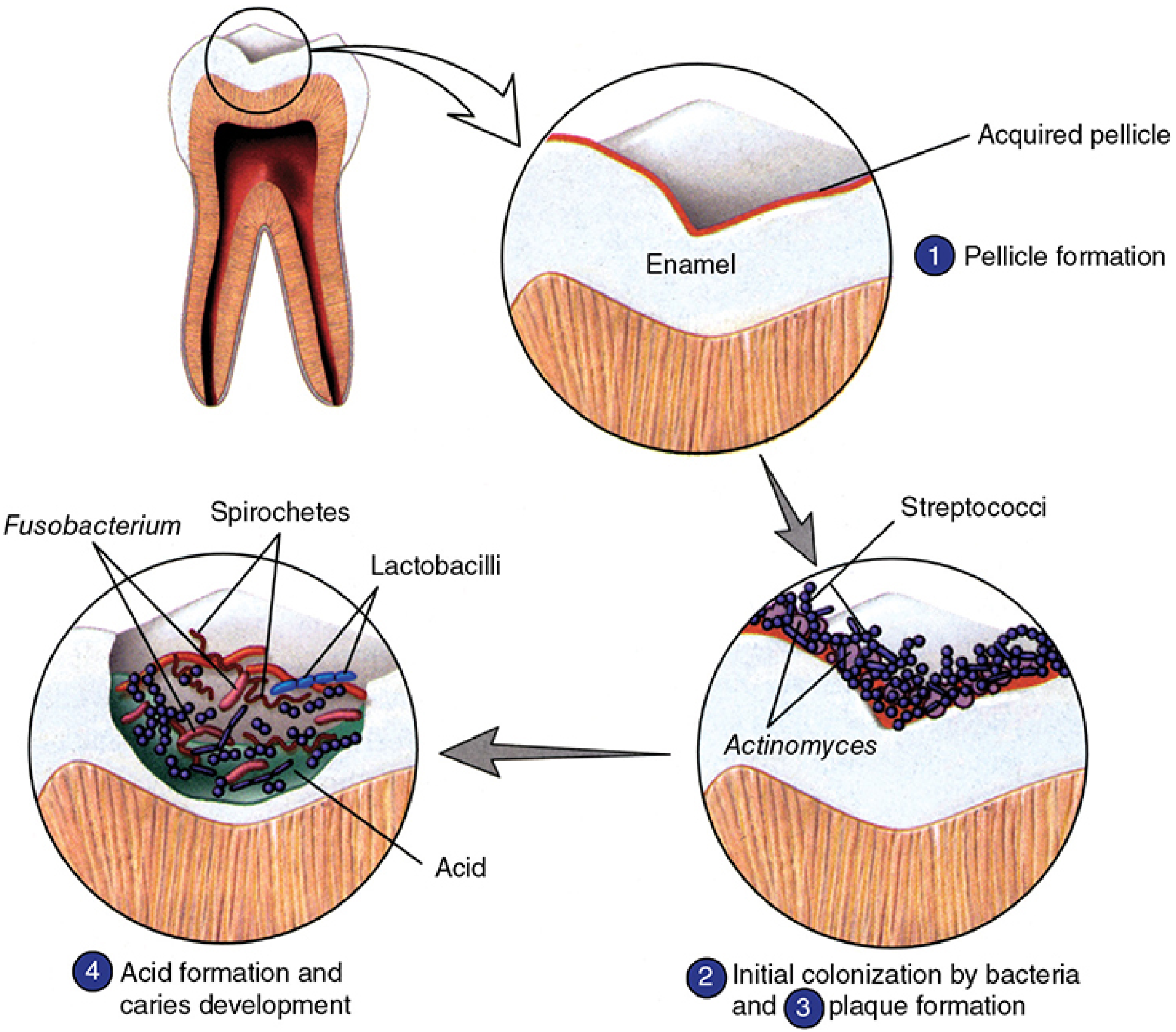
Step 1 — Pellicle formation: Immediately following tooth eruption or cleaning, salivary proteins and glycoproteins adsorb onto the mineralized enamel surface to form an acquired dental pellicle. This thin organic film serves as the substrate to which bacteria initially adhere.
Step 2 — Bacterial colonization: Early colonizers — primarily Streptococcus sanguis, S. mutans, S. mitis, S. salivarius, and Actinomyces species — attach to the pellicle via ionic, hydrophobic, and lectin-like surface interactions.
Step 3 — Plaque biofilm maturation: As the biofilm evolves over 2–4 days, late colonizers appear — predominantly Gram-negative anaerobes (Porphyromonas, Prevotella, Fusobacterium, Veillonella, Treponema denticola). High-molecular-weight extracellular glucan polymers, synthesized mainly by S. mutans in association with Actinomyces species, act as cement binding the biofilm together. Mature dental plaque may harbor 300–400 bacterial species.
Step 4 — Acid production and demineralization: Dietary monosaccharides and disaccharides (glucose, fructose, sucrose, lactose, maltose) permeate the plaque and are metabolized by bacteria — particularly S. mutans — through glycolysis, producing organic acids (chiefly lactic acid). These acids lower the local pH to levels sufficient to dissolve the hydroxyapatite of enamel.
(Jawetz, Melnick & Adelberg's Medical Microbiology; Sherris & Ryan's Medical Microbiology)
Role of Streptococcus mutans
S. mutans is regarded as the dominant organism for the initiation of caries. Its cariogenic superiority stems from several properties:
- Greater efficiency in metabolizing sucrose compared with other oral bacteria
- Regulatory systems that stimulate conversion of dietary carbohydrates to acid and intracellular storage polymers
- Ability to synthesize glucans that enhance plaque adhesion
- Aciduricity — survival and continued acid production at low pH
However, caries is not a single-organism disease. Multiple species including S. salivarius, S. sanguis, S. sobrinus, Lactobacillus acidophilus, L. casei, Actinomyces viscosus, and A. naeslundii participate in the evolution of the carious lesion. In particular, lactobacilli are strongly associated with active lesion progression.
(Sherris & Ryan's Medical Microbiology; Histology: A Text and Atlas)
1. Pit and Fissure Caries
Anatomy of Pits and Fissures
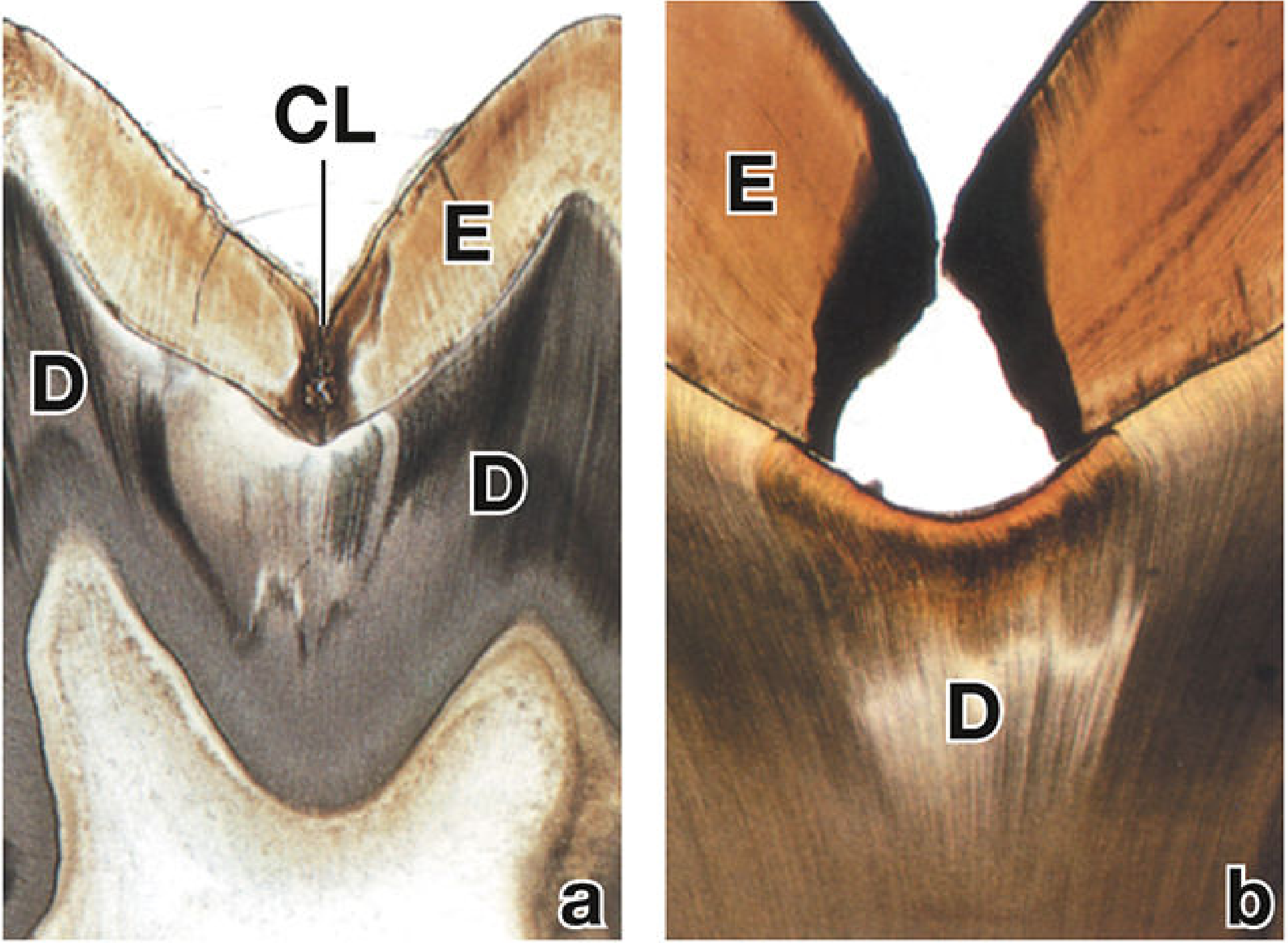
The occlusal surfaces of posterior teeth — particularly molars and premolars — are characterized by complex topographic features including pits, fissures, and grooves. Pits are small, pinpoint depressions at the junction of developmental grooves; fissures are narrow clefts extending into the enamel along developmental lines. The morphology of these structures varies considerably: some are broad and self-cleansing (V-shaped), while others are narrow, deep, and flask-shaped (I- or IK-type), rendering them virtually inaccessible to toothbrush bristles. This anatomical inaccessibility permits the stagnation of food debris and bacteria, creating a microenvironment highly favorable for cariogenesis.
Susceptibility to Caries
Pits and fissures represent the most caries-susceptible sites on the tooth surface for the following reasons:
- Morphological retention — the narrow, constricted orifice traps plaque and substrate that cannot be removed by normal oral hygiene measures
- Reduced enamel thickness — enamel at the base of deep fissures may be extremely thin, offering limited structural resistance
- Limited salivary access — the buffering and remineralizing effects of saliva are diminished within inaccessible fissure depths
- Early plaque colonization — newly erupted teeth with immature, less mineralized enamel are particularly vulnerable
Progression of Pit and Fissure Caries
The carious process in pits and fissures begins at the base of the fissure where bacterial accumulation is greatest. The lesion initiates as subsurface demineralization of enamel — the classic initial enamel lesion or white spot lesion — before cavitation is evident clinically. Microscopically, the lesion progresses in a cone-shaped or triangular pattern with the apex directed toward the enamel-dentin junction (EDJ). Once the advancing front reaches the EDJ, the lesion spreads laterally along this interface, undermining the overlying enamel. Bacterial invasion of the dentinal tubules then follows, progressing toward the pulp.
Preventive Strategies
Pit and fissure sealants constitute the primary preventive intervention. Sealants are resin-based or glass ionomer materials applied to the occlusal surfaces to physically seal pits and fissures, eliminating the retentive plaque habitat and preventing bacterial access to the enamel surface. They are most effective when applied shortly after tooth eruption, before caries initiation.
Fluoride acts through multiple mechanisms to reduce caries susceptibility:
- Incorporation into enamel crystal structure — fluoride substitutes for the hydroxyl ion in hydroxyapatite, forming fluoroapatite (calcium fluorophosphate), which is markedly more resistant to acid dissolution than native hydroxyapatite. This decreases enamel crystal solubility in acid.
- Antimicrobial action — fluoride inhibits bacterial enzymes, reducing acid production by plaque organisms
- Remineralization promotion — fluoride facilitates redeposition of calcium and phosphate ions into early demineralized enamel, reversing incipient lesions before cavitation
Water fluoridation (optimal concentration: 0.5–1.0 ppm) has been largely responsible for the dramatic decline in caries prevalence in high-resource countries.
(Histology: A Text and Atlas; Robbins & Kumar Basic Pathology)
2. Smooth Surface Caries
Etiology and Risk Factors
Smooth surface caries occur on the flat or convex enamel surfaces of the tooth — specifically the interproximal (approximal) surfaces between adjacent teeth and the cervical (gingival) margins. Unlike pit and fissure caries, where anatomical retention drives pathogenesis, smooth surface lesions depend entirely on plaque accumulation secondary to inadequate oral hygiene. Risk factors include:
- Diet — frequency of fermentable carbohydrate intake is the dominant dietary variable; sticky, adherent forms of sucrose are particularly cariogenic because they sustain prolonged acid production
- Reduced salivary flow (xerostomia) — saliva provides buffering of acid, clearance of substrate, and delivery of calcium, phosphate, and fluoride ions essential for remineralization
- High mutans streptococci counts in saliva
- Inadequate fluoride exposure
- Poor oral hygiene — plaque accumulation at interproximal contact areas and along the gingival margin
Clinical Features
The earliest detectable change is the white spot lesion — an opaque, chalky white area representing subsurface demineralization beneath an apparently intact enamel surface. This occurs because the surface enamel undergoes initial remineralization while the subsurface zone undergoes progressive demineralization. With continued acid challenge:
- The surface layer breaks down and a cavity (cavitation) forms
- The lesion surface may appear brown or black due to exogenous pigment incorporation and dentin exposure
- Interproximal lesions may be detected radiographically as a radiolucent shadow within enamel on bitewing radiographs before clinical cavitation is apparent
Role of Fluoride in Prevention
Fluoride remains the cornerstone of smooth surface caries prevention. Topical fluoride (toothpaste, fluoride varnish, professional fluoride application) maintains elevated fluoride concentrations at the tooth surface, promoting the demineralization–remineralization cycle in favor of remineralization. Regular brushing removes plaque from accessible smooth surfaces, and flossing addresses the interproximal regions. Dietary counseling to reduce the frequency of sugar intake further reduces the cariogenic challenge.
3. Root Caries
Etiology and Pathogenesis
Root caries develop on the cementum and root dentin of teeth at or below the cementoenamel junction (CEJ). The fundamental prerequisite is gingival recession or periodontal disease, which exposes the root surface to the oral environment. Unlike the crown, the root surface is covered not by enamel but by the relatively thin and softer cementum, which is far more susceptible to acid demineralization than enamel.
The characteristic of the carious lesion — that it progresses inward from the tooth surface — applies equally to root caries, but the substrate differs:
- Initial attack: Acid demineralization of the cementum surface
- Progression: Bacterial digestion of the protein matrix of exposed cementum and dentin. Subsequent decomposition of cementum and exposed root dentin involves both acid demineralization of the mineral phase and bacterial enzymatic digestion of the organic protein matrix (collagen)
- Advancement: Deep penetration into root dentin toward the pulp
(Jawetz, Melnick & Adelberg's Medical Microbiology; Sherris & Ryan's Medical Microbiology)
Microbiology
The microbial ecology of root caries differs somewhat from coronal caries. Actinomyces viscosus and A. naeslundii play a more prominent role in root surface lesions, alongside S. mutans and lactobacilli. These organisms are well adapted to colonize root surfaces and efficiently metabolize a broad range of carbohydrate substrates.
Clinical Features
- Location: At or just below the CEJ, on exposed root surfaces, commonly in the cervical region
- Appearance: Soft, leathery, light brown discoloration of the root surface in active lesions; darker, harder texture in arrested lesions
- Shape: Root caries characteristically form broad, shallow, crescent-shaped lesions that spread laterally around the circumference of the root rather than penetrating deeply
- Prevalence: Significantly increased in older adults due to cumulative gingival recession and xerostomia associated with polypharmacy; also elevated in patients receiving radiation therapy to the head and neck (radiation-induced xerostomia)
Progression
From the root surface, the carious process involves:
- Demineralization and softening of the cementum
- Breakdown and penetration of the cementum–dentin interface
- Spread through the dentinal tubules toward the pulp chamber
- Possible extension into the periodontal tissues at the root apex, resulting in periapical infection (Sherris & Ryan's Medical Microbiology)
4. Active and Arrested Caries
Definition and Biological Basis
Caries activity reflects the dynamic balance between demineralization (driven by acidogenic bacteria and fermentable substrate) and remineralization (driven by saliva, fluoride, and calcium-phosphate availability). This equilibrium is not fixed; lesions can progress, stabilize, or regress depending on the prevailing oral environment.
Active Caries
An active carious lesion is one in which demineralization is currently occurring and the lesion is advancing. The defining clinical features are:
| Feature | Active Lesion |
|---|---|
| Surface texture | Soft, mushy, or leathery |
| Color | White/yellow-white to light brown |
| Appearance | Dull, opaque, non-lustrous surface |
| Location | In areas with plaque accumulation (at gingival margin, approximal surfaces) |
| Probing | Soft on gentle probing |
Histologically, active lesions show a broad zone of demineralization with an infected outer layer where bacteria are present and an inner affected dentin zone where the mineral is partially dissolved but bacteria have not yet penetrated. The surface may be porous and cavitated.
Clinical significance: Active caries must be intercepted promptly. If remineralization is not achieved:
- Bacterial invasion of dentin progresses
- The lesion extends to the pulp, resulting in pulpitis — initially reversible, then irreversible
- Untreated pulpitis leads to pulp necrosis, periapical abscess, and systemic spread of infection
- Endodontic treatment ("root canal") becomes necessary, followed by crown restoration
Management strategies for active caries include:
- Excavation and restoration: Removal of infected tooth tissue and replacement with amalgam, composite resin, or glass ionomer cement
- Remineralization therapy: Fluoride varnish, casein phosphopeptide-amorphous calcium phosphate (CPP-ACP) for early non-cavitated lesions
- Dietary modification: Reduction of sugar frequency
- Enhanced oral hygiene (Histology: A Text and Atlas)
Arrested Caries
An arrested carious lesion is one in which the demineralization process has ceased and the lesion has stabilized without further progression. Arrest occurs when the cariogenic challenge (plaque accumulation, sugar frequency) is reduced or when remineralization processes dominate. The biological processes underlying arrest include:
- Remineralization of the lesion surface — calcium, phosphate, and fluoride ions redeposit within the demineralized zone, producing a harder, more acid-resistant surface layer
- Sclerosis of dentinal tubules — within arrested dentinal caries, the dentinal tubules become occluded by mineral deposits (dead tracts become sclerotic), effectively sealing off the lesion from further bacterial advance
- Tertiary (reparative) dentin formation — the pulp responds by depositing tertiary dentin along the pulpal wall adjacent to the lesion, increasing distance between the lesion front and pulp tissue
| Feature | Arrested Lesion |
|---|---|
| Surface texture | Hard, shiny |
| Color | Dark brown to black |
| Appearance | Lustrous, polished surface |
| Location | Often self-cleansing areas |
| Probing | Hard on probing |
The dark pigmentation of arrested lesions results from the incorporation of extrinsic stains into the remineralized, porous surface — often compounds from food, beverages, and tobacco. This surface is hard and resistant, and in the absence of symptoms or progression, arrested lesions may not require operative intervention.
Factors Promoting Caries Arrest
- Fluoride application — the single most important factor; fluoride accelerates remineralization and forms an acid-resistant fluoroapatite layer
- Improved oral hygiene — plaque removal eliminates the acidogenic biofilm
- Dietary changes — reduced frequency of fermentable carbohydrate intake lowers the acid challenge
- Increased salivary flow — restoration of adequate salivary function (treatment of xerostomia, cessation of xerogenic medications) enhances buffering and remineralization
- Silver diamine fluoride (SDF) — topically applied SDF arrests active root and coronal carious lesions, particularly in high-risk populations
Summary Table
| Type | Location | Substrate | Key Organisms | Prevention |
|---|---|---|---|---|
| Pit & Fissure | Occlusal pits/fissures | Enamel | S. mutans, lactobacilli | Sealants, fluoride |
| Smooth Surface | Approximal, cervical | Enamel | S. mutans, streptococci | Fluoride, oral hygiene, diet |
| Root Caries | Cementum/root dentin (exposed root) | Cementum, dentin | Actinomyces, S. mutans, lactobacilli | Fluoride, treat gingival recession |
| Active Caries | Any site | Progressing | Acidogenic biofilm | Excavation, remineralization, fluoride |
| Arrested Caries | Any site | Stabilized | — (lesion inactive) | Monitoring, fluoride, OH |
Sources: Robbins & Kumar Basic Pathology | Robbins, Cotran & Kumar Pathologic Basis of Disease | Sherris & Ryan's Medical Microbiology, 8th ed. | Jawetz, Melnick & Adelberg's Medical Microbiology, 28th ed. | Histology: A Text and Atlas with Correlated Cell and Molecular Biology | Rosen's Emergency Medicine: Concepts and Clinical PracticeThe above covers all five learning outcomes (Car1-OD-001 through Car1-OD-005) from your curriculum table, in textbook-style language. Here is what each section addresses:
| Code | Topic | Covered Above |
|---|---|---|
| Car1-OD-001 | Pit and Fissure Caries | Anatomy of pits/fissures, caries susceptibility, sealants, fluoride mechanisms |
| Car1-OD-002 | Smooth Surface Caries | Etiology, risk factors, clinical features, fluoride in prevention |
| Car1-OD-003 | Root Caries | Etiology, pathogenesis, clinical features, progression |
| Car1-OD-004 | Active Caries | Clinical features, significance, management strategies |
| Car1-OD-005 | Arrested Caries | Clinical features, biological processes (remineralization, sclerosis, reparative dentin), contributing factors |
This is a shared conversation. Sign in to Orris to start your own chat.