
Low line placenta
Reading File
Low-lying placenta (also called low-lying placenta or low line placenta) refers to a placenta that is implanted in the lower uterine segment but whose lower edge does not cover the internal cervical os. It is closely related to - but distinct from - placenta previa, where the placenta actually covers the os.
Searching sources...
Finding Sources
Searching PubMed
"low-lying placenta" AND management
Finding Sources
Reading File
Reading File
Reading File
Reading File
Loading Image
Here is a thorough overview of low-lying placenta based on authoritative textbook sources.
Low-Lying Placenta
Definition
A low-lying placenta is diagnosed when the lower edge of the placenta lies within 2 cm of the internal cervical os but does not cover the os. When the placenta actually covers the internal os, it is termed placenta previa. The older sub-classifications - complete, partial, and marginal previa - are no longer recommended. - Creasy & Resnik's Maternal-Fetal Medicine, p. 1017 / Grainger & Allison's Diagnostic Radiology, p. 915
Key cutoff: If the placental edge is >20 mm from the internal os, it is reported as "normal." If it is within 20 mm but not covering the os, it is labeled "low-lying."
Incidence and Epidemiology
- Placenta previa (the more severe end of the spectrum) affects approximately 0.5% of all third-trimester pregnancies.
- Up to 10% of midtrimester sonograms show a low-lying or previa placenta.
- At least 90% of placentas identified as low-lying or previa in early pregnancy resolve by the third trimester via placental migration (trophotropism) - the placenta grows toward the better-vascularized fundus, while the lower segment tissue atrophies. - Creasy & Resnik's, p. 1018
Risk Factors
| Risk Factor | Relative Increase in Risk |
|---|---|
| Previous placenta previa | ~8× |
| Previous cesarean section | 1.5-15× |
| Prior uterine curettage/suction abortion | 1.33× |
| Maternal age >35 years | 4.7× |
| Maternal age >40 years | ~9× |
| Multiparity | 1.1-1.7× |
| Smoking | Increased |
| Prior uterine surgery or scarring | Increased |
Pathogenesis
The underlying cause is not fully understood. There is a clear association between low implantation and prior endometrial damage - from curettage, cesarean scars, or multiple pregnancies. Bleeding occurs when marginal placental vessels in the lower uterine segment are torn as:
-
The lower uterine wall elongates in the third trimester
-
Cervical dilation occurs at or near labor onset
-
Prelabor contractions, intercourse, or cervical examination disturbs the placental margin
-
Creasy & Resnik's, p. 1018; Rosen's Emergency Medicine, p. 3355
Clinical Features
- Painless, bright-red vaginal bleeding is the hallmark - typically in the second half of pregnancy.
- Small "herald bleeds" may precede major hemorrhage.
- In up to 10% of cases, there is no bleeding until labor begins.
- Uterine irritability may be present in ~20% of cases but is generally mild.
- No abdominal pain - this distinguishes it from placental abruption.
Warning: Digital or instrument examination of the cervix is CONTRAINDICATED until placenta previa/low-lying placenta is excluded by ultrasound - it can precipitate catastrophic hemorrhage.
Diagnosis
Ultrasound (Method of Choice)
- Transvaginal ultrasound (TVUS) is the gold standard - more accurate than transabdominal ultrasound for measuring the os-to-placental edge distance.
- The bladder must be emptied before scanning to avoid falsely diagnosing previa.
- Precise measurement of the distance from the placental edge to the internal os is essential.
- A low-lying placenta found in the second trimester may resolve with continued uterine growth; one persisting late in gestation is less likely to improve.
MRI
- Used in equivocal cases or when placenta accreta syndrome is suspected (e.g., if previous cesarean scar is present near the low-lying placenta).
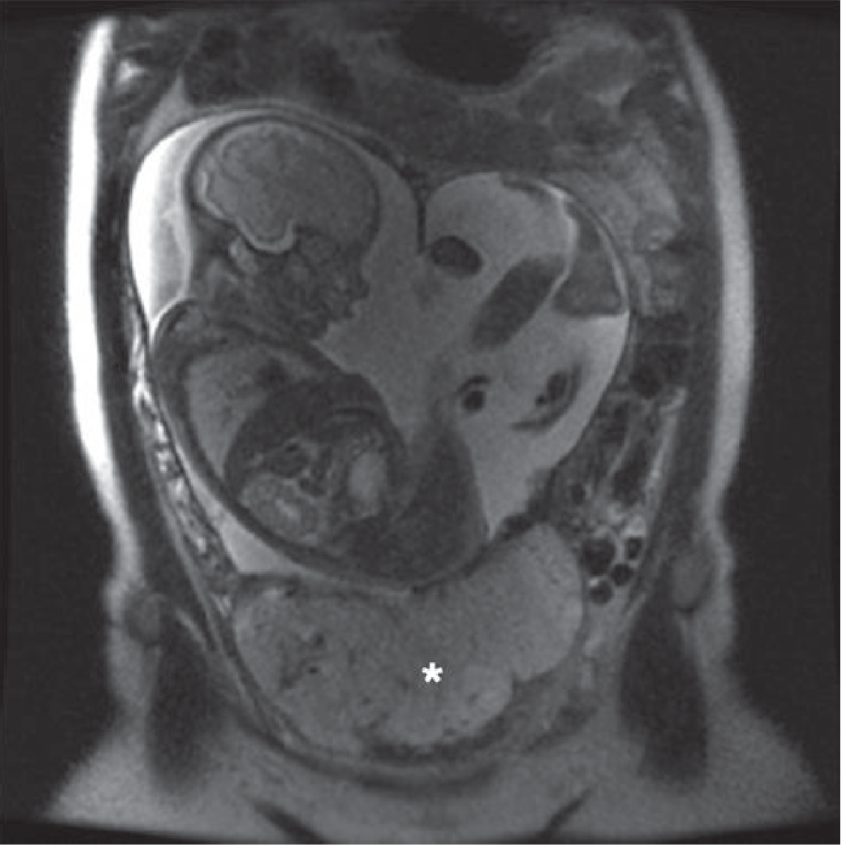
Fig. 35.52 from Grainger & Allison's - Coronal T2-weighted MRI showing a low-lying placenta (asterisk) covering the internal os with the fetus in breech position:
Differential Diagnosis
- Placenta previa (covers the os)
- Localized uterine contraction mimicking a low-lying edge
- Placenta accreta spectrum (invasive placentation - must always be considered)
- Succenturiate lobe
- Vasa previa (cord vessels crossing the os)
Associated Conditions
- Placenta accreta spectrum - particularly with prior cesarean scar
- Vasa previa - fetal vessels running through membranes near the os, risk of fetal exsanguination at rupture of membranes
Management
Antepartum (Stable Patient)
- Serial TVUS to monitor for spontaneous resolution (especially if diagnosed before 28 weeks)
- Pelvic rest (no intercourse, no vaginal exams)
- Corticosteroids for fetal lung maturity if preterm delivery is anticipated
- Rh immune globulin (300 mcg IM within 72 hours) if mother is Rh-negative
If Bleeding Occurs (Emergency)
- Two large-bore IV lines + fluid resuscitation
- Continuous fetal monitoring
- CBC, type and crossmatch
- Coagulation panel (PT, PTT, platelets, fibrinogen - normal in pregnancy: 400-450 mg/dL; levels <300 mg/dL indicate significant coagulopathy)
- Fresh-frozen plasma or fresh whole blood if coagulopathy develops
- Urgent obstetric consultation
Delivery
- Cesarean section is recommended if the placental edge is ≤1 cm from the os or if it is covering the os at the time of delivery planning.
- If placental edge is 1-2 cm from the os, vaginal delivery may be considered on a case-by-case basis with careful monitoring.
- Grainger & Allison's, p. 915
Key Points Summary
| Feature | Low-Lying Placenta | Placenta Previa |
|---|---|---|
| Placental edge location | Within 2 cm of os | Covers the internal os |
| Typical symptom | Painless bleeding | Painless bleeding |
| Diagnosis | TVUS | TVUS |
| Resolution rate | ~90% by 3rd trimester | Less likely to resolve if complete |
| Delivery | May allow vaginal if edge >1 cm from os | Cesarean section |
Recent Evidence
A 2025 systematic review and meta-analysis (PMID 40472373) specifically examined postpartum hemorrhage risk in low-lying placenta, confirming that it carries a significant hemorrhage risk even when the placenta does not fully cover the os - relevant for peripartum planning.
Oligohydraminos
Oligohydramnios is a condition of reduced amniotic fluid volume - defined as an AFI (amniotic fluid index) of ≤5 cm or a single deepest pocket of <2 cm. It is associated with fetal renal anomalies, post-term pregnancy, IUGR, and PPROM, and carries significant fetal risk.
Searching sources...
Finding Sources
Searching PubMed
"oligohydramnios" AND management
Finding Sources
Reading File
Reading File
Reading File
Reading File
Reading File
Loading Image
Here is a comprehensive overview of oligohydramnios from authoritative textbook sources.
Oligohydramnios
Definition
Oligohydramnios is a condition of abnormally low amniotic fluid volume (AFV) for gestational age.
-
AFI (Amniotic Fluid Index) method: AFI <5 cm (significant oligohydramnios)
-
Single Deepest Pocket (SDP) method: <2 cm in both vertical and horizontal planes (score = 0 on biophysical profile)
-
Normal AFI: 8-24 cm from 16 weeks onward
-
"Borderline" oligohydramnios: AFI 5-8 cm
-
Pfenninger & Fowler's Procedures for Primary Care; Creasy & Resnik's Maternal-Fetal Medicine
How Amniotic Fluid is Formed
Amniotic fluid volume reflects a dynamic balance between production and resorption:
| Production | Resorption |
|---|---|
| Fetal urine (major source from ~16 weeks onward) | Fetal swallowing |
| Fetal lung fluid secretion | Intramembranous absorption |
| Transudation across membranes (early pregnancy) |
Since fetal urine is the dominant source from the second trimester onward, any condition impairing fetal urine output will reduce AFV.
Causes (Mnemonic: DRIPPC)
| Category | Examples |
|---|---|
| Drugs | NSAIDs (prostaglandin inhibitors), ACE inhibitors, indomethacin |
| Renal/urinary (fetal) | Bilateral renal agenesis, obstructive uropathy (posterior urethral valves), polycystic kidneys, urethral atresia |
| IUGIR / Uteroplacental insufficiency | Preeclampsia, maternal hypertension, post-term pregnancy |
| PROM (Prelabour Rupture of Membranes) | Most common cause overall (~10% of pregnancies); can cause severe oligohydramnios or anhydramnios |
| Post-dates / Post-term | Placental aging and insufficiency |
| Chromosomal/structural | Trisomies, multiple gestation (twin-twin transfusion) |
Excluding PROM, approximately 83% of patients with oligohydramnios will have fetal IUGR; however, only 16% of patients with IUGR will have oligohydramnios. - Pfenninger & Fowler's
Pathogenesis Diagram (Robbins Pathologic Basis of Disease)
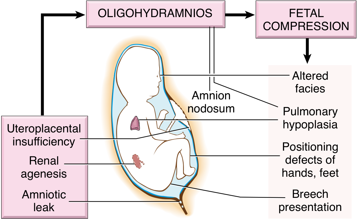
Fig. 10.3 - Pathogenesis of oligohydramnios (Potter) sequence. - Robbins, Cotran & Kumar Pathologic Basis of Disease
Complications - The Potter Sequence
Severe oligohydramnios causes fetal compression against the uterine wall, producing a classic pattern of defects:
1. Pulmonary Hypoplasia (most serious)
- Restricted thoracic expansion prevents normal lung growth
- Reduced hydraulic pressure on lungs affects stretch receptors and Ca²⁺ regulation, impairing lung growth
- Risk increases significantly if oligohydramnios occurs before 26 weeks (canalicular phase of lung development: 16-24 weeks)
- Leads to potentially lethal pulmonary hypoplasia (small, bell-shaped chest)
- The Developing Human - Moore's Embryology; Creasy & Resnik's
2. Facial Compression (Potter's Facies)
- Flattened facies
- Prominent infraorbital (epicanthal) folds
- Beaked/parrot nose
- Low-set ears
3. Musculoskeletal Deformities
- Positional abnormalities of hands and feet (clubfoot / talipes equinovarus)
- Hip dislocation
- Flexion contractures
4. Amnion Nodosum
- Nodules on the amnion surface (squamous cell debris from fetal skin)
- Pathognomonic of prolonged oligohydramnios
5. Umbilical Cord Compression
- Absent fluid cushioning around the cord
- Causes variable decelerations in fetal heart rate during labor
6. Breech Presentation
- Reduced fluid limits fetal movement and rotation
Diagnosis
Ultrasound (Method of Choice)
AFI technique:
- Divide uterus into 4 quadrants
- Measure the deepest vertical fluid pocket in each quadrant (avoiding cord, using color Doppler)
- Sum the 4 values = AFI
- Repeat 3 times and average if AFI <8 cm
Key ultrasound findings to also assess:
- Fetal biometry (estimated fetal weight for IUGR)
- Renal anatomy (bilateral renal agenesis, cystic kidneys, hydronephrosis)
- Bladder filling (small/absent bladder suggests renal cause)
- Amniotic membrane integrity
Additional investigations
- Speculum exam: for pooling of amniotic fluid (PROM)
- Fetal MRI: indicated in severe oligohydramnios when sonographic visualization is limited (e.g., to detect associated anomalies or assess renal structure)
- Fetal urine electrolytes, osmolality, β₂-microglobulin (if obstructive uropathy is suspected - to assess renal function prognosis)
- Fetal karyotype if structural anomalies found
Clinical Features and Intrapartum Complications
During labor, oligohydramnios is associated with:
- Fetal heart rate abnormalities (variable decelerations from cord compression)
- Depressed Apgar scores
- Meconium passage (fetal distress)
- Higher rates of operative/cesarean delivery
Management
Based on Gestational Age and Etiology:
| Situation | Management |
|---|---|
| Idiopathic at term | Delivery is usually indicated |
| PROM at term | Induction of labor |
| Preterm PROM | Conservative management with surveillance; antenatal corticosteroids; antibiotics; delivery at 34-37 weeks depending on risk |
| IUGR-related | Serial growth scans, Doppler velocimetry, BPP; delivery timing based on severity |
| Renal anomaly (severe, bilateral) | Counseling - prognosis generally poor; fetal intervention (vesicoamniotic shunting) in selected cases of obstructive uropathy |
| Drug-induced (NSAIDs) | Stop the offending drug |
Amnioinfusion
- Transcervical infusion of normal saline into the amniotic cavity during labor
- Reduces cord compression, decreases variable decelerations
- Does not improve perinatal survival when used for PROM-associated oligohydramnios
Key rule: In most cases, oligohydramnios at term is an indication for delivery. - Pfenninger & Fowler's
Prognosis (Obstructive Uropathy / LUTO)
- Poor prognosis associated with: earlier gestational age at onset, more severe oligohydramnios, associated structural anomalies, elevated fetal urine β₂-microglobulin/osmolality
- Even with postnatal correction: 25-30% of surviving neonates require dialysis by age 5 - Miller's Anesthesia
Recent Evidence
A 2025 systematic review and meta-analysis (PMID 40693854) examined borderline amniotic fluid levels and adverse perinatal outcome, providing updated data on risk stratification even at the borderline (AFI 5-8 cm) range. A separate 2024 systematic review (PMID 38648897) addressed outcomes of PROM at the limits of viability - the most common cause of severe oligohydramnios in preterm pregnancies.
This is a shared conversation. Sign in to Orris to start your own chat.