
Bone loss and pattern of bone destruction according to carranza laws
Carranza's bone loss patterns bone destruction periodontal disease laws horizontal vertical angular
https://pocketdentistry.com/21-bone-loss-and-patterns-of-bon…
https://dypds.com/study-materials/Perio%20Study%20Material/b…
horizontal vertical angular bone loss periodontal disease pattern diagram
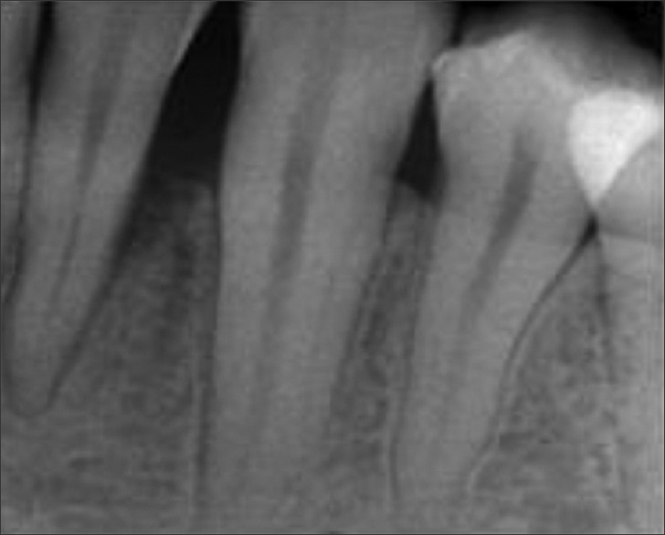
**Imaging Modality:** Intraoral periapical radiograph. **Anatomical Region:** Mandibular posterior segment, involving multiple permanent teeth and their associated supporting structures. **Observed Pathology:** The radiograph demonstrates significant generalized horizontal bone loss. There is a prominent vertical (angular) bony defect distal to the second premolar/mesial to the molar. A radiopaque restorative material is visible on the distal-occlusal aspect of the crown of the most posterior tooth shown. **Characteristic Visual Features:** * **Alveolar Bone:** Reduction in the height of the alveolar crest relative to the cementoenamel junction (CEJ), indicating periodontitis. * **Periodontal Ligament (PDL) Space:** Widening of the PDL space is visible along the root surfaces of the affected teeth. * **Trabecular Pattern:** Normal to slightly decreased radiodensity of the interdental bone, consistent with inflammatory bone resorption. * **Root Anatomy:** The roots appear intact without evidence of external resorption; however, the lack of bone support is evident in the cervical and middle thirds of the roots. **Diagnostic Features:** Horizontal and vertical bone resorption patterns characteristic of chronic periodontal disease. The image captures the baseline state of periodontal destruction prior to therapeutic intervention.
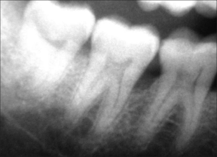
This diagnostic intraoral radiograph (periapical view) illustrates different patterns of periodontal alveolar bone loss in the mandibular right molar region. The image focuses on teeth 46 (first molar) and 47 (second molar). Adjacent to tooth 46, the alveolar bone crest exhibits a horizontal bone loss pattern, characterized by a relatively flat, horizontal reduction in bone height parallel to the cementoenamel junction (CEJ) of adjacent teeth. In contrast, the distal aspect of tooth 47 demonstrates a vertical (angular) bone loss pattern, where the bone defect forms an oblique angle relative to the root surface, creating an intrabony pocket. The radiographic appearance highlights key diagnostic criteria for periodontitis staging, showing the distinction between generalized crestal height reduction and localized vertical osseous defects. Such imaging is critical in dental education for clinical decision-making regarding non-surgical vs. regenerative surgical periodontal therapies.
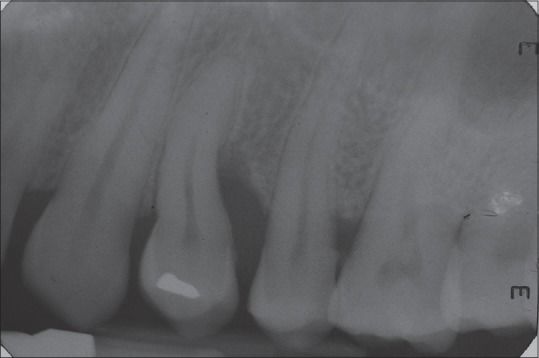
**Imaging Modality:** Intraoral periapical radiograph. **Anatomical Region:** Maxillary posterior quadrant, including premolar and molar regions. **Observed Pathology:** Advanced periodontal bone loss. There is significant localized vertical (angular) bone loss and horizontal bone resorption affecting the interdental alveolar crests. **Characteristic Visual Features:** - **Bone Loss:** A deep, U-shaped radiolucent vertical defect is visible between the second premolar and first molar, extending towards the middle third of the roots. - **Dentition:** The radiograph shows permanent maxillary teeth with visible pulp chambers and root canal morphology. A radiopaque restoration is noted on the occlusal/coronal surface of the second premolar. - **Periodontal Ligament (PDL):** Widening of the PDL space is suggestive of secondary occlusal trauma or active inflammatory periodontal disease. - **Trabecular Pattern:** Normal to slightly decreased radiodensity of the surrounding alveolar bone. **Key Diagnostic Features:** Presence of severe interproximal vertical osseous defects and reduction in the height of the alveolar crest relative to the cemento-enamel junction (CEJ), characteristic of chronic periodontitis.
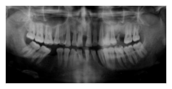
This orthopantomogram (OPG) provides a comprehensive radiographic view of the maxilla and mandible, illustrating advanced generalized periodontal disease. The primary clinical finding is widespread, severe alveolar bone loss affecting both arches. The bone destruction presents as a combination of horizontal resorption and significant vertical (angular) defects. Notably, teeth 13 (maxillary right canine) and 33 (mandibular left canine) demonstrate advanced vertical bone defects characterized by deep, narrow radiolucent areas extending apically along the root surfaces. The alveolar crest appears irregular and uneven throughout, with varying bone density indicative of active resorption and loss of cortical plate integrity. Multiple teeth exhibit increased spacing (pathologic migration) and secondary signs of clinical attachment loss. The maxillary sinuses and mandibular canals are visible as landmarks. This imaging is diagnostic for generalized aggressive periodontitis, emphasizing the pattern of destruction in a potentially young patient where the severity of bone loss is disproportionate to local factors like plaque or calculus.
osseous defects intrabony crater furcation involvement periodontal bone
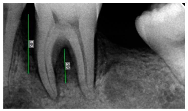
This diagnostic image is a periapical radiovisiography (RVG) radiograph showing the mandibular left first molar (tooth 36) in a pre-operative clinical state. The radiograph demonstrates a significant periodontal bone defect characterized by radiolucency surrounding the mesial and distal roots. Specifically, there is evidence of furcation involvement, where the interradicular bone is diminished. Two vertical measurement lines with digital calipers are visible: one measuring 9.2 units indicating the vertical depth of the mesial osseous defect relative to the cementoenamel junction or bone crest, and another measuring 5.3 units quantifying the height of the furcation defect. The surrounding alveolar bone shows a loss of normal trabecular pattern and increased radiolucency, indicative of inflammatory bone resorption or periodontal disease. The image is intended for dental education regarding the assessment of infrabony defects and the planning of regenerative periodontal therapy.
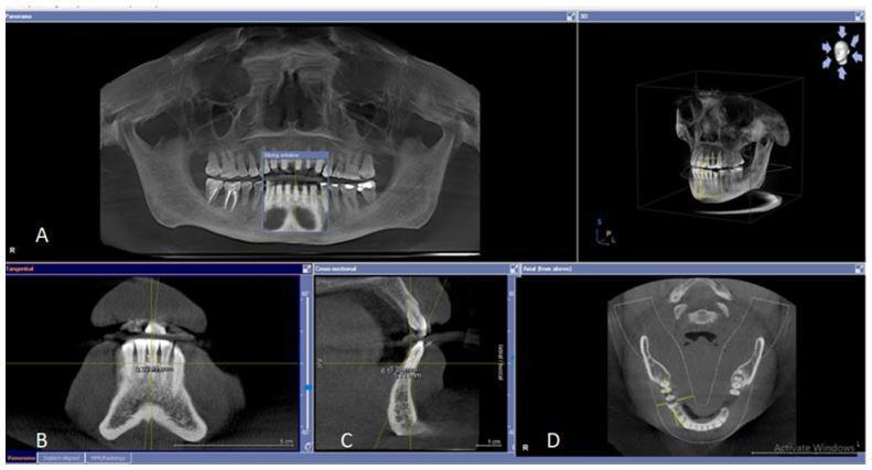
This composite diagnostic image illustrates the application of Cone Beam Computed Tomography (CBCT) and Orthopantomography (OPG) in dental radiology for evaluating periodontal osseous lesions. Panel A displays a panoramic OPG, providing a wide-field view of the maxilla, mandible, and full dentition to identify alveolar crest levels and general bone loss. Panel B presents a tangential CBCT view, optimized for measuring interproximal osseous defects between adjacent teeth. Panel C shows a cross-sectional (buccolingual) slice, used to detect bone loss and cortical plate integrity on the buccal and lingual aspects. Panel D features an axial CBCT view of the mandible, demonstrating the relationship between molar roots and the surrounding alveolar bone, particularly useful for identifying furcation involvement and defects. The top right panel provides a 3D volumetric reconstruction of the skull and dentition. Key educational concepts include the multi-planar assessment of bone height, the detection of vertical and horizontal osseous defects, and the identification of furcation radiolucencies in periodontology.
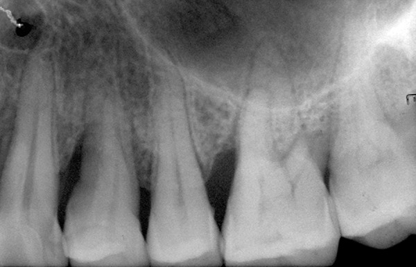
**Imaging Modality:** Intraoral periapical radiograph. **Anatomical Region:** Maxillary posterior dentition, including premolars and molars. **Observed Pathology/Disease Entity:** - **Periodontal Bone Loss:** Evidence of generalized horizontal bone loss and localized vertical (angular) osseous defects. - **Furcation Involvement:** Visible radiolucency in the furcation area of the depicted maxillary molar, indicating Class II or III furcation involvement. - **Dental Caries:** Radiopacities and radiolucencies suggest possible interproximal caries and existing dental restorations. **Characteristic Visual Features:** - **Alveolar Crest:** The crestal bone is apical to the normal level (1–2 mm below the cemento-enamel junction), indicating chronic periodontitis. - **Trabecular Pattern:** Normal to slightly decreased density of the interdental bone. - **Root Morphology:** Clearly visible root canals and periodontal ligament (PDL) spaces; some widening of the PDL space is noted. - **Anatomical Landmarks:** The floor of the maxillary sinus is visible superior to the root apices. **Key Diagnostic Features:** Presence of significant clinical attachment loss as evidenced by the radiographic distance between the CEJ and the alveolar crest, and the involvement of multi-rooted teeth furcations.
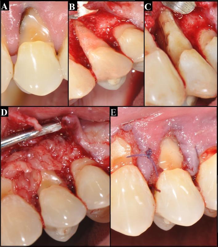
A series of five intraoral clinical photographs (A-E) documenting a periodontal surgical procedure for bone resorption and furcation involvement. Image A shows the preoperative state of a tooth with a deep abfraction—a wedge-shaped, dark-pigmented cervical lesion at the cementoenamel junction. Image B illustrates the vestibular aspect following the elevation of a full-thickness mucoperiosteal flap, revealing extensive alveolar bone resorption. Image C provides a mesial view of the exposed roots, highlighting Class II or III furcation involvement and total bone loss on the mesial-vestibular aspect. Image D demonstrates the application of Leukocyte-Platelet-Rich Fibrin (L-PRF) concentrate, where fibrin membranes are layered over the root surfaces to promote tissue regeneration. Image E shows the final surgical stage with the repositioned soft tissue flap secured by interdental polyglactin (Vicryl) sutures. This sequence illustrates advanced periodontal management using regenerative biomaterials to address significant attachment loss and osseous defects.
Bone Loss and Patterns of Bone Destruction (Carranza's Clinical Periodontology - Chapter 21)
Introduction
The level of bone is a consequence of past pathologic experiences, whereas changes in the soft tissue of the pocket wall reflect the present inflammatory condition. Therefore, the degree of bone loss does not necessarily correlate with the depth of periodontal pockets, the severity of pocket wall ulceration, or the presence or absence of pus.
I. Factors Responsible for Bone Destruction
A. Bacterial (Microbial) Factors
- Bacterial plaque products directly induce differentiation of bone progenitor cells into osteoclasts
- Plaque products stimulate gingival cells to release pro-resorptive mediators
- These products and inflammatory mediators can also inhibit osteoblast function and reduce osteoblast numbers (dual effect: increase resorption + decrease formation)
B. Host-Mediated Factors (Immune-Inflammatory)
- Prostaglandins (especially PGE2) - major mediators of bone resorption
- Interleukin-1 alpha and beta (IL-1a, IL-1b)
- Tumor Necrosis Factor (TNF)
- RANKL/OPG axis - the primary molecular mechanism of osteoclastogenesis in periodontitis
- Matrix metalloproteinases (MMPs) destroy the organic matrix
C. Systemic Factors
- Parathyroid Hormone (PTH): When administered continuously, it increases osteoclastic resorption and suppresses bone formation. When given in low intermittent doses, it has an anabolic (bone-forming) effect.
- Systemic diseases (diabetes, osteoporosis, etc.) modify the local response
- Medications
D. Local Modifying Factors
- Traumatic force from occlusion (TFO)
- Smoking
- Stress
- Subgingival calculus and overhanging restorations
- Food impaction
II. Radius of Action (Key Concept from Carranza)
- Bacterial plaque can induce bone loss within a range of 1.5 mm to 2.5 mm
- Beyond 2.5 mm, there is no effect
- Interproximal angular defects can appear only in spaces wider than 2.5 mm - narrower spaces would be entirely destroyed (no angular defect, just flat horizontal loss)
- Tal (1984) corroborated this with measurements in human patients
- Large defects far exceeding 2.5 mm from the tooth surface may be caused by bacteria in the tissues
III. Mechanisms of Bone Destruction
- Direct effect: Plaque products induce osteoclast differentiation
- Indirect effect: Plaque triggers gingival/immune cells to release IL-1, PGE2, TNF-alpha which activate osteoclasts
- Osteoblast inhibition: Plaque products and inflammatory mediators suppress bone formation
- Episodic destruction: Periodontal destruction occurs in an episodic, intermittent manner - periods of inactivity/quiescence alternate with destructive periods causing collagen loss, bone loss, and pocket deepening
- Onset of destructive periods may coincide with tissue invasion by one or several bacterial species, followed by advanced local host defense that controls the attack
IV. Periods of Destruction (Rates of Bone Loss)
| Group | Proportion | Yearly Attachment Loss | Pattern |
|---|---|---|---|
| Rapid progression | ~8% | 0.1 - 1.0 mm/year | Episodic bursts |
| Moderate progression | ~81% | 0.05 - 0.5 mm/year | Slow continuous |
| Minimal/no progression | ~11% | 0.05 - 0.09 mm/year | Essentially stable |
V. Bone Factor Concept
VI. Patterns of Bone Destruction
1. Horizontal Bone Loss
- Most common pattern in periodontal disease
- Bone is reduced in height, but the bone margin remains roughly perpendicular (at right angles) to the tooth surface
- The interdental septa and facial/lingual plates are affected, but not necessarily to an equal degree around the same tooth
- Associated with suprabony pockets
- Crestal bone is reduced from the normal 1-2 mm below CEJ to greater depth
- Bone loss occurs at an even rate, leading to a symmetrical reduction in alveolar bone height
- Regenerative procedures are not indicated for horizontal bone loss
2. Vertical (Angular) Bone Loss
- Occur in an oblique direction, leaving a hollowed-out trough in the bone alongside the root
- The base of the defect is apical to the level of the surrounding bone
- The bone loss occurs at different rates around adjacent teeth/surfaces
- Primarily affects one tooth and not its adjacent
- Requires adequate volume of alveolar bone for angular defect formation (spaces > 2.5 mm)
- Associated with intrabony (infrabony) pockets
- Can occur interproximally or on the radicular surface
- Increase in frequency with age
- An individual tooth can have combination bone loss (horizontal + vertical components)
| Type | Description | Prognosis with Regeneration |
|---|---|---|
| Three-wall defect (Intrabony/true infrabony) | Bordered by one tooth surface and 3 bony walls | Best prognosis |
| Two-wall defect (Osseous crater - most common 2-wall defect) | Bordered by one tooth surface and 2 bony walls | Intermediate |
| One-wall defect (Hemiseptal defect) | Bordered by one tooth surface and 1 bony wall | Poorest prognosis |
| Circumferential defect | Involves more than one surface of the tooth | Variable |
| Combined defect | Mixed wall numbers at different levels | Variable |
3. Osseous Craters
- Concavities in the crest of the interdental bone confined within the facial and lingual walls
- Most common type of defect in periodontal disease (accounts for ~35% of all defects; ~62% in posterior areas)
- Twice as common in posterior segments as in anterior segments
- Lower jaw craters are more common than upper jaw craters
- Mechanism: Poor accessibility for plaque removal; anaerobic environment favorable for pathogenic bacteria
4. Bulbous Bone Contours
- Bony enlargements caused by exostoses, adaptation to function, or buttressing bone formation
- Result from the body's attempt to reinforce bone under occlusal stress (buttressing bone formation)
- Central buttressing: Within the jaw, causes cupping of the bone crest (endosteal)
- Peripheral buttressing (lipping): At the external surface, can produce shelf-like ledges
5. Reversed Architecture (Reversed Bony Defects)
- Negative bony architecture in which the interdental bone is more apical than the facial/lingual bone
- Occurs when facial and lingual plates are more severely resorbed than the interdental bone
- Normal anatomy is reversed - facial/lingual bone is at a lower level than interdental bone
- Results from destruction of facial and lingual bone without equivalent loss of interdental bone
6. Ledges
- Plateau-like bone margins caused by resorption of thickened bony plates
- Horizontal ledge formations of the alveolar bone
7. Furcation Involvement
- Invasion of the bifurcation and trifurcation of multi-rooted teeth by periodontal disease
- Classified by Glickman into Classes I-IV:
| Class | Description |
|---|---|
| I | Pocket formation into the furcation area but with bone still intact |
| II | Bone destroyed on one or more aspects but not through-and-through |
| III | Through-and-through furcation, but covered by gingiva |
| IV | Through-and-through, clinically visible (gingiva has receded) |
8. Marginal Gutter (Shallow Linear Defect)
- A shallow linear defect between the marginal bone of the radicular cortical plate or interdental crest
- Extends the length of one or more root surfaces
- Usually formed by resorption of the socket side of the plate and deposition on the facial surface
VII. Summary of Key "Laws"/Principles from Carranza
- Bone loss does not equal pocket depth - they are independent measures
- Radius of action = 1.5-2.5 mm - plaque can only destroy bone within this range from the tooth surface
- Angular defects require space > 2.5 mm - narrower spaces yield only horizontal loss
- Episodic destruction - bone loss is not continuous but occurs in bursts
- Horizontal loss is most common; angular loss is associated with intrabony pockets
- 3-wall defects have the best regenerative potential; 1-wall defects the worst
- Osseous craters are the most common defect type (~35% of all defects)