
Give details about type of wounds
Reading File
Now let me search the medical library for wound types.
Finding Sources
Finding Sources
Good results. Let me now read the most relevant sections - the wound classification from surgery textbooks and primary care.
Finding Sources
Reading File
Reading File
Finding Sources
Finding Sources
Finding Sources
Finding Sources
Reading File
Reading File
Reading File
Reading File
Reading File
Reading File
Reading File
Reading File
I now have comprehensive information from multiple authoritative textbooks. Let me compile the complete answer.
Types of Wounds
Wounds are classified by several overlapping systems: their cause/mechanism, depth, degree of contamination (surgical context), and healing trajectory (acute vs. chronic).
1. Classification by Mechanism / Cause
A. Blunt Force (Mechanical) Wounds
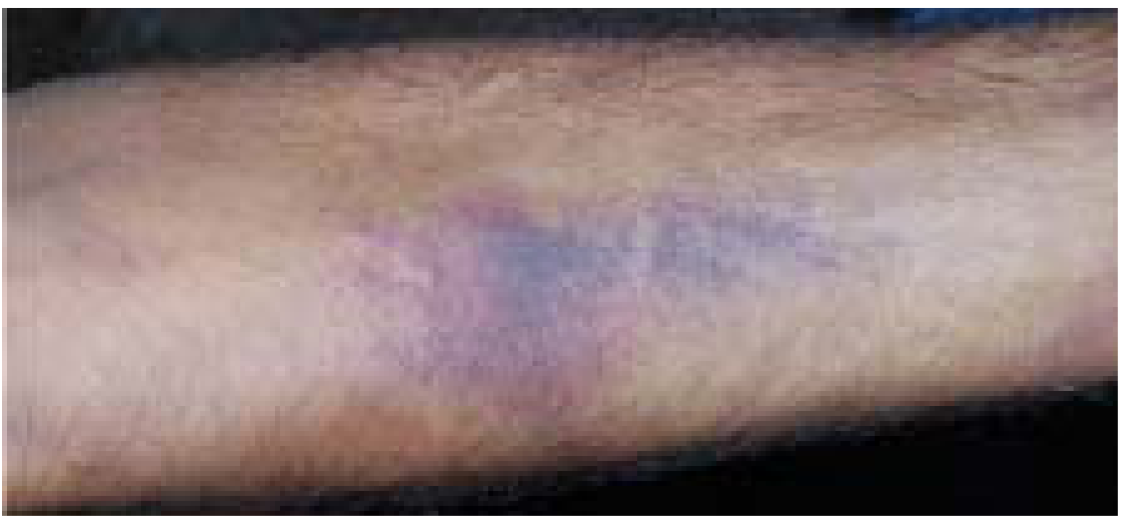
i. Contusion (Bruise)
A contusion is a collection of blood in the tissues due to rupture of blood vessels caused by blunt trauma, without disruption of the external skin surface. Blood extravasates into subcutaneous tissues, producing blurred margins and discoloration. The size varies from a pinhead to a large hematoma, proportional to the force applied.
Factors affecting size/shape:
- Tissue type: Softer tissues bruise more easily
- Age: Elderly and children bruise easily
- Sex: Women bruise more readily due to delicate tissues and greater subcutaneous fat
- Skin color: Visible more easily in fair skin
- Site: Loose tissue areas (eyelids, scrotum) bruise more easily
Medico-legal importance: Bruise color changes help estimate time of injury (red -> blue-black -> green -> yellow over days). Pattern of bruises can indicate weapon type or manner of assault.
ii. Abrasion (Graze/Scratch)
An abrasion is damage to the superficial epithelial covering of skin/mucous membrane due to impact of a hard surface. Typical abrasions remove only the epidermal layer (no full dermal breach). Caused by friction between weapon and body surface.
Types of abrasion:
- Scratch abrasion - caused by a sharp point moving over skin (e.g., fingernail scratches)
- Graze abrasion - caused by sliding contact with a rough surface (e.g., road rash)
- Impact/pressure abrasion - caused by direct impact of a blunt object pressing on skin
- Patterned abrasion - mirrors the shape of the causative weapon (forensically important)
Significance: Abrasions indicate the point of application of force, the direction of force, and the type of weapon. They heal without scarring unless infected.
iii. Laceration
A laceration is a tear or disruptive stretching of tissue caused by application of blunt force. Key features: irregular, jagged edges with intact bridging blood vessels across the wound (distinguishes from incised wounds), often with tissue bruising and crushing at wound margins.
Subtypes of lacerations:
| Subtype | Mechanism |
|---|---|
| Split laceration | Skin crushed between two hard objects (scalp over skull, shin, eyebrow). May look clean-cut, mimicking incised wounds - these are called "incised-looking lacerated wounds" |
| Stretch laceration | Excessive stretching beyond skin tensile limit; seen in run-over accidents, entrapment in machinery; may form skin flaps |
| Avulsion laceration | Separation/tearing of skin from underlying tissue; seen in high-velocity trauma |
| Cut laceration | Combination type |
B. Sharp Force Wounds
iv. Incised Wound
Inflicted by a sharp-edged instrument (knife, scalpel, glass) drawn across the skin. Features: clean, straight edges; longer than it is deep; bridging vessels are severed (not intact, unlike lacerations); bleeds profusely. Typically deeper at the start ("heel") and shallower at the end ("tail").
v. Stab / Puncture Wound
Caused by a pointed instrument thrust into the body. A puncture wound is caused by a long, narrow instrument. It is termed:
- Penetrating - instrument pierces tissue but has no exit
- Perforating - instrument traverses tissue with both entry and exit wounds
Stab wounds are deeper than long (opposite of incised wounds). The shape of the wound can suggest the weapon used (single-edged vs. double-edged blade).
C. Firearm Wounds (Special Puncture Wounds)
Gunshot wounds are special forms of puncture wounds. Features that vary by range:
- Close-range: soot/powder burns, muzzle abrasion, stellate laceration at entry
- Distant-range (>4-5 feet): no powder burns; entry wound smaller than exit
- Entry wound: circular with abraded collar; Exit wound: larger, ragged, irregular, no abraded collar
D. Thermal Wounds
| Type | Cause |
|---|---|
| Burns | Heat (flame, contact, radiation); classified by depth (first, second, third degree) |
| Scalds | Hot liquids/steam |
| Frostbite / Trench foot | Cold injury |
Burn depth classification:
- First degree (superficial): Epidermal layer only - red, painful, no blistering
- Second degree (partial thickness): Epidermis + superficial dermis - blistering, very painful; regeneration possible from dermal appendages
- Third degree (full thickness): Entire dermis destroyed including appendages - anesthetic (nerve endings destroyed), requires grafting
E. Chemical Wounds
- Corrosives (strong acids, alkalis): cause coagulative or liquefactive necrosis
- Irritants (weak acids, alkalis): cause inflammation and partial-thickness damage
F. Other Wound Types
- Electrical wounds - entry and exit burns at current path
- Radiation wounds - from X-rays, radioactive substances
- Defense wounds - on hands/forearms, sustained while fending off an attack
- Fabricated (self-inflicted) wounds - for medicolegal purposes
2. Classification by Depth (Pressure Ulcer Staging - NPUAP)
Applicable primarily to pressure ulcers but used as a general tissue depth framework:
| Stage | Description |
|---|---|
| Stage I | Intact skin; non-blanching redness over bony prominence |
| Stage II | Partial-thickness dermis loss; shallow open ulcer with red-pink wound bed; may present as serum-filled blister |
| Stage III | Full-thickness tissue loss; subcutaneous fat visible; bone/tendon NOT exposed |
| Stage IV | Full-thickness loss with exposed bone, tendon, or muscle |
| Unstageable | Full-thickness wound covered by slough/eschar; depth cannot be determined until base is exposed |
(NPUAP 2007, as cited in Pfenninger and Fowler's Procedures for Primary Care)
3. Surgical Wound Classification (CDC / Current Surgical Therapy)
Used to predict risk of surgical site infection (SSI):
| Class | Definition | Infection Risk |
|---|---|---|
| Class I - Clean | No breach of respiratory, GI, GU, or genital tracts; no inflammation (e.g., hernia repair, thyroidectomy, joint replacement) | 1.8-2.6% |
| Class II - Clean-contaminated | Controlled entry into respiratory/GI/GU tract without major break in sterile technique (e.g., bowel resection, hysterectomy) | 4.8-6.7% |
| Class III - Contaminated | Open/fresh accidental wound (<4 hours), major sterile technique break, or gross GI spillage; acute non-purulent inflammation (e.g., non-perforated appendicitis) | 5.6-8.6% |
| Class IV - Dirty/Infected | Old wound (>4 hours), existing infection, perforated viscus (e.g., peritonitis, perforated appendicitis, open fracture repair) | 8.5-11.8% |
(Current Surgical Therapy 14th Ed.)
4. Classification by Healing Trajectory
| Type | Description |
|---|---|
| Acute wound | Heals in an orderly, well-organized fashion through the standard phases: hemostasis → inflammation → proliferation → epithelialization |
| Chronic wound | Fails to progress through normal healing stages; most commonly arrested in the inflammatory phase. Examples: diabetic foot ulcers, venous leg ulcers, pressure ulcers |
Chronic wound characteristics (from Pfenninger & Fowler):
| Characteristic | Features |
|---|---|
| Necrotic | Devitalized, blackened, dry, retracted epidermis |
| Sloughy | Viscous, yellow, adherent slough; wet; potentially malodorous |
| Granulating | Beefy red / deep pink; highly vascularized granulation tissue |
| Epithelializing | Pink wound margins; isolated pink islands on wound surface |
Summary Table
| Category | Main Types |
|---|---|
| Blunt force | Contusion, Abrasion, Laceration (split, stretch, avulsion) |
| Sharp force | Incised wound, Stab/Puncture wound |
| Firearm | Gunshot wound (entry/exit) |
| Thermal | Burns (1st/2nd/3rd degree), Scalds, Frostbite |
| Chemical | Corrosive, Irritant |
| By depth | Stage I-IV (pressure staging), Superficial/Partial/Full thickness |
| Surgical | Clean, Clean-contaminated, Contaminated, Dirty |
| By healing | Acute, Chronic |
Sources:
- PC Dikshit Textbook of Forensic Medicine and Toxicology, Classification of Injuries chapter
- Robbins & Kumar Basic Pathology (Robbins Pathology), Morphology of mechanical injury
- Pfenninger and Fowler's Procedures for Primary Care, 3rd Ed., Wound Types and Healing Stages
- Current Surgical Therapy 14e, Surgical Wound Classification Table 4
- Rosen's Emergency Medicine, Abrasions and Lacerations chapter
This is a shared conversation. Sign in to Orris to start your own chat.