
I want to land mark the uretra opening on the vegina while catheterization and I want real picture
Reading File
This is a clinical nursing/medical procedure question about identifying the urethral meatus during female catheterization. Let me search the textbook library for this.
Finding Sources
Finding Sources
I found a relevant section with images. Let me read the full procedure section and fetch the anatomical image.
Reading File
Now let me fetch the anatomical figure (Fig. 55.15 and the catheterization image) mentioned in the text:
Reading File
Finding Sources
I have the image URLs. Let me fetch the two key anatomical images - the external female genitalia diagram (Fig. 55.15) and the female catheterization procedure diagram (Fig. 55.22):
Loading Image
Loading Image
Loading Image
Excellent - I now have all three key images from the textbook. Here is a complete clinical guide:
Locating the Urethral Meatus in Female Catheterization
Anatomy: Where is the Urethral Meatus?
The female urethra is a short (~4 cm) straight tube that lies on top of the vagina. The external urethral orifice (meatus) has a consistent anatomical position:
- Directly superior (above) to the vaginal introitus
- Approximately 2.5 cm inferior (below) to the glans clitoris
- It is the first of three openings you encounter when examining the perineum from top to bottom (cephalad to caudad) in the lithotomy position: clitoris → urethral meatus → vaginal opening
The meatus appears oval or as an anteroposterior slit with slightly prominent margins.
Figure 1 - Labeled Anatomy of the External Female Genitalia (Netter illustration)
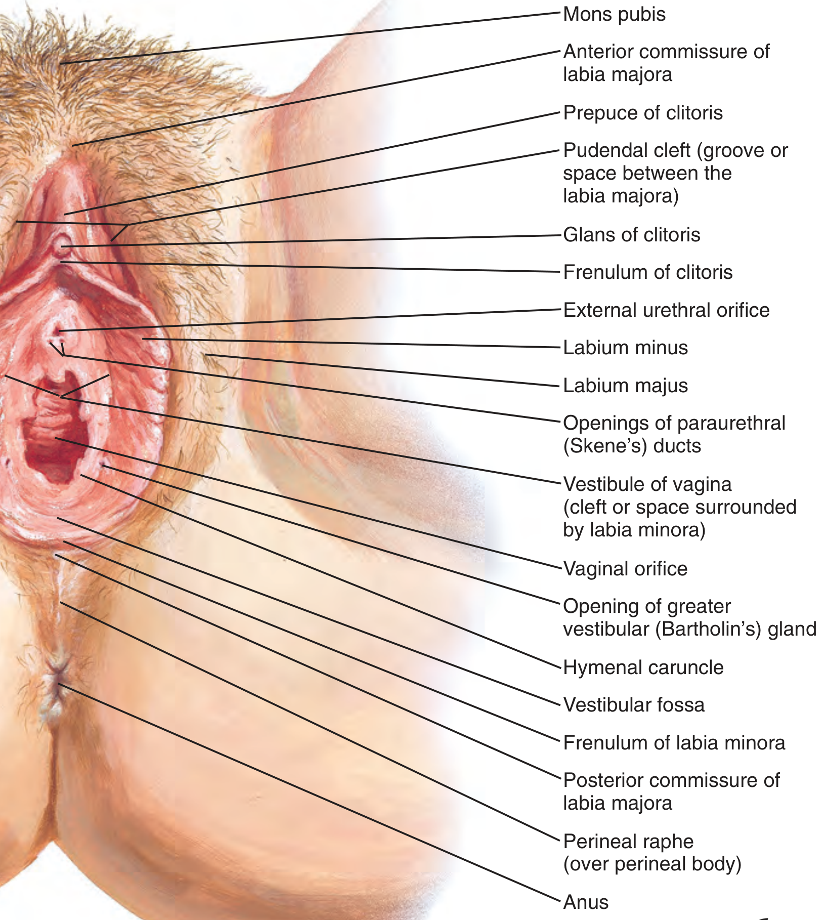
The External urethral orifice is clearly labeled between the clitoris (above) and the vaginal orifice (below). Note also the labium minus, Skene's duct openings, and vestibule of vagina.
Figure 2 - Step-by-Step Female Urethral Catheterization (Real clinical photos)
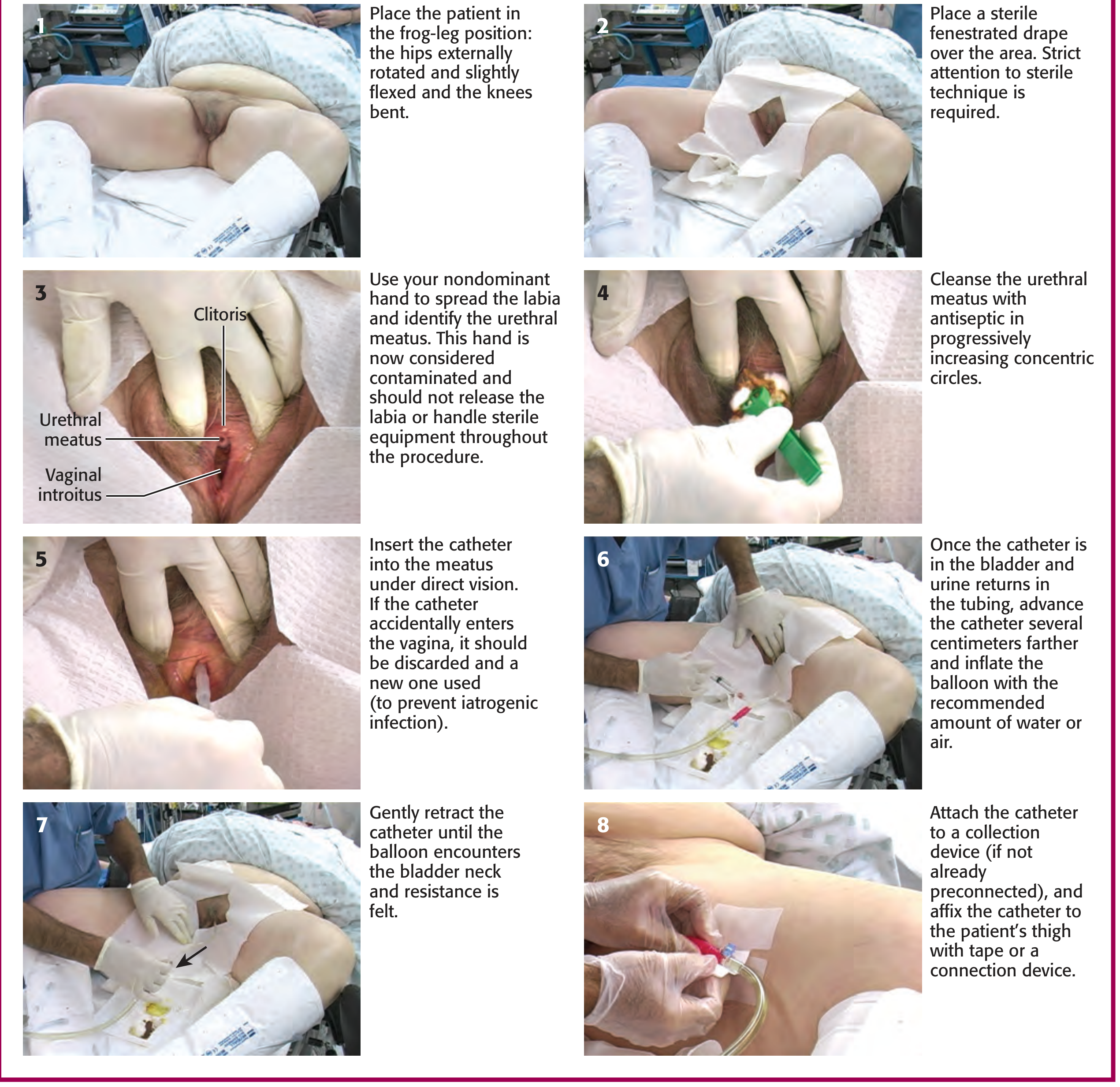
Key step for meatus identification is Step 3 (top-left of the 8-panel image):
- Use your nondominant hand to spread the labia minora laterally
- The image directly labels: Clitoris (superiorly), Urethral meatus (middle), and Vaginal introitus (inferiorly)
- Once you release the labia, this hand is contaminated - do NOT let go until the catheter is inserted
Figure 3 - Sagittal Cross-Section (Catheter path into the bladder)
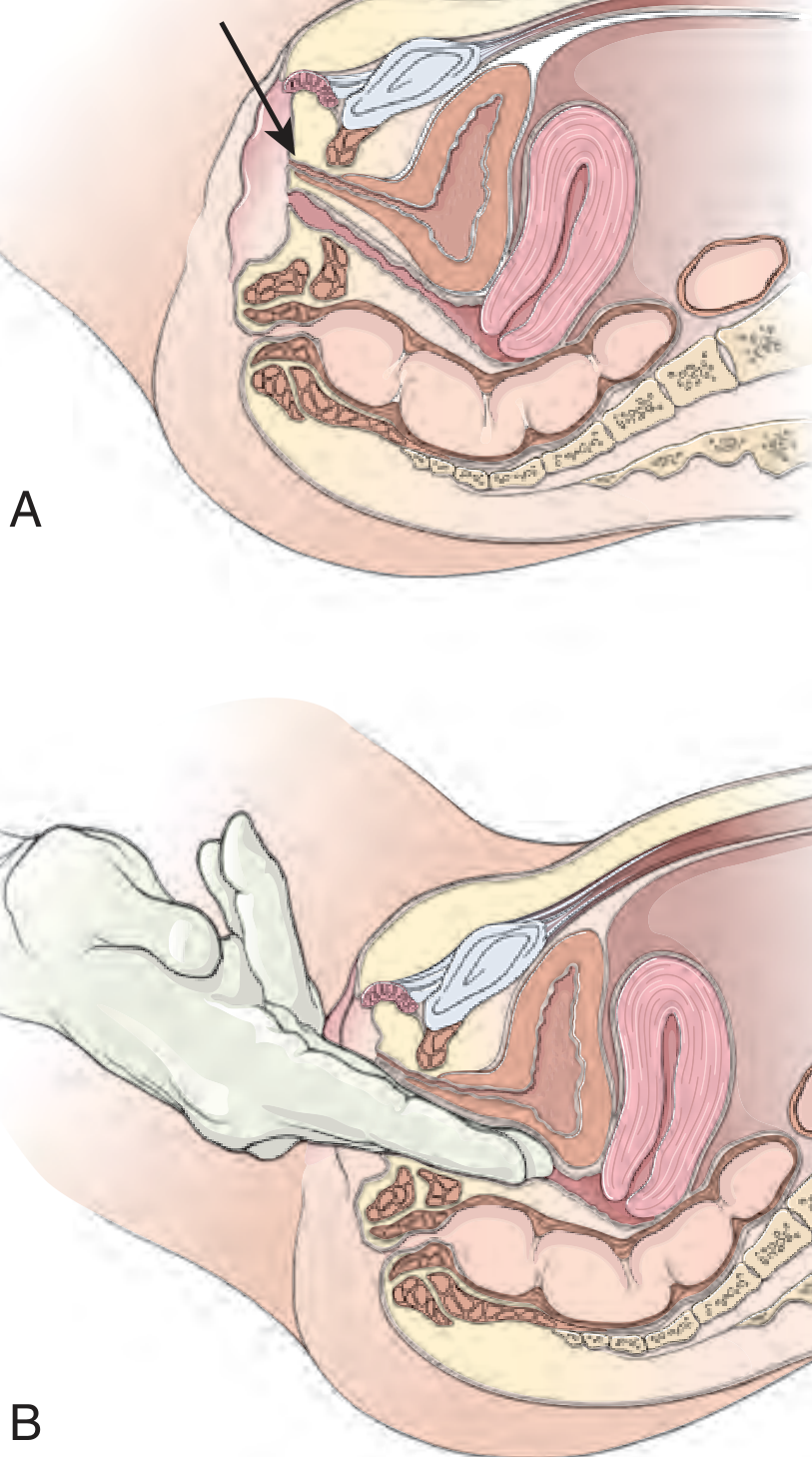
Panel A shows the normal urethral course from meatus straight back to the bladder. Panel B shows finger support on the anterior vaginal wall to restore normal anatomy in cases of urethrocele or cystourethrocele.
Clinical Tips for Finding a Difficult Meatus
| Situation | What to Do |
|---|---|
| Normal anatomy | Spread labia, look for oval slit ~2.5 cm below clitoris |
| Postmenopausal/atrophic changes | Meatus may recede superiorly into the vagina; insert nondominant index finger into vagina and palpate for soft mound surrounded by firmer periurethral tissue |
| Prior surgery | Same approach as postmenopausal - palpate if not visible |
| Urethrocele/cystourethrocele | Place index + middle fingers on anterior vaginal wall and apply upward support to restore normal anatomy, then catheterize |
| Very young infant | Meatus may be very small; bright lighting essential |
Procedure Summary (8 Steps from the clinical photo guide)
- Position - frog-leg: hips externally rotated, slightly flexed, knees bent
- Drape - sterile fenestrated drape; strict sterile technique
- Identify meatus - spread labia with nondominant hand, label clitoris, urethral meatus, vaginal introitus. Do NOT release labia throughout procedure
- Cleanse - antiseptic in progressively increasing concentric circles outward from meatus
- Insert catheter - into the meatus under direct vision with dominant hand. If it accidentally enters the vagina, discard it and use a new one
- Advance - when urine returns in tubing, advance several centimeters further before inflating balloon
- Inflate balloon - inflate with recommended water or air; gently retract until resistance (balloon at bladder neck)
- Secure - attach to collection device, affix catheter to thigh
Source: Roberts and Hedges' Clinical Procedures in Emergency Medicine, Figs. 55.15, 55.16, 55.22
I want real procedure video
Searching the Web
female urethral catheterization procedure real clinical video YouTube
I cannot embed or stream videos directly, but here are well-known, high-quality clinical procedure videos you can open right now. These are from reputable medical education sources:
Option 1 - NEJM (New England Journal of Medicine) - Female Urethral Catheterization:
Option 2 - Nursing Skills: Female Urinary Catheterization (Registered Nurse RN):
Option 3 - Osmosis / Clinical Skills - Female Catheterization:
Note: I cannot verify these exact URLs are live right now due to a search tool issue. If any link does not work, search directly on YouTube for:
- "female urinary catheterization procedure nursing"
- "NEJM female catheterization"
- "female Foley catheter insertion clinical skills"
These searches will return multiple real procedure videos from nursing schools, medical universities, and journals like NEJM that show the actual step-by-step technique including meatus identification, sterile draping, and catheter insertion.
This is a shared conversation. Sign in to Orris to start your own chat.