
How to approach chronic sacroilitis
sacroiliitis management treatment
sacroiliitis MRI pelvis imaging findings
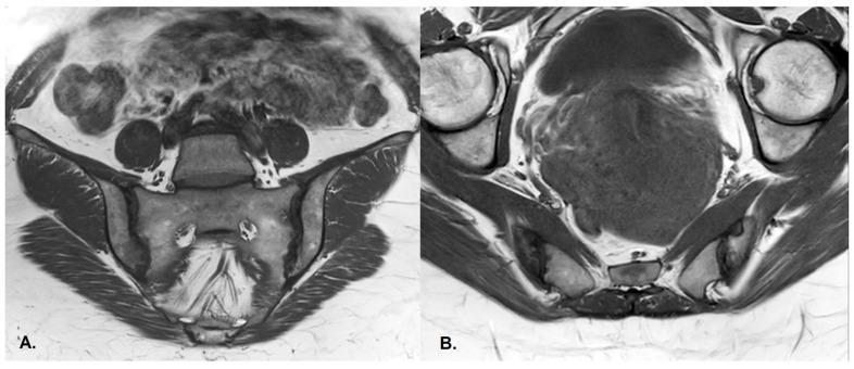
This diagnostic image consists of two T1-weighted MRI sequences of the pelvis focusing on the sacroiliac (SI) joints: a coronal view (A) and an axial view (B). The imaging reveals clear structural manifestations of chronic inflammatory sacroiliitis. Key findings include prominent subchondral sclerosis, visualized as areas of low signal intensity (darkening) within the bone marrow adjacent to the articular surfaces. There are also visible cortical disruptions and irregularities consistent with subchondral erosions, particularly affecting the iliac side of the joint. These changes are asymmetric and more pronounced on the right sacroiliac joint compared to the left. The constellation of findings, including the degree of sclerosis and erosion without complete ankylosis, is characteristic of Grade III sacroiliitis, often associated with axial spondyloarthritis such as psoriatic arthritis. The anatomical context clearly identifies the sacrum, iliac bones, and hip joints (visible in the axial plane), providing a comprehensive view of the pelvic girdle pathology.
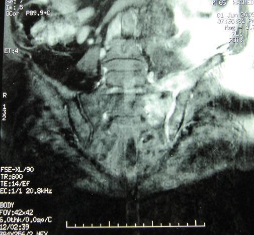
This diagnostic image is a Short Tau Inversion Recovery (STIR) sequence MRI of the pelvis, focusing on the sacroiliac (SI) joints in a coronal plane. The image illustrates classic radiological signs of infectious sacroiliitis, predominantly localized to the left side. Key visual findings include a significant widening of the left sacroiliac joint space, accompanied by cortical erosions and pronounced irregularity of the articular surfaces. There is evidence of hyperintense signal within the left joint region, characteristic of bone marrow edema or inflammatory effusion. The right sacroiliac joint appears relatively preserved in comparison. This imaging modality is essential for identifying early inflammatory changes and structural damage in the axial skeleton, aiding in the diagnosis of pyogenic, brucellar, or tuberculous sacroiliitis. The educational focus is on distinguishing normal sacroiliac anatomy from pathological erosion and joint space alterations in the context of infectious or rheumatological disease.
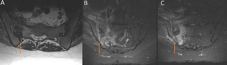
Diagnostic Magnetic Resonance Imaging (MRI) of the sacrum and pelvis, presented in axial (A) and coronal (B, C) views, demonstrating pathology of the right sacroiliac (SI) joint. The images reveal significant signal intensity abnormalities centered on the right SI joint compared to the healthy left side. A prominent high T2-weighted signal focus (indicated by orange arrows) is visible, signifying localized edema or an inflammatory collection. This hyperintensity extends from the joint space into the adjacent sacral alar marrow and anteriorly towards the right iliacus muscle, suggesting infectious sacroiliitis or a similar inflammatory process. The surrounding pelvic anatomy, including the sacrum and iliac wings, provides anatomical context for the extent of marrow involvement. These visual findings are characteristic of pyogenic or inflammatory sacroiliitis, highlighting the importance of MRI in assessing soft tissue and marrow changes in pelvic pathology.
Approaching Chronic Sacroiliitis
1. Understanding the Lesion
- Subchondral bone marrow edema (active inflammation, seen on MRI STIR)
- Erosions and sclerosis
- Joint space narrowing
- Eventual bony ankylosis
| Grade | Finding |
|---|---|
| 0 | Normal |
| 1 | Suspicious (pseudo-widening) |
| 2 | Minimal change (small erosions/sclerosis) |
| 3 | Definite change (erosions, sclerosis, widening/narrowing) |
| 4 | Total ankylosis |
2. Clinical Assessment
- Inflammatory back pain: insidious onset, age < 45, duration > 3 months, morning stiffness > 30 min, improves with exercise, not rest, nocturnal pain
- Associated features: uveitis, psoriasis, inflammatory bowel disease, peripheral arthritis, enthesitis, dactylitis
- Family history of spondyloarthropathy
- Direct sacroiliac joint palpation for tenderness
- Provocative tests: FABERE (Patrick's test), Gaenslen's test, pelvis rock - note these lack specificity/sensitivity and may be negative in early disease or late-stage ankylosis
- Lumbar flexion (Schober's test), chest expansion, tragus-to-wall distance
- Enthesitis examination (heel, Achilles)
- HLA-B27: positive in ~90-95% of AS; useful for screening but present in 4-8% of healthy White individuals; a negative result does not exclude diagnosis
- ESR, CRP: elevated in only 50-60% of axSpA; CRP correlates with treatment response and radiographic progression; normal CRP/ESR does not exclude active disease
- Fecal calprotectin if IBD suspected (>90% specificity and sensitivity)
- Mild normochromic anemia in up to 15% of patients
- Firestein & Kelley's Textbook of Rheumatology
3. Imaging
- First-line imaging; graded per Modified New York Criteria
- Insensitive in early disease - definite radiographic changes appear late
- STIR sequences show active bone marrow edema (hyperintense signal)
- T1 sequences show structural damage (erosions, sclerosis, fat metaplasia)
- Active sacroiliitis on MRI predicts later radiographic sacroiliitis and supports early diagnosis
4. Classification Criteria (ASAS)
5. Disease Activity Monitoring
- BASDAI (Bath Ankylosing Spondylitis Disease Activity Index) - patient-reported; score ≥4 indicates active disease
- ASDAS (Ankylosing Spondylitis Disease Activity Score) - includes CRP; preferred by ASAS. Score ≥2.1 = high disease activity; cut-off for biologic initiation
- BASFI - Bath Ankylosing Spondylitis Functional Index for tracking physical function
6. Treatment - ASAS/EULAR Phased Approach
Phase I (All Patients)
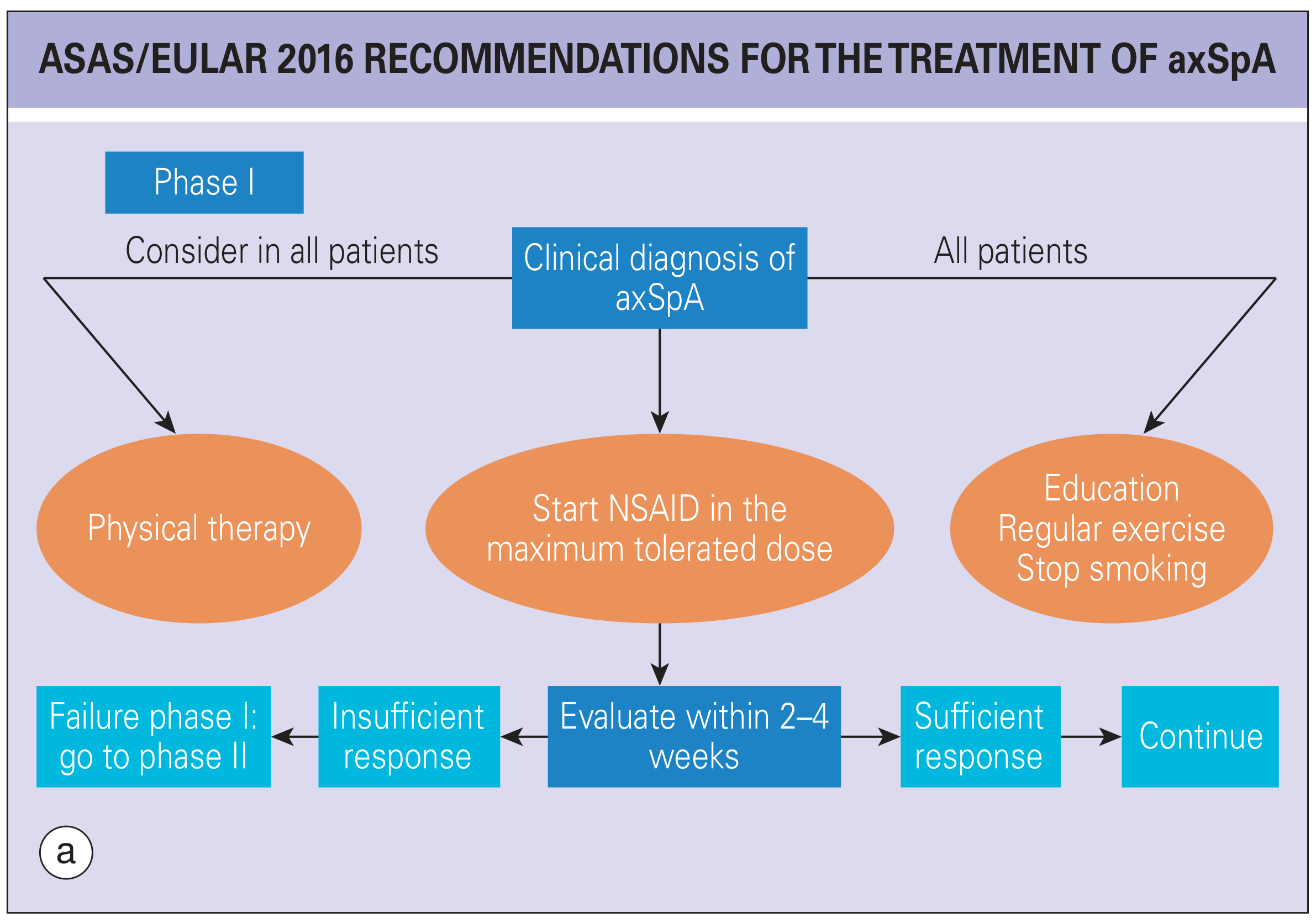
- Physical therapy - exercise, posture training, hydrotherapy. This is the non-pharmacological backbone and remains important throughout the disease course, including established disease (lung function may be adversely affected if neglected)
- NSAIDs at maximum tolerated dose - first-line pharmacologic treatment; continuous use preferred over on-demand in active disease; evaluate response in 2-4 weeks
- Education + regular exercise + smoking cessation - for all patients
Phase II (If Phase I Fails)
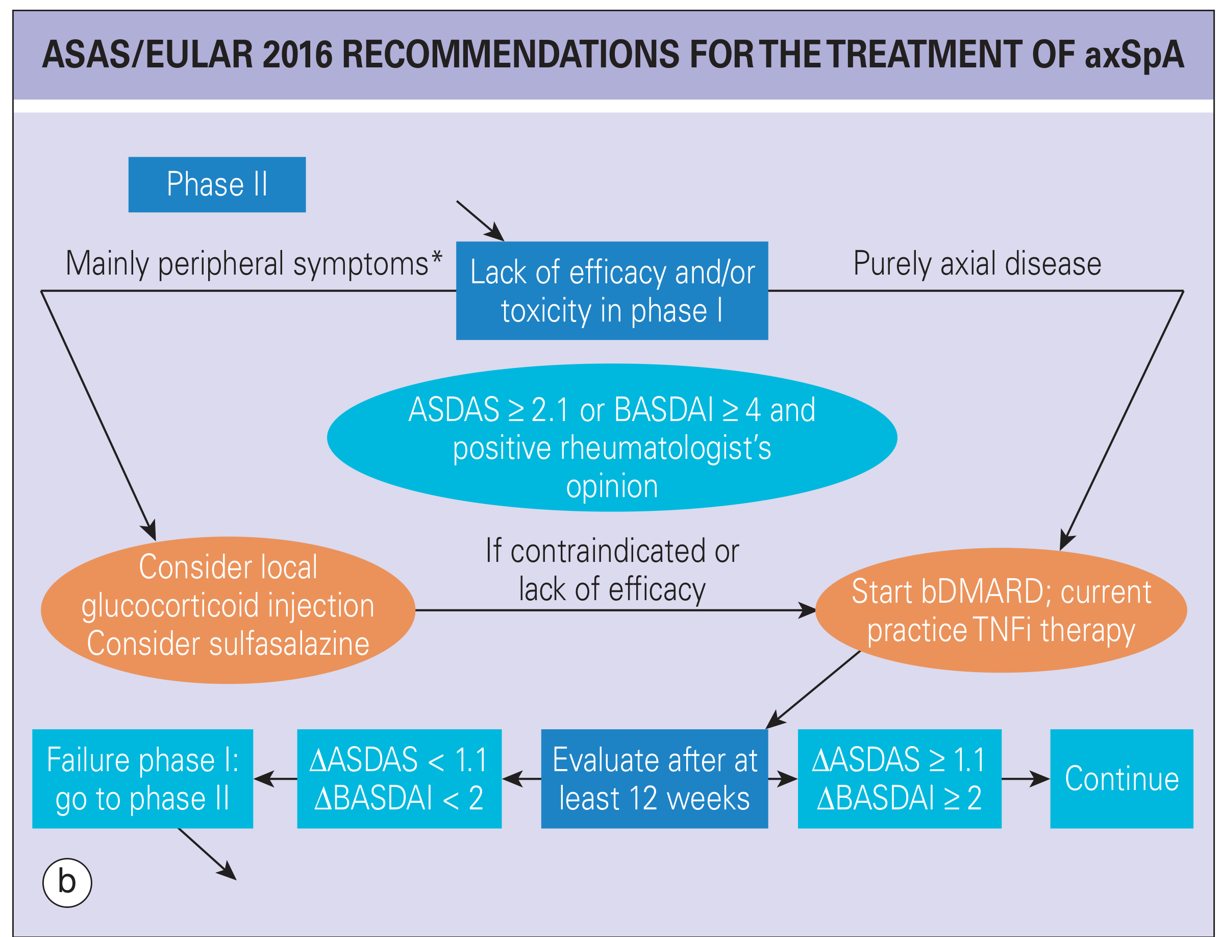
- Purely axial disease: Start a biologic DMARD (bDMARD) - current first practice is a TNF inhibitor (TNFi)
- Peripheral-predominant disease: Consider local glucocorticoid injection into SI joint (fluoroscopy- or US-guided) and/or sulfasalazine (for peripheral arthritis, less evidence for axial disease)
- If peripheral measures fail or are contraindicated: escalate to TNFi
- Etanercept, infliximab, adalimumab, certolizumab, golimumab
Phase III (TNFi Failure / Second-line Biologics)
- IL-17A inhibitors: Secukinumab, ixekizumab - approved for axSpA, especially useful where TNFi has failed or for patients with prominent skin psoriasis
- JAK inhibitors (tsDMARDs): Tofacitinib, upadacitinib - approved in radiographic axSpA; option when biologics fail or are contraindicated
- Switching between TNFi agents is an option before moving to a different class
7. Specific Scenarios
Non-radiographic axSpA (nr-axSpA)
- Same treatment algorithm as radiographic AS
- TNFi and IL-17 inhibitors are effective when there is objective evidence of inflammation (elevated CRP or active SI joint inflammation on MRI)
SI Joint Injections
- Fluoroscopy- or CT-guided corticosteroid injection into the SI joint provides short-to-medium-term relief
- A 2025 systematic review (PMID 40397173) on sacroiliac joint pain management outlines current interventional approaches
PRP vs. Corticosteroid Injections
- A 2026 systematic review (PMID 42332419) evaluated PRP versus corticosteroid injections for chronic low back pain secondary to sacroiliitis - emerging evidence for PRP as an alternative to steroids
Pyogenic (Septic) Sacroiliitis
- A different entity - acute, with fever, severe pain; seen in postpartum women and IV drug users
- Diagnosis by blood cultures, fluoroscopic fine-needle aspiration, or open biopsy
- Antibiotic therapy for 6 weeks is usually adequate; surgical drainage rarely needed
- Rheumatology, 2-Volume Set (Elsevier, 2022)
Surgery
- Reserved for severe structural damage with functional impairment
- Total hip arthroplasty for hip joint ankylosis
- Spinal osteotomy for severe kyphotic deformity (> 10-15 years of progressive disease)
- Controversy exists about role of bone grafts in surgical SI joint debridement
8. Monitoring and Treat-to-Target
- Target: remission (ASDAS < 1.3) or low disease activity (ASDAS 1.3-2.1)
- Treat-to-target trials have not consistently shown benefit over standard care, though maintaining a disease activity target remains clinically useful for individual patients
- Regular reassessment of ASDAS/BASDAI, function (BASFI), imaging progression
- Watch for extra-articular manifestations: uveitis (refer ophthalmology), IBD, psoriasis
- Cardiovascular risk monitoring (NSAIDs, chronic inflammation both increase CV risk)
- Bone density monitoring - chronic inflammation + limited mobility = osteoporosis risk
Quick Summary Table
| Step | Intervention |
|---|---|
| All patients | NSAIDs (max dose), physical therapy, education, exercise, smoking cessation |
| NSAID failure (axial) | TNF inhibitor (etanercept, infliximab, adalimumab) |
| NSAID failure (peripheral) | SI joint injection, sulfasalazine; if fails → TNFi |
| TNFi failure | Switch TNFi, or move to IL-17i (secukinumab/ixekizumab) or JAK inhibitor |
| Structural damage | Hip replacement, spinal osteotomy |
| Septic sacroiliitis | 6 weeks antibiotics, rarely surgical drainage |