
Scrub typus
"scrub typhus"[MeSH Terms] AND treatment
scrub typhus eschar rash skin
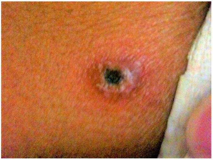
This clinical photograph displays a characteristic eschar on the skin, a pathognomonic sign of scrub typhus (Orientia tsutsugamushi infection). The lesion features a necrotic, centrally depressed black crust (eschar) approximately 0.5 to 1 cm in diameter. Surrounding the dark central necrotic core is a distinct, slightly raised, pale grayish-white rim. This is further encircled by a prominent area of erythema (redness) and mild edema, indicating a localized inflammatory response at the site of the larval mite (chigger) bite. The skin texture within the erythematous zone appears slightly irregular. Such lesions are typically found in areas where skin surfaces are in contact, such as the axilla, groin, or back, and are critical for the early clinical diagnosis of rickettsial diseases when other symptoms like fever, myalgia, and rash are present.
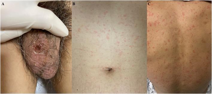
This composite of clinical photographs illustrates the dermatological manifestations of scrub typhus (Orientia tsutsugamushi infection). Panel A displays a characteristic eschar located on the scrotum. The lesion is approximately 1 x 1 cm, presenting as an erythematous to black ulcerative area with a central necrotic crust. Panels B and C show the associated systemic skin involvement, characterized by a diffuse, blanching maculopapular rash distributed across the trunk (B) and the back (C). The rash consists of multiple small, pink-to-red macules and papules that are non-confluent. These findings are diagnostic hallmarks of rickettsial diseases, where the eschar represents the site of the chigger bite and the generalized rash indicates hematogenous dissemination. This clinical presentation, combined with a history of outdoor exposure, is used to identify vector-borne infectious diseases in endemic regions.
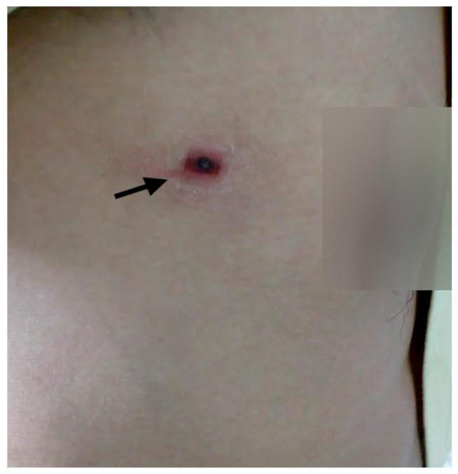
A clinical photograph of a human chest area demonstrating a pathognomonic eschar, a classic skin lesion associated with scrub typhus. The lesion, indicated by a black arrow, features a small, dark, necrotic central crust (eschar) that is circular to oval in shape. This necrotic center is surrounded by a prominent, well-demarcated erythematous halo and a slightly raised, indurated rim, characteristic of an inflammatory reaction at the site of a mite bite (chigger). The surrounding fair skin appears otherwise unremarkable without diffuse rash or additional lesions. This image serves as a key diagnostic educational tool for infectious disease and dermatology, illustrating the hallmark physical finding used in the clinical diagnosis of Orientia tsutsugamushi infection.
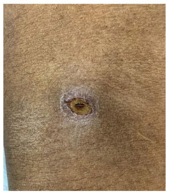
A clinical photograph showing a characteristic eschar associated with scrub typhus located on the right antecubital fossa. The lesion is approximately 7 x 5 mm, featuring a prominent, oval-shaped, necrotic central area that is dark brown to black in color and slightly depressed. This central necrotic core is surrounded by a well-demarcated, yellowish-tan intermediate ring and a peripheral zone of erythema and mild inflammation. The surrounding skin exhibits a normal texture with visible hair follicles and no evidence of diffuse rash in the immediate field of view. This visual finding is a hallmark diagnostic sign of Orientia tsutsugamushi infection, typically occurring at the site of a larval mite (chigger) bite. The image serves as a critical educational tool for identifying rickettsial-like infections in patients presenting with undifferentiated fever, particularly in endemic regions.
Scrub Typhus (Tsutsugamushi Disease)
Causative Agent
Epidemiology
- Geographic range: The "scrub typhus triangle" - eastern Russia, northern Japan, eastern Australia, and everything in between: South/Southeast Asia, China, the Indian subcontinent, and Pacific/Indian Ocean islands. Emerging cases have been reported in Chile, sub-Saharan Africa, and UAE.
- ~1 million cases annually; up to 1 billion people at risk
- One of the three most common causes of prolonged fever in rural Asia
- Seasonality follows mite larval emergence: autumn and spring in temperate zones, wet season in tropical zones
- Immunity to the homologous strain wanes within 1-3 years; cross-protection can be lost in as little as 1 month
Mite (transovarial) → Rodents (rats, mice, shrews) → Mite larvae (chiggers) → Humans
- The larval stage (chigger) is the only stage that feeds on vertebrates and transmits disease
- Reservoir: Trombiculid mites (Leptotrombidium deliense, L. akamushi) - infection maintained transovarially
- Person-to-person transmission does NOT occur
Pathogenesis
Clinical Features
| Feature | Details |
|---|---|
| Eschar | Punched-out ulcer with a blackened scab at the chigger bite site; found in <50% of returning travelers |
| Maculopapular rash | Appears day 4-6 of illness; seen in <40% of cases |
| Regional lymphadenopathy | Tender nodes draining the eschar; generalized lymphadenopathy also occurs |
- Sudden-onset fever (104-105°F), headache, myalgia, malaise, cough, GI symptoms
- Relative bradycardia (pulse-temperature dissociation)
- Lymphocytosis
- Leukopenia, thrombocytopenia, elevated liver enzymes
- Encephalitis / meningismus (neuromeningeal involvement is relatively common)
- Interstitial pneumonia
- Septic shock
- Cardiac involvement
- Movement disorders (rare; recognized increasingly - 2026 systematic review)
- Acute pancreatitis (rare complication; 2026 review, PMID 41187339)
Characteristic Eschar
Diagnosis
| Test | Notes |
|---|---|
| Serology (IFA, ELISA) | Mainstay; uses major serotypes (Kato, Karp, Gilliam, Kawasaki); antibody rise needed |
| PCR | Effective on eschar biopsy; less sensitive on blood; allows early diagnosis before seroconversion |
| Weil-Felix reaction | Positive with Proteus OX-K; less specific, still used in resource-limited settings |
| Skin biopsy | Immunohistochemical staining (days 4-8); available in reference labs |
| Culture | Technically difficult, requires BSL-3; rarely done clinically |
Treatment
| Drug | Regimen | Notes |
|---|---|---|
| Doxycycline (first-line) | 100 mg PO twice daily × 7-15 days; 200 mg IV daily for severe cases | Some resistance in Thailand |
| Azithromycin | 500 mg PO daily × 3 days | Preferred in pregnant women; useful for doxycycline-resistant cases |
| Chloramphenicol | 500 mg QID × 7-15 days (or 2 g/day) | Alternative |
| Rifampin | 600 mg PO daily | For doxycycline/chloramphenicol-resistant cases |
| Doxycycline + Azithromycin | Combination | Beneficial in severe scrub typhus vs. monotherapy |
- Sulfonamides and quinolones are contraindicated (sulfonamides enhance the disease; quinolones should be avoided)
- Single-day doxycycline is followed by relapses - treat for the full course
- Response to early treatment is rapid; delay worsens outcome
Prognosis
- Untreated: case-fatality rate 6% (range 0-30% depending on population and strain)
- With doxycycline treatment: mortality <1%
- Relapses may occur; immunity to homologous strain wanes within 1-3 years
- Abortion commonly occurs in pregnant women
Prevention & Control
- Repellents (DEET) applied to skin and clothing
- Protective clothing in endemic areas (covering skin reduces chigger access)
- Rodent and mite control in endemic foci
- No licensed vaccine available (antigenic diversity of O. tsutsugamushi makes this challenging)
- Prophylactic doxycycline (200 mg weekly) has been used in high-risk exposures
Recent evidence note (2025-2026): A large systematic review and meta-analysis on scrub typhus in India (PMID 40754340) highlights that the disease is underdiagnosed and associated with significant morbidity. Neurological complications including movement disorders are now increasingly recognized (PMID 41939484).