
Just list the questions first
- Basics of anesthesia (slides)
- GA, LA, regional anesthesia (class notes)
- Anesthesia drugs and doses
- Anesthesia equipments
- Anesthesia work station
- Airway management
- Muscle relaxants
- CPR
Leave the first two topics there. Give me basic anesthesia drugs and their doses. Drugs that are used from induction to maintenance, everything. Uh, mainly give, uh, give me the drugs that are commonly used, you know, drugs like midazolam, then... So commonly you would use... Give me the general order first, you know, what, uh, all drugs are given in anesthesia first. Uh, just, uh, make me understand the basics. And then general anesthesia, like, um, we give someone midazolam first, then propofol, then, um, I don't know, vecuronium or pancuronium or, uh, rocuronium, something like that. Um... Then also, uh, regional anesthetic drugs and their uses, pre-anesthetic drugs and their uses. Uh, give me these in separate, uh, tables. And some anesthesia equipments, their parts. Their users, also the anesthesia workstation and its details, parts, user, uh, parts, and, uh, various things I need to know about it, and also, um, details of airway management. So yeah, give me comprehensive notes on these topics.
anesthesia workstation machine components diagram
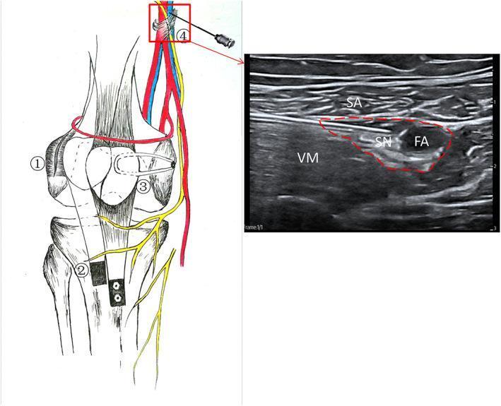
This composite educational image illustrates a '3-in-1' surgical procedure for recurrent patellar dislocation (RPD) alongside an ultrasound-guided adductor canal block (ACB). The left anatomical diagram depicts the surgical components: (1) lateral patellofemoral ligament release to loosen the lateral retinaculum; (2) distal realigning via lower-inner transfer of the tibial tuberosity, shown fixed with two screws; and (3) reconstruction of the medial patellofemoral ligament (MPFL) using a graft tendon. Step (4) demonstrates the needle placement for an adductor canal block relative to the femoral neurovascular bundle. The right panel is a transverse ultrasound scan of the medial mid-thigh, labeled for regional anesthesia. It identifies the sartorius (SA) muscle superficially and the vastus medialis (VM) muscle laterally. Within the adductor canal (outlined in red), the hypoechoic saphenous nerve (SN) and the pulsatile femoral artery (FA) are clearly visible. This figure serves as a clinical guide for orthopedic surgeons and anesthesiologists managing patellar instability through combined surgical correction and regional analgesia.
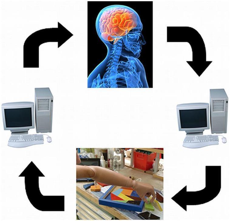
This infographic illustrates the conceptual framework of a closed-loop Brain-Computer Interface (BCI) system for prosthetic control. The diagram is organized in a circular flow using four main components connected by thick black arrows. At the apex, a sagittal anatomical diagram of a human head shows a brain with highlighted orange cortical activity, representing neural intent or motor planning. Following the clockwise flow, a computer workstation symbolizes the signal processing and decoding of neural data. Below this, a clinical photograph shows a skin-toned prosthetic arm and hand performing a fine motor task: manipulating geometric puzzle pieces on a workbench. This represents the effector output and sensory feedback acquisition. Completing the loop, a second computer workstation symbolizes the encoding of feedback signals back to the user. The illustration demonstrates the integration of neural engineering, neurorehabilitation, and embodiment in advanced prosthetics, highlighting the bidirectional communication required for sophisticated bionic limb function.
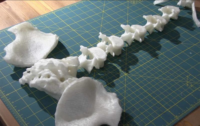
Anatomical Diagram/Specimen: This clinical photograph displays a collection of 3D-printed anatomical components intended for the construction of a lumbar neuraxial block task trainer. The specimens, printed in white polylactic acid (PLA) using fused filament fabrication (FFF), include individual lumbar vertebral bodies (T10 through L5), bilateral iliac crests, and the sacrum. The vertebral bodies are arranged linearly on a green cutting mat, showcasing detailed posterior elements including spinous and transverse processes. The iliac crests exhibit characteristic complex, irregular morphology and a porous surface texture typical of layer-by-layer 3D printing. These high-fidelity models, derived from CT DICOM data, serve as realistic palpable landmarks for medical simulation. The educational focus is on the spatial orientation of the lumbar spine, sacrum, and pelvis, highlighting their clinical relevance in anesthesiology for identifying entry points for epidural or spinal anesthesia.
airway management laryngoscopy intubation LMA

This sequence of clinical images demonstrates the four steps of endotracheal intubation (ETI) using a polycarbonate barrier box on an airway trainer manikin. The setup is designed to minimize aerosol exposure during airway management. Step 1: An operator wearing personal protective equipment (PPE), including blue gloves, inserts hands through the rear access ports of the transparent box to begin laryngoscopy. Step 2: The operator uses a Macintosh blade laryngoscope to visualize the glottis while an assistant provides a 7.0 mm cuffed endotracheal tube through a side port. Step 3: The operator inserts the tracheal tube into the trachea. Step 4: The tube is secured, the cuff is inflated, and an Ambu-Bag (manual resuscitator) is connected to begin positive-pressure ventilation. The images highlight the ergonomic constraints and procedural adjustments required when performing direct laryngoscopy within a confined barrier enclosure, commonly utilized in protocols for highly infectious respiratory diseases such as COVID-19.
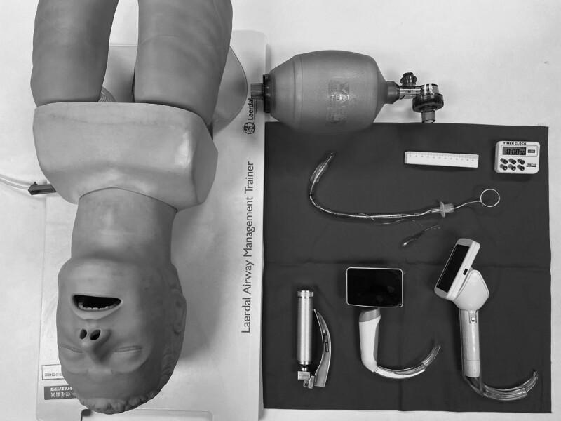
A high-angle clinical photograph displays standard and advanced airway management equipment for endotracheal intubation, positioned alongside a Laerdal Airway Management Trainer mannequin. The equipment includes three types of laryngoscopes: a conventional Macintosh-style laryngoscope with a metal handle and curved blade, and two video laryngoscopes featuring integrated digital screens and specialized curved blades for enhanced glottic visualization. Also present is a Bag Valve Mask (BVM) for manual ventilation. Centrally located is a clear cuffed endotracheal tube (ETT) with an internal stylet bent into a 'hockey-stick' configuration to facilitate insertion; the ETT includes a visible pilot balloon, connector, and depth markings. Ancillary procedural tools include a ruler for measurement and a digital timer for monitoring intubation time. This setup is typical for anesthesia training, emergency medicine simulation, or comparative studies between direct and video laryngoscopy techniques.
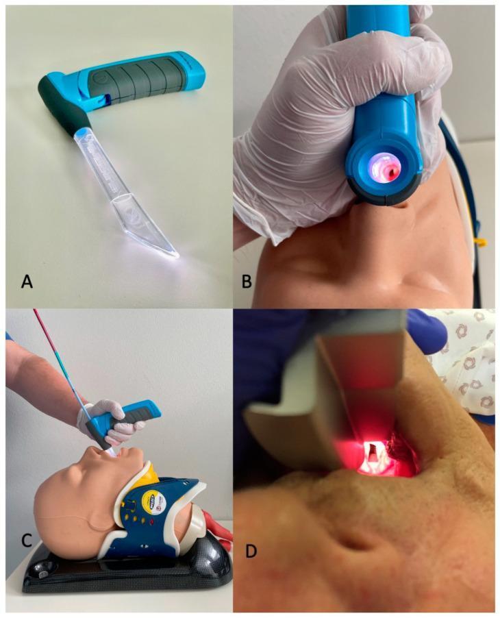
This composite educational image illustrates airway management techniques using the VieScope, a novel Miller-shaped (straight) laryngoscope, in comparison to conventional methods. Panel A displays the device, featuring a transparent acrylic glass tube blade that provides 360-degree circular LED illumination and a blue ergonomic handle with a textured grip. Panel B shows the device being used on a medical manikin, demonstrating the line-of-sight visualization through the lumen of the laryngoscope tube. Panel C illustrates the clinical technique for intubation with this device, where a bougie is first introduced through the VieScope to facilitate subsequent placement of an endotracheal tube; the manikin is shown with a cervical collar, simulating a trauma or difficult airway scenario. Panel D provides a contrast with a traditional Macintosh curved-blade laryngoscope, showing the visualization of the glottic opening during direct laryngoscopy. The content focuses on anesthesiology, emergency medicine, and advanced airway management education, highlighting the structural differences and procedural workflow between straight-tube and curved-blade laryngoscopy.
COMPREHENSIVE ANESTHESIA NOTES
OVERVIEW: Order of Drug Administration in General Anesthesia
- Pre-anesthetic medication (night before / 1-2 hrs before) - anxiolysis, analgesia, antisecretion
- Induction (IV or inhalational) - render patient unconscious
- Intubation facilitator (muscle relaxant) - facilitate laryngoscopy and intubation
- Maintenance (volatile agent + opioid ± N₂O) - keep patient asleep and pain-free
- Reversal (anticholinesterase + anticholinergic) - reverse neuromuscular blockade
- Emergence - wake patient up
TABLE 1: PRE-ANESTHETIC (PREMEDICATION) DRUGS
| Drug | Class | Route | Adult Dose | Purpose |
|---|---|---|---|---|
| Midazolam | Benzodiazepine | Oral / IV | 7.5-15 mg oral; 0.025-0.1 mg/kg IV | Anxiolysis, amnesia, sedation. Most commonly used premedicant |
| Diazepam | Benzodiazepine | Oral | 5-10 mg oral | Anxiolysis, sedation |
| Lorazepam | Benzodiazepine | Oral / IV | 2-4 mg oral (2h pre-op) | Prolonged anxiolysis (used in cardiac surgery) |
| Morphine | Opioid | IM | 0.1-0.2 mg/kg | Analgesia, sedation |
| Pethidine (Meperidine) | Opioid | IM | 1-2 mg/kg | Analgesia, sedation |
| Fentanyl | Opioid | IV | 1-2 mcg/kg | Analgesia |
| Atropine | Anticholinergic | IM / IV | 0.01-0.02 mg/kg | Antisecretion (dries secretions), prevents bradycardia |
| Glycopyrrolate | Anticholinergic | IM / IV | 0.2-0.4 mg | Antisecretion (no CNS penetration, preferred) |
| Ranitidine / Pantoprazole | H2 blocker / PPI | Oral / IV | Ranitidine 150 mg; Pantoprazole 40 mg | Reduce gastric acidity (aspiration prophylaxis) |
| Metoclopramide | Prokinetic | IV / IM | 10 mg | Reduce gastric volume, antiemetic |
| Dexmedetomidine | α2 agonist | IV infusion | 0.5-1 mcg/kg over 10 min | Anxiolysis, analgesia, sedation without respiratory depression |
| Clonidine | α2 agonist | Oral | 3-5 mcg/kg | Anxiolysis, reduce anesthetic requirements |
| Ondansetron | 5-HT3 antagonist | IV | 4-8 mg | PONV prophylaxis |
TABLE 2: INDUCTION AGENTS
| Drug | Class | IV Induction Dose | Onset | Duration | Key Notes |
|---|---|---|---|---|---|
| Propofol | Alkylphenol | 1.5-2.5 mg/kg IV (2-2.5 mg/kg in healthy adults) | 45 sec | 5-10 min | Most widely used. Can cause hypotension. Pain on injection. Antiemetic property. |
| Thiopentone (Thiopental) | Barbiturate | 4-6 mg/kg IV | 30-60 sec | 5-15 min | Classic agent. Still used in many countries. Can cause laryngospasm. Contraindicated in porphyria. |
| Ketamine | NMDA antagonist | 1-2 mg/kg IV; 4-6 mg/kg IM | 60 sec (IV) | 15-20 min | Dissociative anesthesia. Maintains BP and HR. Bronchodilator. Causes emergence delirium, hypersalivation. Ideal for hemodynamic compromise and asthmatics. |
| Etomidate | Imidazole | 0.3 mg/kg IV | 30-60 sec | 5-10 min | Minimal cardiovascular effects - ideal in hemodynamically unstable patients. Causes myoclonus, PONV. Inhibits cortisol synthesis (single dose effect brief). |
| Midazolam | Benzodiazepine | 0.1-0.3 mg/kg IV | 2-3 min | 30-60 min | Slower induction. Used for sedation more than full induction. |
TABLE 3: INTRAOPERATIVE OPIOIDS (Analgesia during and after surgery)
| Drug | IV Dose | Duration | Notes |
|---|---|---|---|
| Fentanyl | 1-3 mcg/kg bolus | 30-60 min | Most commonly used intraoperative opioid. Fast onset. |
| Morphine | 0.05-0.2 mg/kg IV | 4-6 hrs | Longer acting. Good for postoperative analgesia. |
| Remifentanil | 0.05-2 mcg/kg/min infusion | Ultra-short (context insensitive) | Metabolized by plasma esterases. Excellent for procedures requiring rapid offset. |
| Pethidine (Meperidine) | 0.5-1 mg/kg IV | 2-4 hrs | Also used for shivering post-op. |
| Sufentanil | 0.1-0.5 mcg/kg | 30-60 min | 5-10x more potent than fentanyl. |
TABLE 4: NEUROMUSCULAR BLOCKING AGENTS (Muscle Relaxants)
A. Depolarizing (Succinylcholine)
| Drug | Dose | Onset | Duration | Notes |
|---|---|---|---|---|
| Succinylcholine (Suxamethonium) | 1-1.5 mg/kg IV (1.5 mg/kg for RSI) | 60-90 sec | 10-15 min | Only depolarizing agent in use. Fastest onset - gold standard for RSI and emergency intubation. Causes fasciculations, hyperkalemia, malignant hyperthermia, bradycardia. Contraindicated in burns, crush injuries, upper motor neuron lesions. |
B. Non-depolarizing (Competitive)
| Drug | Duration | Intubating Dose | Maintenance Dose | Notes |
|---|---|---|---|---|
| Vecuronium | Intermediate (25-40 min) | 0.1 mg/kg | 0.01-0.015 mg/kg | Clean hemodynamic profile. No histamine release. Hepatic metabolism. |
| Rocuronium | Intermediate (30-60 min) | 0.6 mg/kg (RSI: 1.2 mg/kg) | 0.1-0.2 mg/kg | Fastest onset among non-depolarizing agents (90 sec). Reversed by Sugammadex. Alternative to succinylcholine for RSI. |
| Atracurium | Intermediate (20-35 min) | 0.5 mg/kg | 0.1-0.2 mg/kg | Hoffman elimination (independent of liver/kidney). Safe in organ failure. Releases histamine. |
| Cisatracurium | Intermediate (40-60 min) | 0.15-0.2 mg/kg | 0.03 mg/kg | Similar to atracurium but NO histamine release. Preferred in ICU patients. |
| Pancuronium | Long (60-120 min) | 0.1 mg/kg | 0.01-0.02 mg/kg | Causes tachycardia (vagolytic). Now less commonly used. |
| Mivacurium | Short (12-20 min) | 0.15-0.2 mg/kg | - | Metabolized by plasma cholinesterase. |
Reversal Agents
| Drug | Dose | Used With | Notes |
|---|---|---|---|
| Neostigmine | 0.05 mg/kg IV (max 5 mg) | Glycopyrrolate 0.2 mg per 1 mg neostigmine | Anticholinesterase. Reverses non-depolarizing block. Must co-administer anticholinergic. |
| Sugammadex | 2-16 mg/kg IV (dose depends on depth of block) | Nothing needed | Selectively encapsulates rocuronium/vecuronium. Rapid complete reversal. |
TABLE 5: MAINTENANCE AGENTS
Inhalational Agents (Volatile Anesthetics)
| Agent | MAC (%) | Properties | Notes |
|---|---|---|---|
| Sevoflurane | 2% | Non-pungent, rapid induction | Most used for inhalational induction (especially children). Pleasant smell. |
| Isoflurane | 1.15% | Pungent, cardiovascular stable | Most widely used volatile agent globally. Coronary steal risk (controversial). |
| Desflurane | 6% | Least soluble, fastest emergence | Pungent (not for induction). Fastest wake-up. Requires heated vaporizer. |
| Halothane | 0.75% | Non-pungent, sweet smell | Older agent. Causes halothane hepatitis, sensitizes heart to catecholamines. |
| Nitrous Oxide (N₂O) | 104% (IMAC) | Analgesic, carries other agents | Used as carrier gas with O₂ (usually 50:70% mix). Weak anesthetic alone. Contraindicated in pneumothorax, bowel obstruction. |
IV Maintenance Agents
| Drug | Infusion Rate | Notes |
|---|---|---|
| Propofol | 4-10 mg/kg/hr (TIVA) | Total intravenous anesthesia (TIVA). Antiemetic. Fast emergence. |
| Midazolam | 0.25-1 mcg/kg/min | For hypnosis and amnesia in maintenance. |
| Dexmedetomidine | 0.2-0.7 mcg/kg/hr | Sedation in ICU/procedural. Reduces opioid and anesthetic requirements. |
| Ketamine | 0.5-2 mg/kg/hr infusion | Sub-anesthetic dose for analgesia, especially chronic pain surgery. |
| Remifentanil | 0.05-0.5 mcg/kg/min | Opioid infusion for intraoperative analgesia. |
TABLE 6: REGIONAL ANESTHETIC DRUGS
Local Anesthetic Agents
| Drug | Class | Onset | Duration (plain) | Duration (with epinephrine) | Max Safe Dose | Common Uses |
|---|---|---|---|---|---|---|
| Lignocaine (Lidocaine) | Amide | Fast | 1-2 hrs | 2-3 hrs | 3 mg/kg plain; 7 mg/kg with epi | Spinal, epidural, infiltration, peripheral nerve blocks, topical |
| Bupivacaine | Amide | Medium | 4-8 hrs | 6-10 hrs | 2 mg/kg (max 175 mg) | Spinal (most common), epidural, peripheral blocks. NOT for IV regional (Bier's block - cardiotoxic) |
| Ropivacaine | Amide | Medium | 4-8 hrs | - | 3 mg/kg (max 200 mg) | Epidural, peripheral nerve blocks. Less cardiotoxic than bupivacaine. Vasoconstrictor property. |
| Levobupivacaine | Amide | Medium | 4-8 hrs | - | 2-3 mg/kg | S-enantiomer of bupivacaine. Safer cardiac profile. |
| Cocaine | Ester | Fast | 1 hr | - | 3 mg/kg (max 200 mg) | ONLY local anesthetic that is a vasoconstrictor. Used for nasal/ENT procedures only. |
| Procaine | Ester | Medium | 30-60 min | 1-2 hrs | 7 mg/kg | Spinal, infiltration. Metabolized by plasma cholinesterase. |
| Tetracaine (Amethocaine) | Ester | Slow | 2-4 hrs | 4-6 hrs | 1.5 mg/kg | Spinal, topical ophthalmic. |
| Chloroprocaine | Ester | Very fast | 30-45 min | - | 11 mg/kg | Epidural. Short duration. Rapid metabolism - safe in obstetrics. |
- Epinephrine (Adrenaline) 1:200,000 - prolongs block, reduces systemic absorption, acts as marker for intravascular injection
- Sodium bicarbonate - alkalinizes solution, speeds onset
- Clonidine / Dexmedetomidine - prolongs duration
- Opioids (Fentanyl, Morphine) - intrathecal/epidural for enhanced analgesia
Neuraxial Anesthesia Doses
| Technique | Drug | Dose | Level |
|---|---|---|---|
| Spinal (intrathecal) | Bupivacaine 0.5% heavy | 1.5-3 ml (7.5-15 mg) | T4-T10 |
| Spinal | Lignocaine 5% heavy | 1.5-2.5 ml | T4-T10 |
| Epidural | Bupivacaine 0.25-0.5% | 15-20 ml | - |
| Epidural | Lignocaine 2% | 15-20 ml | - |
ANESTHESIA EQUIPMENT
Key Equipment and Their Uses
| Equipment | Description | Use |
|---|---|---|
| Laryngoscope | Handle + blade (Macintosh = curved; Miller = straight) | Direct visualization of vocal cords for intubation |
| Endotracheal Tube (ETT) | Cuffed/uncuffed tube | Secures airway, allows ventilation and delivery of volatile agents |
| Laryngeal Mask Airway (LMA) | Supraglottic device - sits over larynx | Airway maintenance without intubation. Used for short procedures, also as rescue device |
| Oropharyngeal Airway (Guedel) | Curved rigid device | Maintains airway in unconscious patient - prevents tongue falling back |
| Nasopharyngeal Airway | Soft rubber tube via nose | Airway maintenance in semi-conscious/awake patients |
| Bag-Valve-Mask (Ambu Bag) | Self-inflating bag + mask | Manual positive pressure ventilation |
| Video Laryngoscope | Camera-equipped laryngoscope | Improved glottic view, especially in difficult airways |
| Fiberoptic Bronchoscope | Flexible scope | Gold standard for anticipated difficult airway - awake fiberoptic intubation |
| Stylet / Bougie | Stiff wire / gum elastic introducer | Aid ETT placement when cords not fully visible |
| Vaporizer | Agent-specific device on machine | Delivers precise concentrations of volatile anesthetic |
| Capnograph (ETCO₂ monitor) | Waveform CO₂ monitor | Confirms ETT placement, monitors ventilation adequacy |
| Pulse Oximeter | SpO₂ probe | Continuous oxygen saturation monitoring |
| Invasive arterial line | Intra-arterial catheter | Continuous BP monitoring, blood gas sampling in major surgery |
ANESTHESIA WORKSTATION
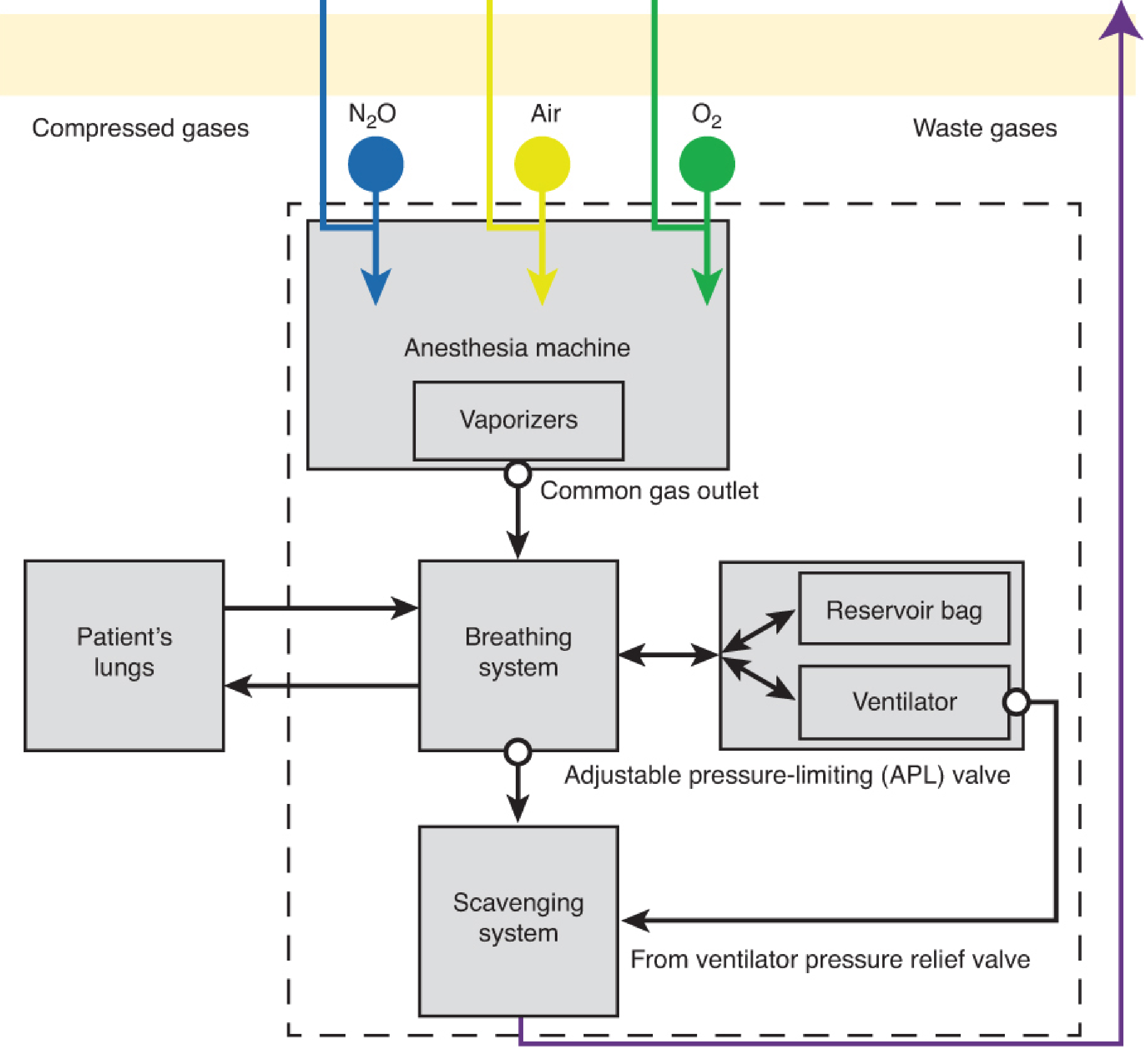
Basic Configuration Diagram
Detailed Machine Diagram
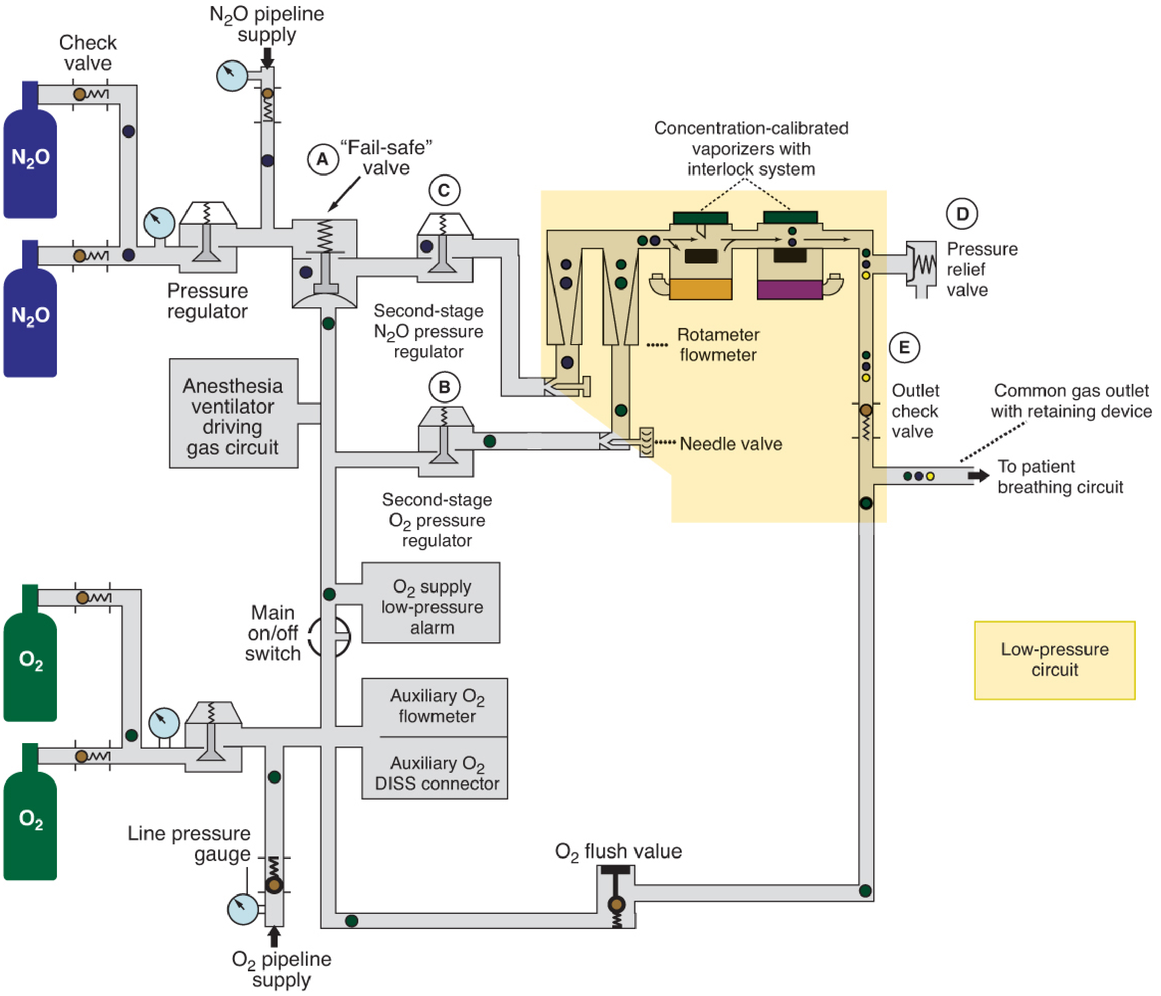
Pressure Circuits in the Workstation
| Circuit | Pressure Range | Components |
|---|---|---|
| High-Pressure Circuit | O₂: up to 2,200 psig → regulated to 45 psig; N₂O: 750 psig → 45 psig | Gas cylinders, cylinder primary pressure regulators |
| Intermediate-Pressure Circuit | 45-55 psig | Pipeline inlets, second-stage regulators, O₂ flush valve, ventilator driving gas |
| Low-Pressure Circuit (LPC) | Below 15 psig | Flow control valves → rotameters → vaporizers → common gas outlet |
Key Components of the Workstation
| Component | Function |
|---|---|
| Gas cylinders | O₂ (green), N₂O (blue), Air (yellow) - backup supply |
| Pipeline supply | Primary gas source from hospital wall at ~50 psig |
| Pressure regulator | Reduces high cylinder pressure to working pressure |
| Fail-safe valve | Shuts off N₂O if O₂ supply pressure drops - prevents hypoxic mixture |
| Rotameter (flowmeter) | Calibrated glass tube with bobbin - controls and measures gas flow |
| Vaporizer | Agent-specific device that delivers precise % of volatile anesthetic (e.g., sevoflurane, isoflurane, desflurane). Has interlock system - only one vaporizer can be on at a time. |
| O₂ flush valve | Delivers 100% O₂ directly to breathing circuit at 35-75 L/min. Bypasses vaporizer - washes out anesthetic agent. |
| Common gas outlet | Final point where all gases and vapor mix before going to the breathing circuit |
| Breathing circuit (circle system) | Delivers gas to patient. Contains: inspiratory limb, expiratory limb, CO₂ absorber (soda lime), APL valve, reservoir bag, Y-piece connector |
| APL valve (Adjustable Pressure Limiting) | Pops off excess gas - prevents barotrauma during spontaneous breathing |
| CO₂ absorber (Soda lime) | Absorbs exhaled CO₂ in a rebreathing system |
| Reservoir bag (Breathing bag) | 1-3L bag - for manual ventilation and as a reservoir; also monitors breathing |
| Ventilator | Automatically ventilates the patient when required |
| Scavenging system | Collects and removes waste anesthetic gases to prevent operating room pollution |
| Oxygen analyzer | Only monitor that checks the LPC integrity - monitors actual O₂ delivered to patient |
Pre-use Check (Key Points)
- Check oxygen analyzer - calibrate to 21% in room air
- Low-pressure circuit leak test
- Circle system/breathing circuit test
- Check backup O₂ cylinder and SIRB (self-inflating resuscitation bag)
- Check vaporizer fill levels
- Check alarm limits and monitors
AIRWAY MANAGEMENT
Assessment Before Intubation (Predictors of Difficult Airway)
| Assessment | Test | Difficult Airway Prediction |
|---|---|---|
| Mallampati score | Visualize mouth opening in sitting position | Class III-IV = difficult laryngoscopy |
| Thyromental distance | Chin to thyroid cartilage | < 6 cm = difficult |
| Mouth opening (IID) | Interincisor distance | < 3 cm = difficult |
| Neck movement | Atlanto-occipital extension | Restricted = difficult |
| ULBT | Upper lip bite test | Unable to bite upper lip = difficult |
| BMI / Obesity | - | Higher difficulty; use ramped position |
Airway Devices
| Device | Description | When Used |
|---|---|---|
| Oropharyngeal Airway (OPA) | Guedel airway - rigid curved device | Unconscious patients only - causes gag reflex if awake |
| Nasopharyngeal Airway (NPA) | Soft rubber tube via nostril | Can be used in semi-conscious patients. Contraindicated in basal skull fracture |
| LMA (Classic) | Cuff-sealed supraglottic airway | Short procedures, spontaneous ventilation, rescue device. Sizes 1-5 by weight. |
| LMA ProSeal | Modified LMA with gastric drainage port | Better seal for positive pressure ventilation; allows gastric tube passage |
| ETT (Endotracheal Tube) | Cuffed tube into trachea | Definitive airway. Protects against aspiration. Required for PPV and major surgery. |
| Double-lumen tube | Two-lumen tube (left/right) | One-lung ventilation in thoracic surgery |
| Tracheostomy tube | Tube through surgical tracheal opening | Long-term airway management, failed intubation |
Rapid Sequence Induction (RSI)
- Pre-oxygenate with 100% O₂ for 3-5 minutes (denitrogenation)
- Apply cricoid pressure (Sellick's maneuver) - compresses esophagus
- IV Induction: Propofol 2 mg/kg (or Ketamine/Thiopentone)
- Succinylcholine 1.5 mg/kg IV (or Rocuronium 1.2 mg/kg if succinylcholine contraindicated)
- Intubate as soon as fasciculations cease (≈ 60 seconds)
- Confirm placement with capnography and chest auscultation
- Inflate cuff, release cricoid pressure
Laryngoscope Blades
| Blade | Shape | Technique | Best For |
|---|---|---|---|
| Macintosh | Curved | Tip in vallecula (between epiglottis and tongue base) | Adults - most common |
| Miller | Straight | Tip lifts epiglottis directly | Neonates, infants; anterior larynx |
Grading of Laryngeal View (Cormack-Lehane)
| Grade | View |
|---|---|
| Grade I | Full view of vocal cords |
| Grade II | Partial view of cords |
| Grade III | Only epiglottis visible |
| Grade IV | Not even epiglottis visible |
Failed Intubation Drill (Can't Intubate - Can't Oxygenate)
- Call for help
- Attempt bag-mask ventilation
- Insert LMA as rescue device
- Fiberoptic intubation through LMA / awake fiberoptic intubation
- Surgical airway (cricothyrotomy / emergency tracheostomy) as last resort
QUICK SUMMARY CARD: Drugs at a Glance
| Phase | Drug | Dose |
|---|---|---|
| Pre-med | Midazolam | 7.5-15 mg oral OR 0.025-0.1 mg/kg IV |
| Pre-med (antisecretion) | Glycopyrrolate | 0.2-0.4 mg IM |
| Induction | Propofol | 1.5-2.5 mg/kg IV |
| Induction (emergency/hemodynamic instability) | Ketamine | 1-2 mg/kg IV |
| Induction (hemodynamically unstable) | Etomidate | 0.3 mg/kg IV |
| Intubation (RSI) | Succinylcholine | 1.5 mg/kg IV |
| Intubation (elective) | Rocuronium | 0.6 mg/kg IV |
| Intraop analgesia | Fentanyl | 1-3 mcg/kg IV bolus |
| Maintenance (volatile) | Sevoflurane | 1-2% (with N₂O/O₂) |
| Maintenance (TIVA) | Propofol infusion | 4-10 mg/kg/hr |
| Reversal of block | Neostigmine + Glycopyrrolate | 0.05 mg/kg + 0.01 mg/kg IV |
| Reversal of rocuronium | Sugammadex | 2-4 mg/kg IV |
| Spinal anesthesia | Bupivacaine 0.5% heavy | 2-3 ml (10-15 mg) |
Can you also... Explain various fluids given to patients. Crystalloids, colloid, their differences, um, in table and, uh, that. But, and also some other things like... CPR. And airway management, if you have not given that. Which I believe you have. And spinal and epidural anesthesia, if you have not given that. That is it.
ADDITIONAL ANESTHESIA NOTES
TOPIC 3: INTRAVENOUS FLUIDS
What Are IV Fluids?
- Replace fluid deficits (fasting, bleeding, losses)
- Maintain normovolemia intraoperatively
- Correct electrolyte imbalances
- Deliver drugs
TABLE 1: CRYSTALLOIDS vs COLLOIDS - Key Differences
| Feature | Crystalloids | Colloids |
|---|---|---|
| Definition | Aqueous solutions of ions (salts) ± glucose | Contains high-molecular-weight substances (proteins / large glucose polymers) |
| Distribution | Distribute throughout entire extracellular fluid space (intravascular + interstitial) | Mostly remain intravascular - maintain oncotic pressure |
| Volume needed | 3-4x more volume needed compared to colloids | Smaller volume required for same effect |
| Onset | Slower expansion of intravascular volume | Faster intravascular expansion |
| Cost | Cheap | Expensive |
| Risk of edema | Yes - large volumes (>4-5L) cause tissue edema | Less risk of tissue edema |
| Examples | Normal saline, Ringer's lactate, PlasmaLyte, D5W | Albumin, Gelatin, Hetastarch, Dextran |
| First-line use | Yes - initial resuscitation in most cases | Added after initial crystalloid, or in severe deficit |
TABLE 2: CRYSTALLOID SOLUTIONS IN DETAIL
| Fluid | Na⁺ (mmol/L) | Cl⁻ (mmol/L) | K⁺ | Other | Osmolarity (mOsm/L) | pH | Uses / Notes |
|---|---|---|---|---|---|---|---|
| Normal Saline (0.9% NaCl) | 154 | 154 | - | - | 308 | 4.5-7.0 | Most versatile. Use for hypochloremic alkalosis, diluting PRBCs. Large volumes → hyperchloremic metabolic acidosis. |
| Ringer's Lactate (RL / Hartmann's) | 130 | 109 | 4 | Lactate 28 mmol/L, Ca²⁺ | 273 | 6.0-7.5 | Closest to plasma. Preferred balanced solution for most surgeries. Lactate metabolized to bicarbonate. Do not use with blood transfusion (Ca²⁺ chelates citrate). |
| PlasmaLyte | 140 | 98 | 5 | Acetate + Gluconate | 295 | 7.4 | Best balanced crystalloid. pH of 7.4 matches plasma. No lactate. Preferred in liver disease. |
| 5% Dextrose (D5W) | 0 | 0 | - | Glucose 50g/L | 252 | - | For pure water deficit and sodium-restricted patients. Glucose metabolized rapidly - equivalent to free water. Hypotonic - do NOT use for resuscitation. |
| 3% NaCl (Hypertonic saline) | 513 | 513 | - | - | 1026 | - | Severe symptomatic hyponatremia, raised ICP. Use carefully - risk of central pontine myelinolysis. |
| 0.45% NaCl (Half normal) | 77 | 77 | - | - | 154 | - | Hypotonic - maintenance fluids, free water replacement. |
TABLE 3: COLLOID SOLUTIONS IN DETAIL
| Colloid | Type | Molecular Weight | Duration of Effect | Notes / Cautions |
|---|---|---|---|---|
| Human Albumin (4-5%) | Natural protein colloid | 69,000 Da | 12-24 hrs | Most physiologic colloid. Expensive. Used in burns, liver failure, SBP, nephrotic syndrome. 4-5% = iso-oncotic (replaces lost albumin). 20-25% = hyperoncotic (mobilizes edema fluid). |
| Human Albumin (20-25%) | Natural protein colloid | 69,000 Da | Longer | Hyperoncotic - draws fluid from interstitium. Used in cerebral edema, ascites management. |
| Gelatin (Gelofusine, Haemaccel) | Synthetic - degraded gelatin | ~30,000 Da | 2-4 hrs | Cheapest synthetic colloid. Risk of histamine release/anaphylaxis. Not approved in all countries. |
| Hydroxyethyl Starch (HES) - Voluven, Volulyte | Synthetic - starch polymer | 130,000 Da | 4-6 hrs | Effective but controversial - associated with renal impairment and coagulopathy. Contraindicated in sepsis and renal failure (EMA/FDA restrictions). |
| Dextran (Dextran 40, Dextran 70) | Synthetic - glucose polymer | 40,000-70,000 Da | 4-6 hrs | Dextran 40: improves microcirculation (used in flap surgery). Risk of anaphylaxis, coagulopathy at high doses. |
Practical Rules for Fluid Choice
| Situation | Preferred Fluid |
|---|---|
| Routine surgical maintenance | Ringer's Lactate or PlasmaLyte |
| Blood loss replacement (< 500 mL) | Ringer's Lactate (3:1 ratio) |
| Blood loss replacement (large / rapid) | Colloid or blood products |
| Hypochloremic alkalosis (vomiting, NG suction) | Normal saline (0.9% NaCl) |
| Head injury (raised ICP) | Normal saline or hypertonic saline (avoid hypotonic fluids) |
| Diabetic patients / children fasting | Dextrose-containing solutions |
| Severe hyponatremia | 3% NaCl (hypertonic saline) - slowly |
| Diluting packed red blood cells | Normal saline only (RL has Ca²⁺ which chelates citrate) |
Maintenance Fluid Formula (Holliday-Segar Rule)
| Weight | Rate |
|---|---|
| First 10 kg | 4 mL/kg/hr |
| Next 10 kg (10-20 kg) | 2 mL/kg/hr |
| Each kg above 20 kg | 1 mL/kg/hr |
TOPIC 4: CPR (Cardiopulmonary Resuscitation)
Definition
Chain of Survival (AHA 2020)
- Recognition of arrest and activation of EMS
- Immediate high-quality CPR
- Rapid defibrillation
- Advanced resuscitation (drugs, airway)
- Post-resuscitation care
- Recovery
TABLE 4: AHA BLS (Basic Life Support) Guidelines - Adult CPR
| Parameter | Guideline |
|---|---|
| Check for response | Tap shoulders, shout "Are you okay?" |
| Call for help | Activate EMS / call code blue |
| Check pulse | Carotid pulse - not more than 10 seconds |
| Compression rate | 100-120 compressions/minute |
| Compression depth | 5-6 cm (2-2.4 inches) in adults |
| Chest recoil | Full recoil between compressions (don't lean on chest) |
| Compression fraction | At least 80% of time spent doing compressions |
| Compression:Ventilation ratio | 30:2 (without advanced airway); Continuous compressions (10 breaths/min) with advanced airway |
| Ventilation | 1 breath per 5-6 seconds = 10-12 breaths/min |
| Hand position | Lower half of sternum, heel of both hands |
| Defibrillation | Shock as soon as AED available - minimize interruption |
| CPP target | Coronary perfusion pressure ≥ 15 mmHg for ROSC |
| ETCO₂ target | ≥ 10 mmHg - below this, enhance CPR quality |
TABLE 5: ACLS - Cardiac Arrest Rhythms and Management
| Rhythm | Action |
|---|---|
| VF / pulseless VT | Shockable - immediate defibrillation + CPR |
| PEA (Pulseless Electrical Activity) | Non-shockable - CPR + treat reversible causes (4H's & 4T's) |
| Asystole | Non-shockable - CPR + treat reversible causes |
Reversible Causes of Cardiac Arrest - The 4H's and 4T's
| 4 H's | 4 T's |
|---|---|
| Hypovolemia | Tension pneumothorax |
| Hypoxia | Tamponade (cardiac) |
| Hypo/Hyperkalemia + metabolic | Toxins (drug overdose) |
| Hypothermia | Thrombosis (PE or MI) |
TABLE 6: ACLS Drugs During Cardiac Arrest
| Drug | Dose | Indication | Timing |
|---|---|---|---|
| Epinephrine (Adrenaline) | 1 mg IV every 3-5 minutes | All cardiac arrest rhythms (VF, pVT, PEA, Asystole) | After 1st or 2nd shock in VF/pVT; immediately in PEA/Asystole |
| Amiodarone | 300 mg IV bolus (repeat 150 mg once) | Shock-refractory VF / pulseless VT | After 3rd shock |
| Lidocaine | 1-1.5 mg/kg IV (repeat 0.5-0.75 mg/kg) | Shock-refractory VF/pVT (if amiodarone not available) | After 3rd shock |
| Atropine | No longer recommended for PEA/asystole (2010 guidelines removed it) | Previously used for bradycardia arrest | - |
| Sodium bicarbonate | 1 mEq/kg IV | Known hyperkalemia, tricyclic antidepressant OD, severe acidosis | Selected cases - not routine |
| Calcium chloride | 10 mL of 10% IV | Hyperkalemia, hypocalcemia, calcium channel blocker OD | Selected cases |
| Magnesium sulfate | 1-2 g IV | Torsades de pointes | Specific arrhythmia only |
| Vasopressin | 40 IU IV (single dose replaces 1st or 2nd epi) | VF/pVT; not superior to epinephrine | Removed from 2015 AHA guidelines |
Post-ROSC (Return of Spontaneous Circulation) Care
- 12-lead ECG - rule out STEMI (immediate PCI if found)
- Targeted temperature management (TTM): 32-36°C for 24 hours in comatose survivors - improves neurological outcome
- BP target: Mean arterial pressure ≥ 65 mmHg
- Avoid hyperoxia - target SpO₂ 94-98%
- Avoid hypo/hyperglycemia
- CT brain/coronary angiography as indicated
CPR Monitoring Tools
| Monitor | What It Shows |
|---|---|
| ETCO₂ (capnography) | Best monitor during CPR - ETCO₂ ≥10 mmHg indicates adequate CPR; sudden rise signals ROSC |
| ECG | Rhythm only - does not confirm mechanical activity |
| Pulse check | Carotid / femoral - unreliable if rapid |
| Coronary perfusion pressure (CPP) | Needs arterial line + CVP; CPP ≥ 15 mmHg needed for ROSC |
| Bedside ultrasound | Detects cardiac movement, identifies reversible causes (tamponade, PE) |
TOPIC 5: SPINAL AND EPIDURAL ANESTHESIA (Neuraxial Anesthesia)
Comparison Table
| Feature | Spinal (Intrathecal) | Epidural |
|---|---|---|
| Space entered | Subarachnoid space (CSF) | Epidural space (outside dura) |
| Needle used | Spinal needle: 25-27G (pencil-point: Whitacre, Sprotte; cutting: Quincke) | Epidural needle: 16-18G Tuohy needle |
| Confirmation | Free flow of CSF | Loss of resistance to saline/air |
| Drug dose | Small (2-3 mL) | Large (15-25 mL) |
| Onset | Fast (3-5 min) | Slow (15-20 min) |
| Duration | Fixed / limited | Adjustable via catheter - can top up |
| Catheter | Usually no catheter (single shot) | Catheter left in place for continuous/repeated dosing |
| Block level | Predictable | More controllable |
| Risk of post-dural puncture headache (PDPH) | Yes (lower with pencil-point needles) | No (unless accidental dural puncture) |
| Uses | Lower abdominal, perineal, lower limb surgery; C-section | Major abdominal surgery, labour analgesia, thoracic surgery, post-op pain |
Spinal Anesthesia - Step by Step
- Clean and drape with full sterile precautions
- Infiltrate skin with local anesthetic (1-2% lignocaine)
- Insert spinal needle at L3-L4 or L4-L5 (below L1 - spinal cord ends at L1-L2 in adults)
- Two "pops" felt: (1) ligamentum flavum, (2) dura-arachnoid membrane
- Remove stylet - confirm free flow of CSF
- Inject local anesthetic (e.g., bupivacaine 0.5% heavy 2-3 mL)
- Patient is then positioned appropriately
- Bupivacaine 0.5% heavy (hyperbaric) - most common; 1.5-3 mL
- Lignocaine 5% heavy - shorter duration
- Additives: Fentanyl 25 mcg or Morphine 0.1-0.2 mg intrathecal to prolong analgesia
Factors Affecting Level of Spinal Block
| Most Important | Other Factors |
|---|---|
| Baricity of solution (heavy/light/isobaric) | Patient height |
| Patient position during and after injection | Age |
| Drug dosage | Level of injection |
| Curvature of spine |
- Hyperbaric (heavy) solution - goes DOWN in sitting position, goes UP in head-down (Trendelenburg)
- Hypobaric (light) solution - goes UP (opposite direction)
- Isobaric solution - stays at level of injection
Epidural Anesthesia - Step by Step
- Loss of Resistance (LOR) technique - most common: advance needle through ligamentum flavum, gentle pressure on plunger. When tip enters epidural space, sudden loss of resistance to injection. Can use saline or air.
- Hanging drop technique - drop of saline at needle hub gets sucked in when epidural space is entered (negative pressure). Less reliable.
- Position patient - sitting or lateral
- Identify interspace (thoracic/lumbar depending on surgery)
- Insert Tuohy needle - LOR technique
- Thread catheter 3-5 cm into epidural space
- Test dose: 3 mL of 1.5% lignocaine + 15 mcg epinephrine
- If intrathecal: rapid dense block (too much)
- If intravascular: heart rate rises >20% within 30 sec
- If test dose negative → give drug in incremental 5 mL boluses
- Lignocaine 2%: 15-20 mL (faster onset)
- Bupivacaine 0.25-0.5%: 15-25 mL (longer duration)
- Ropivacaine 0.2-0.75%: 15-25 mL (less motor block - preferred in obstetrics)
- Additives: Fentanyl 50-100 mcg, Morphine 2-4 mg, Clonidine 75-150 mcg
TABLE 7: Complications of Neuraxial Anesthesia
| Complication | Spinal | Epidural | Treatment |
|---|---|---|---|
| Hypotension | Common (sympathetic block) | Common | IV fluids, Ephedrine 6-9 mg IV, Phenylephrine |
| Bradycardia | Yes (high block → Bezold-Jarisch reflex) | Less common | Atropine 0.6 mg IV |
| Post-dural puncture headache (PDPH) | Yes - especially with larger/cutting needles | Only if accidental dural puncture | Bed rest, hydration, caffeine; Epidural blood patch if severe |
| High/Total spinal | Yes (overdose or prone positioning) | If inadvertent intrathecal injection of full epidural dose | Immediate intubation, vasopressors |
| Urinary retention | Yes | Yes | Urinary catheter |
| Epidural hematoma | Rare | Rare but serious | Emergency MRI, neurosurgery |
| Cauda equina syndrome | Rare (neurotoxicity - lignocaine) | - | Supportive |
| Infection / Meningitis | Rare | Rare | Antibiotics |
Contraindications to Neuraxial Anesthesia
| Absolute | Relative |
|---|---|
| Patient refusal | Coagulopathy / anticoagulation |
| Local infection at injection site | Hypovolemia |
| Raised ICP | Sepsis |
| Uncorrected coagulopathy | Severe aortic/mitral stenosis |
| Pre-existing neurological disease | |
| Inability to cooperate |
QUICK SUMMARY: Spinal vs Epidural at a Glance
| Spinal | Epidural | |
|---|---|---|
| Needle | 25-27G Whitacre/Quincke | 16-18G Tuohy |
| Space | Subarachnoid (CSF) | Epidural (outside dura) |
| Confirmation | CSF flows freely | Loss of resistance |
| Drug dose | Small (2-3 mL) | Large (15-25 mL) |
| Onset | 3-5 min | 15-20 min |
| Duration | Fixed (2-4 hrs) | Indefinite via catheter |
| PDPH risk | Yes | No (unless accidental) |
| Common use | LSCS, lower limb, perineal surgery | Labour analgesia, thoracic/abdominal surgery |
Explain local anesthetics a bit more, you know, the mechanism of action, the side effects, and all that, uh, particularly lignocaine and bupivacaine. Also explain spinal anest- uh, spinal anesthesia and re- uh, epidural anesthesia a bit more.
local anesthetic mechanism sodium channel blockade diagram
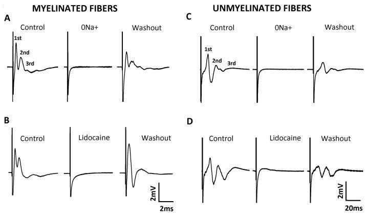
This physiological diagram illustrates extracellular recordings of compound action potentials (CAPs) from myelinated and unmyelinated nerve fibers, categorized into four panels (A-D). Panels A and B display CAP traces for myelinated fibers, characterized by high conduction velocities (scale bar: 2 ms). Panels C and D show unmyelinated fibers with slower conduction (scale bar: 20 ms). In the 'Control' conditions, three distinct components (1st, 2nd, and 3rd) are identified, representing different fiber populations. The experiment demonstrates the sodium-dependency of these potentials: removing extracellular sodium (0Na+) in panels A and C or applying the local anesthetic Lidocaine (1 mM) in panels B and D results in the total abolition of the CAP signals. The 'Washout' phases show partial or near-complete recovery of the action potential waveforms, confirming the viability of the nerve preparation and the reversible nature of the blockade. This visual provides clinical relevance for understanding nerve conduction, the mechanism of action of sodium channel blockers, and the electrophysiological differences between fiber types.
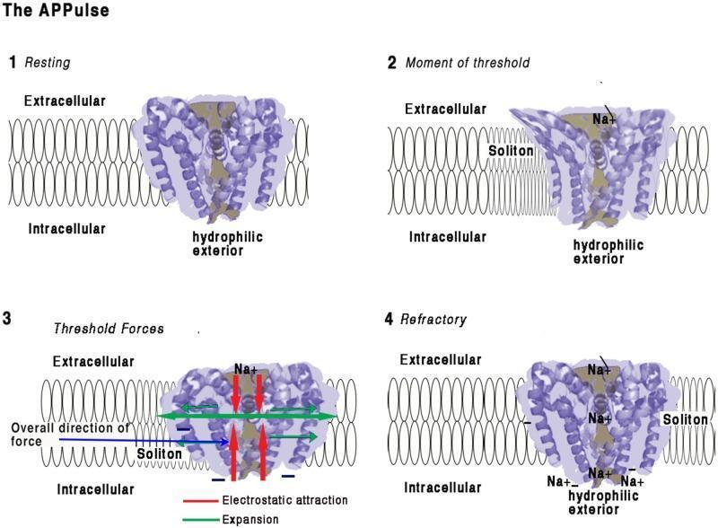
This pathophysiology diagram illustrates the four-stage mechanism of a voltage-gated sodium (Na+) ion channel embedded in a lipid bilayer, focusing on the mechanical and electrostatic changes during depolarization. (1) Resting: The channel's coiled helices are closed, maintaining an electrostatic insulating seal across the 10 nm membrane. (2) Moment of Threshold: An approaching membrane soliton (mechanical wave) exerts pressure, displacing helices and disrupting the electrical insulation. This allows Na+ ions to enter the pore and interact with hydrophilic, negatively charged interior surfaces. (3) Threshold Forces: Inward Na+ influx creates electrostatic attraction (red arrows) toward the intracellular space. Mechanical 'iris-like' opening and contraction occur, causing lateral membrane expansion (green arrows) and a net intracellular force vector (blue arrow). (4) Refractory: Positive charge equalization occurs on the intracellular side, leading to pore closure. The channel remains in a refractory state with localized Na+ accumulation until charges are cleared by diffusion, preventing immediate reactivation.
LOCAL ANESTHETICS - IN DEPTH
1. BASIC STRUCTURE
Lipophilic aromatic ring  ---  Intermediate chain  ---  Ionizable amine group
       (benzene ring)          (ESTER or AMIDE)          (tertiary amine)
- Ester link = unstable, hydrolyzed in plasma by pseudocholinesterase (short duration, more allergy)
- Amide link = stable, metabolized in the liver (longer duration, less allergy)
2. MECHANISM OF ACTION
Step-by-Step Mechanism
-
Local anesthetic is injected - exists in two forms based on tissue pH and its own pKa:
- Charged (cationic) form - water-soluble, cannot cross membrane
- Uncharged (base) form - lipid-soluble, crosses the nerve membrane
-
The uncharged form crosses the lipid cell membrane (axon membrane) into the axoplasm
-
Inside the axoplasm, it re-equilibrates - the charged cation form binds to a receptor at the inner vestibule of the voltage-gated Na⁺ channel (from inside - the "specific receptor theory")
-
This blocks Na⁺ influx - the channel is inactivated
-
Without Na⁺ influx:
- Threshold for excitation increases
- Rate of rise of action potential decreases
- Action potential amplitude decreases
- Eventually - action potential is completely abolished
-
No action potential = no impulse transmission = local anesthesia
Why Local Anesthetics Fail in Infected Tissue
3. KEY PHYSICOCHEMICAL PROPERTIES
| Property | Determines | Details |
|---|---|---|
| pKa | Speed of onset | Lower pKa = more uncharged base at physiological pH = faster membrane penetration = faster onset. Lidocaine pKa 7.9 (fast), bupivacaine pKa 8.1 (slightly slower) |
| Lipid solubility | Potency | More lipid-soluble = more potent = lower dose required. Bupivacaine 16x more potent than procaine; lidocaine 4x |
| Protein binding | Duration of action | Higher protein binding = stays bound to channel longer = longer duration. Bupivacaine 95% protein bound (long); lidocaine 65% (medium) |
| Molecular size / diffusion | Onset | Affects penetration through nerve sheath and tissue |
4. ORDER OF NERVE FIBER BLOCKADE
| Order Blocked | Fiber Type | What Is Lost |
|---|---|---|
| First | B fibers (autonomic) | Sympathetic block - vasodilation, BP drop |
| Second | C fibers (unmyelinated) | Pain (slow) |
| Third | Aδ fibers (small myelinated) | Pain (fast), temperature, touch |
| Fourth | Aβ fibers | Touch, pressure, proprioception |
| Last | Aα fibers (large myelinated motor) | Motor block |
- In spinal anesthesia, the sympathetic block is 2 segments higher than the sensory block
- The sensory block is 2 segments higher than the motor block
- This is why patients can lose pain sensation before motor function, and why BP drops even before the patient is numb to touch
5. LIGNOCAINE (LIDOCAINE) - IN DETAIL
Profile
| Feature | Detail |
|---|---|
| Class | Amide |
| pKa | 7.9 |
| Protein binding | 65% |
| Onset | Fast (5-10 min) |
| Duration (plain) | 1-2 hours |
| Duration (+ epinephrine) | 2-4 hours |
| Elimination half-life | 1.6 hours |
| Metabolism | Liver (CYP1A2, CYP3A4) |
| Potency | 4x procaine |
| Introduced | 1948 - first amide local anesthetic. Still one of the most versatile. |
Uses of Lignocaine
| Use | Concentration | Dose |
|---|---|---|
| Infiltration | 0.5-1% | 3 mg/kg (7 mg/kg with epi) |
| Peripheral nerve block | 1-2% | 3-4 mg/kg |
| Epidural | 2% | 15-20 mL |
| Spinal (intrathecal) | 5% heavy | 1.5-2.5 mL |
| IV Regional (Bier's block) | 0.5% | 3 mg/kg |
| Topical (airway) | 4% spray / gel | - |
| IV anti-arrhythmic | 1% | 1-1.5 mg/kg bolus |
| IV analgesic (opioid-sparing) | - | 1.5 mg/kg loading + 1-3 mg/kg/hr infusion |
Maximum Safe Doses
- Plain: 3 mg/kg (max 200 mg)
- With epinephrine: 7 mg/kg (max 500 mg)
Side Effects / Toxicity of Lignocaine
| Stage | Serum Level | Symptoms |
|---|---|---|
| CNS excitation (early) | ~3 mcg/mL | Circumoral numbness, tingling of lips and tongue, metallic taste, tinnitus, dizziness, visual disturbances |
| CNS excitement | ~5 mcg/mL | Restlessness, tremors, slurred speech, muscle twitching |
| CNS depression / seizures | ~7-8 mcg/mL | Grand mal seizures, LOC |
| CVS toxicity | >10 mcg/mL | Hypotension, bradycardia, arrhythmia, cardiac arrest |
Treatment of LAST
- Stop injection immediately
- Call for help
- Airway - 100% O₂, hyperventilate (raising pH favors uncharged form, reduces channel binding)
- Seizures - Benzodiazepine (midazolam) or propofol; avoid succinylcholine (masks convulsions)
- MOST IMPORTANT: 20% Intralipid (lipid emulsion) - 1.5 mL/kg IV bolus, then 0.25 mL/kg/min infusion - acts as a "lipid sink" - absorbs the lipophilic local anesthetic out of cardiac tissue
- Standard ACLS / CPR if cardiac arrest occurs
6. BUPIVACAINE - IN DETAIL
Profile
| Feature | Detail |
|---|---|
| Class | Amide |
| pKa | 8.1 |
| Protein binding | 95% (very high - long duration) |
| Onset | Medium (10-20 min) |
| Duration (plain) | 4-8 hours |
| Duration (+ epinephrine) | 6-10 hours |
| Elimination half-life | 3.5 hours |
| Metabolism | Liver |
| Potency | 16x procaine (4x lignocaine) |
| Introduced | Filled the need for longer-acting block than lidocaine |
Uses of Bupivacaine
| Use | Concentration | Dose |
|---|---|---|
| Spinal (intrathecal) - most common | 0.5% heavy (hyperbaric) | 1.5-3 mL (7.5-15 mg) |
| Epidural | 0.25-0.5% | 15-25 mL |
| Labour epidural (analgesia) | 0.0625-0.125% (low conc.) | 10-15 mL |
| Peripheral nerve block | 0.25-0.5% | Up to 2 mg/kg |
| Infiltration | 0.25-0.5% | Up to 2 mg/kg (max 175 mg) |
Maximum Safe Doses
- Plain: 2 mg/kg (max 175 mg)
- With epinephrine: 2.5 mg/kg (max 175 mg - not much increase)
Why Bupivacaine is Cardiotoxic
- Bupivacaine binds to cardiac Na⁺ channels with very high affinity (high lipid solubility + high protein binding)
- It binds during systole and does NOT readily unbind during diastole (unlike lidocaine which quickly unbinds - "fast in, fast out")
- Bupivacaine is "slow in, slow out" in cardiac channels
- This leads to: prolonged QRS widening → VT/VF → cardiac arrest
- Critically: cardiovascular collapse can occur almost simultaneously with CNS toxicity - there is much less warning
- Once bupivacaine-induced cardiac arrest occurs, it is extremely difficult to resuscitate
- ACLS drugs are often ineffective
- Lipid emulsion (20% Intralipid) is the key rescue treatment
Stereoisomers - Why Levobupivacaine and Ropivacaine Were Developed
| Drug | Isomer | Cardiac toxicity |
|---|---|---|
| Bupivacaine (racemic) | 50:50 R and S | High - R(+) isomer is the culprit |
| Levobupivacaine | Pure S(-) isomer | Less cardiotoxic |
| Ropivacaine | Pure S(-) isomer (propyl group instead of butyl) | Less cardiotoxic, slightly less potent, also causes vasoconstriction |
7. LIGNOCAINE vs BUPIVACAINE - COMPARISON TABLE
| Feature | Lignocaine | Bupivacaine |
|---|---|---|
| Class | Amide | Amide |
| pKa | 7.9 | 8.1 |
| Protein binding | 65% | 95% |
| Onset | Fast | Medium |
| Duration | 1-2 hours | 4-8 hours |
| Potency | 4x procaine | 16x procaine |
| Max dose (plain) | 3 mg/kg | 2 mg/kg |
| Cardiotoxicity | Less - CVS collapse comes after CNS | High - CVS and CNS toxicity can coincide |
| Cardiac binding | Fast in, fast out | Slow in, slow out (persistent) |
| Safe for IV regional? | YES | NO (cardiotoxic) |
| Main uses | Infiltration, epidural, IV anti-arrhythmic, topical airway, spinal | Spinal, epidural, peripheral nerve blocks |
| Distinguishing feature | Most versatile LA; also an anti-arrhythmic | Longest acting; best for spinals |
8. EPINEPHRINE AS AN ADDITIVE
| Effect | Mechanism | Clinical Use |
|---|---|---|
| Prolongs duration (by 50-100%) | Vasoconstriction reduces vascular absorption | Longer blocks |
| Reduces peak plasma levels | Less systemic absorption | Safer - allows higher total dose |
| Reduces bleeding | Vasoconstriction | Field clarity in minor surgery |
| Acts as intravascular marker | IV injection → sudden tachycardia (>20 bpm) | Test dose for epidurals |
9. SPINAL ANESTHESIA - IN DEPTH
Anatomy Relevant to Spinal
Spinal Needles
| Type | Design | Advantage |
|---|---|---|
| Quincke (cutting tip) | Beveled sharp tip | Easy CSF aspiration; but higher PDPH rate |
| Whitacre (pencil-point) | Side-opening port, non-cutting | Lower PDPH rate - spreads dural fibers rather than cutting |
| Sprotte (pencil-point) | Larger side port | Lower PDPH rate |
Baricity and Positioning
| Solution | Density vs CSF | Behavior | Clinical Use |
|---|---|---|---|
| Hyperbaric (heavy) | Denser than CSF (glucose added) | Sinks in CSF - follows gravity | Turn patient to operate in dependent position. Most commonly used. |
| Hypobaric (light) | Less dense than CSF (water/fentanyl added) | Floats - moves to non-dependent side | Used for hip surgery in lateral position - blocks operative side only |
| Isobaric | Same density as CSF | Stays at level of injection | Predictable level, position-independent |
Dermatomal Levels Required for Surgery
| Surgery | Block Level Required |
|---|---|
| Lower limb surgery | T12 (for thigh: T10) |
| Hip surgery | T10 |
| Lower abdominal surgery (appendix, hernia) | T6 |
| Cesarean section (C-section) | T4 (level of nipples) |
| Perineal/anal surgery | S2-S4 |
| Knee surgery | T12 |
Complications of Spinal Anesthesia - In Depth
| Complication | Cause | Prevention | Treatment |
|---|---|---|---|
| Hypotension | Sympathetic block → vasodilation → decreased venous return → decreased CO | Pre-load with 500-1000 mL IV fluids; left lateral tilt in pregnant women | IV Ephedrine 6-9 mg (acts on α and β - preferred in obstetrics); Phenylephrine 50-100 mcg IV; Mephentermine 6-15 mg |
| Bradycardia | High sympathetic block (T1-T4) removes cardiac accelerator fibers (Bezold-Jarisch reflex) | Atropine in pre-med | Atropine 0.6 mg IV; Ephedrine; if severe - Epinephrine 0.5-1 mg IV |
| Post-dural puncture headache (PDPH) | CSF leak through dural hole → reduced CSF pressure → intracranial traction on pain-sensitive structures | Use small gauge pencil-point needles; limit patient movement post-procedure | Bed rest, oral hydration, caffeine (100-300 mg), NSAIDs; if severe/persistent: Epidural blood patch (15-20 mL autologous blood injected epidurally - seals the hole) |
| High / Total spinal | Accidental overdose or excessive spread - reaches cervical cord | Correct baricity and patient positioning | Immediate: 100% O₂, intubation, vasopressors (ephedrine/epinephrine). Reassure patient - wears off! |
| Urinary retention | Sacral nerve block (S2-S4) | - | Urinary catheterization |
| Nausea and vomiting | Hypotension → cerebral ischemia; or high block (vagus unopposed) | Treat hypotension | Treat hypotension; Ondansetron 4 mg IV; Atropine if bradycardia |
| Cauda equina syndrome | Neurotoxicity from concentrated hyperbaric lignocaine (transient neurological symptoms - TNS) | Use bupivacaine; avoid high concentrations of lignocaine intrathecally | Supportive |
10. EPIDURAL ANESTHESIA - IN DEPTH
Key Differences from Spinal
The Tuohy Needle
- 16-18 gauge - large bore to allow catheter passage
- Curved (Huber) tip - directs the catheter cephalad
- Has depth markings - epidural space is typically 3.5-5 cm from the skin in adults (less in thin patients, more in obese)
Epidural Space Identification
- Attach 10 mL syringe (saline or air) to Tuohy needle
- Advance slowly through interspinous ligament and ligamentum flavum
- Continuous gentle pressure on plunger - resistance felt while in ligament
- When tip enters epidural space: sudden loss of resistance - plunger moves freely
- Fill hub of Tuohy needle with saline so a drop hangs
- As needle enters epidural space, negative pressure sucks the drop inward
- Less reliable - blocked needles give false negatives
Epidural Catheter Placement
- Thread a flexible catheter 3-5 cm into the space through the Tuohy needle
- Withdraw needle over catheter
- Aspirate to check: no CSF (intrathecal?) and no blood (intravascular?)
- Give test dose: 3 mL of 1.5% lignocaine + 15 mcg epinephrine
- Intrathecal injection: Dense spinal block within 3-5 min (too much drug for epidural)
- Intravascular injection: HR increases >20 bpm within 30 sec (epinephrine marker)
- If test dose negative, give drug incrementally - 5 mL at a time (incremental dosing)
Epidural Drugs
| Drug | Concentration | Volume | Use |
|---|---|---|---|
| Bupivacaine | 0.25-0.5% | 15-25 mL | Surgical anesthesia |
| Bupivacaine | 0.0625-0.125% | 10-15 mL | Labour analgesia (low concentration - more sensory than motor block) |
| Lignocaine | 2% | 15-20 mL | Faster onset epidural |
| Ropivacaine | 0.2-0.75% | 15-25 mL | Labour (less motor block), thoracic epidural |
| Fentanyl | 50-100 mcg added | - | Enhances quality of epidural analgesia, reduces LA dose needed |
| Morphine | 2-4 mg added | - | Prolonged post-op analgesia |
| Clonidine | 75-150 mcg added | - | Prolongs block, reduces LA dose |
| Epinephrine | 1:200,000 added | - | Prolongs duration, detects intravascular placement |
Complications of Epidural - In Depth
| Complication | Details | Management |
|---|---|---|
| Accidental dural puncture (ADP) | 0.5-2% incidence; Tuohy needle punctures dura → large hole → severe PDPH | If recognized: thread catheter intrathecally (run as continuous spinal) OR remove and try another level. Treat PDPH with epidural blood patch. |
| Intravascular injection | Catheter migrates into epidural vein | Detected by test dose (epinephrine → tachycardia). Prevent with aspiration + test dose + incremental dosing. Treat LAST if occurs. |
| Accidental total spinal | Full epidural dose injected intrathecally | Immediate airway management, intubation, vasopressors. |
| Epidural hematoma | Rare but can cause paraplegia - especially with anticoagulants | Urgent MRI; neurosurgical decompression within 6-8 hours for chance of recovery |
| Epidural abscess | Breach of sterile technique or bacteremia | MRI; IV antibiotics; surgical drainage |
| Hypotension | Sympathetic block | IV fluids, vasopressors (same as spinal) |
| Motor block | Bupivacaine in too high concentration | Use lower concentration (0.0625-0.125%) for labour analgesia |
| Urinary retention | Sacral block | Catheterization |
| Catheter migration / kinking | Catheter moves in/out of space | Check position; re-site if needed |
Epidural vs Spinal - When to Choose Which
| Choose SPINAL when: | Choose EPIDURAL when: |
|---|---|
| Short, defined surgery (C-section, TURP, hip replacement) | Long procedures needing adjustable anesthesia |
| Lower extremity / perineal surgery | Labour analgesia (continuous infusion) |
| No catheter needed | Post-operative pain management needed |
| Rapid dense reliable block needed | Thoracic surgeries |
| Elderly patients (lower doses work well) | Repeat top-ups needed |
Combined Spinal-Epidural (CSE)
- Spinal component gives fast, reliable, dense block for surgery
- Epidural catheter provides option to extend duration, give top-ups, or post-op analgesia
Okay, so just two questions. Um, give me one final table of the most important drugs, just, like, maybe one or two drugs, two drugs from each group needed, especially for general anesthesia. Just give me, like, doses simply and, uh, mechanism of all the drugs necessary, like, just the important ones in one table finally. And what is the combination of lignocaine and adrenalin, adrenalin? Lignocaine.
MASTER TABLE: Key Anesthesia Drugs
| # | Drug | Group | Dose | Mechanism of Action |
|---|---|---|---|---|
| 1 | Midazolam | Pre-med / Benzodiazepine | 0.025-0.1 mg/kg IV; 7.5-15 mg oral | Enhances GABA-A receptor activity → ↑ Cl⁻ influx → CNS depression → anxiolysis, sedation, amnesia |
| 2 | Glycopyrrolate | Pre-med / Anticholinergic | 0.2-0.4 mg IM/IV | Blocks muscarinic receptors → reduces secretions, prevents vagal bradycardia |
| 3 | Propofol | Induction agent | 1.5-2.5 mg/kg IV | Potentiates GABA-A receptor → sedation/hypnosis. Also inhibits NMDA receptors |
| 4 | Ketamine | Induction agent | 1-2 mg/kg IV; 4-6 mg/kg IM | NMDA receptor antagonist → dissociative anesthesia. Maintains BP (stimulates SNS, inhibits norepinephrine reuptake) |
| 5 | Succinylcholine | Depolarizing muscle relaxant | 1-1.5 mg/kg IV (RSI: 1.5 mg/kg) | Mimics ACh at nicotinic NMJ receptor → persistent depolarization (Phase I block) → fasciculations then flaccid paralysis |
| 6 | Rocuronium | Non-depolarizing muscle relaxant | 0.6 mg/kg IV (RSI: 1.2 mg/kg) | Competitive antagonist at nicotinic NMJ receptor → blocks ACh → flaccid paralysis without fasciculations |
| 7 | Fentanyl | Opioid analgesic | 1-3 mcg/kg IV bolus | Agonist at µ (mu) opioid receptors in CNS and periphery → analgesia, sedation, suppresses stress response |
| 8 | Sevoflurane | Volatile maintenance agent | 1-2% (MAC 2%) | Potentiates GABA-A and NMDA antagonism at multiple CNS sites → general anesthesia. Also direct cardiac depression. |
| 9 | Neostigmine | Reversal agent | 0.05 mg/kg IV (max 5 mg) | Inhibits acetylcholinesterase → ACh accumulates at NMJ → displaces non-depolarizing blocker → reverses paralysis |
| 10 | Sugammadex | Reversal agent (selective) | 2-4 mg/kg IV | Encapsulates rocuronium/vecuronium molecule directly → forms complex → removed from NMJ → instant reversal |
| 11 | Lignocaine | Local anesthetic | 3 mg/kg plain; 7 mg/kg with epi | Blocks voltage-gated Na⁺ channels from inside the axon → prevents action potential → local anesthesia |
| 12 | Bupivacaine | Local anesthetic | 2 mg/kg plain (max 175 mg) | Same as above but high protein binding → long duration. Binds cardiac Na⁺ channels persistently → risk of arrhythmia |
| 13 | Epinephrine (ACLS) | Vasopressor / Cardiac arrest | 1 mg IV every 3-5 min | α1 agonist → vasoconstriction → ↑ aortic diastolic pressure → ↑ coronary perfusion pressure during CPR |
| 14 | Atropine | Anticholinergic | 0.6 mg IV | Blocks muscarinic receptors at SA node → ↑ heart rate → treats bradycardia |
Lignocaine + Adrenaline Combination
What Adrenaline (Epinephrine) Does When Added
| Effect | Mechanism | Benefit |
|---|---|---|
| Prolongs duration | α1-mediated vasoconstriction at injection site → reduces blood flow → slows absorption of lignocaine from the site | Duration increases from 1-2 hrs to 2-4 hrs |
| Reduces systemic toxicity | Slower absorption = lower peak plasma levels of lignocaine | Allows you to use a higher total dose safely (3 mg/kg → 7 mg/kg) |
| Reduces bleeding | Vasoconstriction at the site | Useful in surgeries like dental, ENT, scalp procedures |
| Acts as intravascular marker | If accidentally injected IV, epinephrine causes sudden tachycardia (HR rises >20 bpm in 30 sec) | Used as test dose in epidurals |
Concentrations Used
| Preparation | Adrenaline Concentration | Common Use |
|---|---|---|
| 1:80,000 | 12.5 mcg/mL | Dental anesthesia |
| 1:100,000 | 10 mcg/mL | Infiltration, dental |
| 1:200,000 | 5 mcg/mL | Epidural, peripheral nerve blocks |
Where NOT to Use Lignocaine + Adrenaline
- Digits (fingers, toes), penis, nose tip, ear lobes - end arteries - risk of ischemia and gangrene
- Patients with severe cardiac disease or on MAO inhibitors
- Areas with compromised vascular supply