
Indications of blood transfusion in ent
Indications for Blood Transfusion in ENT
General Surgical Indications (applicable in ENT)
- Acute blood loss - to replace circulating volume and maintain oxygen delivery
- Perioperative anaemia - to ensure adequate oxygen delivery during the perioperative phase
- Symptomatic chronic anaemia - without haemorrhage or impending surgery
ENT-Specific Indications
1. Post-tonsillectomy Haemorrhage
- Prolonged the patient's stay in hospital
- Required blood transfusion
- Required return to the operating theatre
- Resulted in readmission for secondary haemorrhage
2. Double-jaw (Bimaxillary Orthognathic) Surgery
3. Massive/Torrential ENT Haemorrhage
- Severe post-operative bleeds (e.g., after neck dissection, major head and neck resections)
- Uncontrolled epistaxis (e.g., in hereditary haemorrhagic telangiectasia/Osler-Weber-Rendu)
- Trauma to head and neck vessels
- Intraoperative haemorrhage during major ENT/head and neck surgery
4. Haematological Disorders Presenting to ENT
- Symptomatic anaemia from chronic disease (e.g., carcinoma of the larynx, hypopharynx, or nasopharynx causing chronic blood loss)
- After chemotherapy for head and neck malignancies
- Chronic haematological conditions (sickle cell disease, thalassaemia) where patients may present to ENT with complications
Transfusion Trigger (Haemoglobin Thresholds)
| Hb level (g/dL) | Recommendation |
|---|---|
| < 6 | Transfusion will probably benefit |
| 6-8 | Transfusion unlikely to benefit in absence of active bleeding or impending surgery |
| > 8 | No indication in absence of other risk factors |
| < 7 | Widely used restrictive trigger in surgical ICU patients |
- Historically, patients were transfused to achieve Hb >10 g/dL - this is now shown to be unnecessary and associated with increased morbidity/mortality.
- Patients with ischaemic heart disease or other cardiovascular comorbidities may require a higher trigger due to limited cardiac reserve.
Blood Components Used in ENT
| Component | Indication in ENT |
|---|---|
| Red Blood Cells (RBC) | Massive haemorrhage, symptomatic anaemia, post-chemotherapy, chronic haematological conditions |
| Fresh Frozen Plasma (FFP) | Coagulopathy, massive haemorrhage, DIC, liver disease (12-15 ml/kg dose) |
| Cryoprecipitate | Massive haemorrhage with fibrinogen < 1.5 g/L; rich in fibrinogen, Factor VIII, vWF |
| Platelets | Thrombocytopenia with active bleeding, or pre-operatively if platelets <50 x 10⁹/L |
Alternatives to Blood Transfusion in ENT
- Autologous pre-donation: Patient pre-donates their own blood up to 3 weeks before elective ENT surgery
- Intraoperative cell salvage (cell saver): Blood collected, washed, and returned intraoperatively
- Tranexamic acid (TXA): Shown to be effective in epistaxis, especially when conventional therapy fails and in hereditary haemorrhagic telangiectasia
- Erythropoietin: Used preoperatively to optimise Hb in anaemic patients before major surgery
- Iron supplementation: Treat iron deficiency anaemia before elective surgery
Key Principle (BOX 24.1, Scott-Brown's)
"Ensure the right blood is transfused to the right patient at the right time in the right place and for the right indication."
Management of dizziness in emergency setting as a ent surgeon
HINTS exam head impulse test nystagmus test of skew vertigo
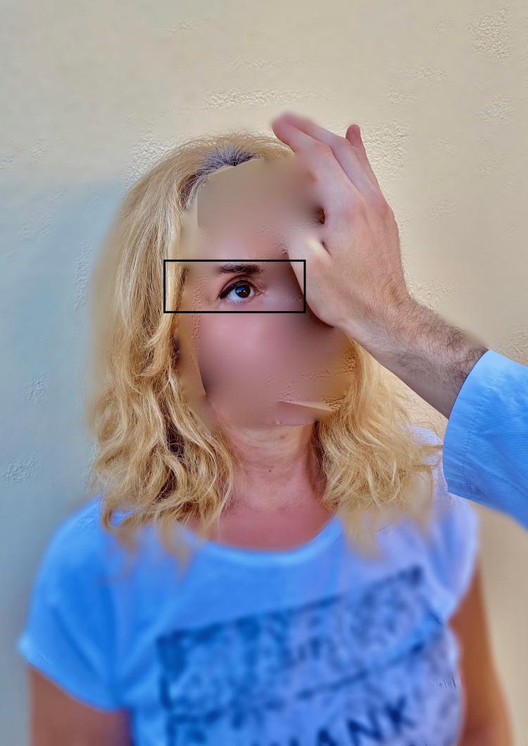
A clinical photograph demonstrating the 'test of skew,' a component of the HINTS (Head Impulse, Nystagmus, Test of Skew) examination used to differentiate central from peripheral causes of acute vestibular syndrome. The image shows an examiner’s hand placed over a patient's left eye while the patient's right eye remains uncovered and is highlighted by a rectangular box to indicate observation. During this maneuver, the examiner performs an alternate cover test while the patient maintains fixation on a central target (typically the examiner’s nose). The clinical focus is to detect skew deviation—a vertical ocular misalignment. In central vestibular disorders, such as a brainstem stroke, uncovering an eye may reveal a corrective vertical shift (refixation saccade). Conversely, the absence of vertical deviation, as seen in this clinical demonstration, is more characteristic of peripheral vestibular conditions like vestibular neuritis. This diagnostic procedure is essential for evaluating patients presenting with continuous vertigo, nystagmus, and nausea.
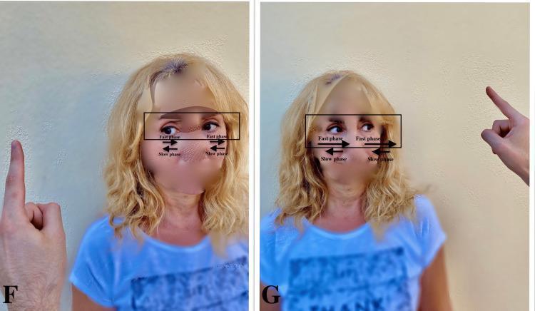
Clinical photograph consisting of two side-by-side images (labeled F and G) demonstrating the assessment of vestibular nystagmus in a patient with acute vestibular loss. In panel F, the examiner's finger is positioned to the patient's left; directional arrows indicate a nystagmus with a slow phase drifting toward the patient's right (the pathologic side) and a fast corrective phase toward the left (the healthy side). In panel G, the examiner's finger moves to the patient's right; annotations illustrate that the nystagmus intensity increases when looking in the direction of the fast phase, consistent with Alexander's Law. This visual educational tool demonstrates the HINTS (Head Impulse, Nystagmus, Test of Skew) examination component used to differentiate peripheral vestibular neuritis from central causes of vertigo, emphasizing that peripheral nystagmus typically has a unidirectional fast phase and follows a specific pattern of slow-phase drift toward the affected ear.
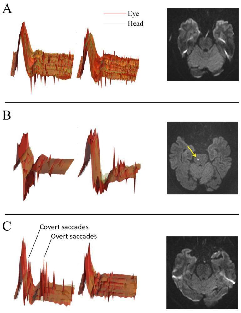
This composite educational graphic correlates Video Head Impulse Test (vHIT) data with axial brain Magnetic Resonance Imaging (MRI) to demonstrate the clinical evaluation of acute vestibular syndrome. The image is divided into three panels (A, B, and C). On the left, vHIT traces show head velocity (grey) and eye velocity (red) during rapid head movements. On the right, corresponding Diffusion-Weighted Imaging (DWI) MRI slices through the brainstem and cerebellum are displayed. Panel A shows relatively symmetric head and eye traces with a normal DWI MRI. Panel B highlights a central lesion, where the vHIT shows significant discrepancies between head and eye traces, and the MRI reveals a focal, hyperintense punctate area of restricted diffusion in the right paramedian pons (yellow arrow), diagnostic of an acute pontine infarction. Panel C illustrates a peripheral pattern; the vHIT shows labeled compensatory 'covert' and 'overt' saccades (distinct red spikes following the initial movement) indicative of a vestibulo-ocular reflex (VOR) deficit, alongside an unremarkable MRI. The image serves as a diagnostic guide for differentiating between peripheral vestibulopathy and central cerebrovascular events using the HINTS (Head Impulse, Nystagmus, Test of Skew) plus protocol and neuroimaging.
Management of Dizziness in the Emergency Setting - ENT Surgeon's Perspective
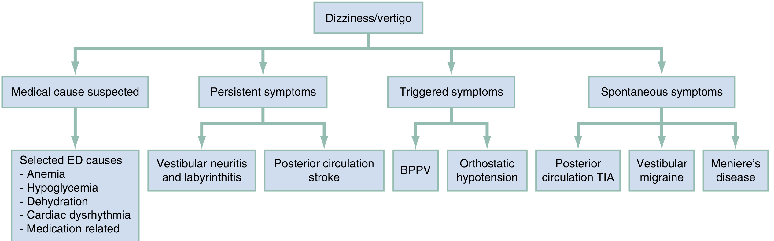
Step 1: Initial Triage and Classification
| Symptom Pattern | Likely Cause |
|---|---|
| Triggered (occurs with head movement, brief <1 min) | BPPV |
| Persistent (constant, hours to days + nystagmus) | Vestibular neuritis / labyrinthitis vs. cerebellar/brainstem stroke |
| Spontaneous (episodic, not movement-triggered) | Meniere's disease, vestibular migraine, posterior circulation TIA |
| Medical cause suspected | Anaemia, hypoglycaemia, dehydration, dysrhythmia, medication toxicity |
Step 2: History - Red Flag Features
- Sudden onset of severe persistent vertigo in older patients
- Associated dysarthria, dysphagia, diplopia, facial numbness/weakness
- Truncal/limb ataxia - unable to stand or walk unaided
- Headache or neck pain (vertebral artery dissection)
- New onset of any focal neurological symptoms
- Vascular risk factors: hypertension, diabetes, AF, prior stroke, male sex, elderly
| Site of Lesion | Key Associated Symptoms |
|---|---|
| Peripheral (labyrinth/VIII nerve) | Tinnitus, hearing loss, ear fullness |
| CP angle | Cranial nerves V, VI, VII involvement |
| Ramsay Hunt | Ear canal vesicles + VII palsy |
| Brainstem | Diplopia, facial numbness, dysarthria, dysphagia, bilateral limb weakness/numbness |
| Cerebellum | Ataxia, dysmetria, inability to walk |
| Cerebral hemisphere | Unilateral weakness, sensory loss, hemianopia |
Step 3: Physical Examination
A. Vital Signs
- Check for orthostatic hypotension (cardiovascular cause of dizziness)
- Blood pressure in both arms if subclavian steal suspected (>40 mmHg difference is significant)
- (Rosen's Emergency Medicine)
B. Oculomotor Examination
| Feature | Peripheral | Central |
|---|---|---|
| Direction | Unidirectional (horizontal/rotatory) | Bidirectional, direction-changing with gaze, or purely vertical |
| Fixation suppression | Suppressed | Not suppressed |
| Pattern | Fast phase always to same side (Alexander's Law) | Variable, may be downbeating or purely torsional |
| Fatigability | Fatigable | Non-fatigable |
C. Dix-Hallpike Test (DHT) - for TRIGGERED symptoms
- Turn head 45 degrees to one side, lay patient rapidly into head-hanging position
- Positive test: upbeating torsional nystagmus (fast phase toward eyebrows) with latency, fatigable - confirms posterior canal BPPV
- Purely horizontal or downbeating nystagmus on positional testing = may indicate central cause
- Do NOT perform DHT if patient has ongoing constant vertigo with spontaneous nystagmus - perform HINTS instead
D. HINTS Exam - for PERSISTENT symptoms (Acute Vestibular Syndrome)
- Rapidly rotate the head horizontally ~20 degrees
- Positive (corrective saccade) = vestibulo-ocular reflex (VOR) deficit = PERIPHERAL cause (reassuring - likely vestibular neuritis)
- Negative (no saccade) = VOR intact = CENTRAL cause (concerning for stroke)
- Unidirectional horizontal nystagmus = peripheral
- Direction-changing nystagmus (fast phase changes side with gaze direction) = CENTRAL
- Alternate cover test looking for vertical ocular misalignment
- Vertical skew deviation = CENTRAL (brainstem pathology)
Interpretation: CENTRAL pattern = "I" negative head impulse + direction-changing nystagmus + skew deviation = HIGH suspicion for stroke
Step 4: Emergency Management Algorithm
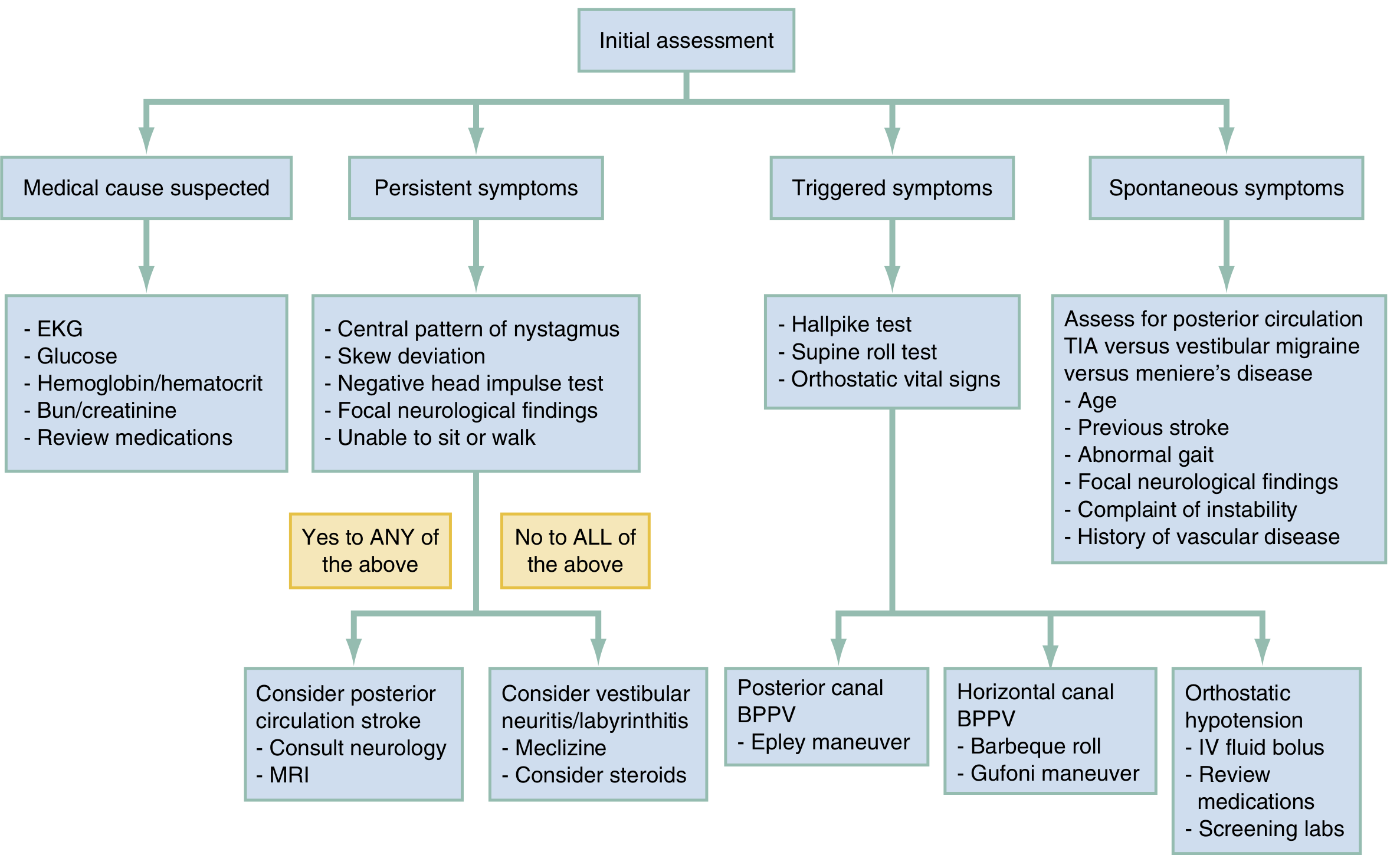
A. BPPV (Triggered, brief vertigo)
- Epley Maneuver - first-line treatment, performs in ED
- 4-5 sequential head positions held 30 seconds each (or until nystagmus resolves)
- After 10 minutes, repeat DHT - if negative, patient can be discharged
- If Epley fails twice, teach patient home Epley twice daily and arrange follow-up
- Do NOT prescribe vestibular suppressants for typical BPPV (episodes <30 sec do not warrant suppressants)
- No imaging required for typical positive DHT
- Gufoni maneuver or Barbeque (360 degree) roll maneuver
- Rarer, harder to assess; refer to specialist if treatment fails
B. Vestibular Neuritis / Labyrinthitis (Persistent, constant vertigo)
- HINTS exam: positive head impulse test + unidirectional nystagmus = reassuring peripheral cause
- Symptomatic treatment with short course of antiemetics/vestibular suppressants
- Steroids: Evidence supports oral prednisolone to accelerate recovery (though Cochrane review shows modest benefit). Consider 1 mg/kg/day for 5 days tapering
- Antivirals (valacyclovir): Not routinely recommended despite presumed viral aetiology
- Vestibular rehabilitation - refer on discharge; do NOT prescribe long-term benzodiazepines (interfere with vestibular compensation)
- Most recover over weeks to months
C. Posterior Circulation Stroke / Cerebellar Infarction (EMERGENCY)
- Immediate CT head (to exclude haemorrhage)
- MRI brain with DWI - modality of choice for posterior fossa; note early MRI may be falsely negative in first 24-48 hours
- Neurology/neurosurgery consultation urgently
- Admission for monitoring - risk of cerebral oedema and obstructive hydrocephalus
- Changing/rapidly progressive symptoms = impending posterior circulation occlusion - consider MR angiography ± anticoagulation after haemorrhage excluded
- Secondary stroke prevention commenced during admission
D. Meniere's Disease / Vestibular Migraine (Spontaneous, episodic vertigo)
- Acute attack: vestibular suppressants + antiemetics
- Distinguish by history (Meniere's: triad of vertigo + hearing loss + tinnitus; vestibular migraine: migraine history, no hearing loss)
- Refer to ENT/otoneurology for definitive investigation and long-term management
E. Medical Causes
- ECG, blood glucose, FBC (anaemia), BUN/creatinine, medication review
- Orthostatic vital signs, fluid bolus for dehydration/orthostatic hypotension
Step 5: Pharmacological Treatment
| Drug | Route/Dose | Antiemetic Effect | Notes |
|---|---|---|---|
| Ondansetron (Zofran) | 4 mg IV/IM/SL | Prominent | First-line IV agent; fewer side effects |
| Promethazine (Phenergan) | 12.5-25 mg IM/PO/PR | Moderate | Most effective parenteral suppressant; IV route has FDA boxed warning - use IM only |
| Prochlorperazine (Compazine) | 5-10 mg IV/IM/PO | Prominent | |
| Dimenhydrinate (Dramamine) | 50-100 mg IM/IV/PO | Moderate | |
| Metoclopramide (Reglan) | 5-10 mg IV/IM/PO | Prominent | |
| Meclizine (Antivert) | 12.5-50 mg PO q4-6h | Mild | Onset ~1 hour; for BPPV after failed Epley or vestibular neuritis |
| Lorazepam (Ativan) | 1-2 mg IV/IM | Mild | Reserve for intractable vomiting unresponsive to antiemetics |
| Diazepam (Valium) | 1 mg IV | Mild | |
| Transdermal scopolamine | Patch | - | Third-line; disappointing results for peripheral vertigo |
Do NOT discharge patients with oral benzodiazepines in vestibular neuritis/labyrinthitis - these interfere with the process of vestibular compensation/habituation.
Step 6: ENT-Specific Considerations
| Condition | ENT-Specific Action |
|---|---|
| Acute labyrinthitis with hearing loss | Urgent pure-tone audiogram; oral steroids; consider MRI to exclude retrocochlear pathology |
| Ramsay Hunt syndrome | Ear vesicles + VII palsy + vertigo = acyclovir + prednisolone |
| Temporal bone trauma + vertigo | CT temporal bones; consider perilymphatic fistula; bed rest + head elevation |
| Perilymphatic fistula | Suspect after barotrauma/Valsalva; bed rest, no straining; may need surgical exploration |
| Sudden SNHL + vertigo | Urgent audiogram; oral steroids; rule out AICA infarct with MRI (HINTS+ hearing loss) |
| Bilateral vestibular failure | Oscillopsia + gait ataxia; aminoglycoside toxicity? Refer vestibular rehabilitation |
Disposition Summary
| Diagnosis | Disposition |
|---|---|
| BPPV (treated with Epley) | Discharge if post-Epley DHT negative; follow-up if recurs |
| Vestibular neuritis/labyrinthitis | Discharge with antiemetics; vestibular rehab referral; no benzodiazepines |
| Meniere's/vestibular migraine | Discharge after acute treatment; ENT/neurology follow-up |
| Posterior circulation stroke/TIA | Admit; urgent neurology consultation |
| Medical cause (e.g., arrhythmia, severe anaemia) | Treat underlying cause; appropriate admission/observation |
| Unable to ambulate; bilateral neurological signs | Admit for investigation and monitoring |