
General and obstetric examination
Leopold maneuvers obstetric abdominal examination pregnancy
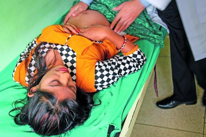
This clinical photograph captures an obstetric physical examination in a community or public health setting. A female patient is lying supine on an examination table with her abdomen exposed. The gravid abdomen is clearly visible, demonstrating significant distension consistent with pregnancy. The hands of a healthcare provider are positioned on the uterine fundus and lateral aspects of the abdomen, performing Leopold's maneuvers or a similar abdominal palpation to assess fetal position, presentation, or fundal height. The patient is wearing colorful traditional clothing, and the clinician is dressed in professional attire, including a white lab coat. The image illustrates a core procedural skill in maternal healthcare and prenatal screening, highlighting the clinical assessment of gestation within the obstetric and gynecological specialty. It serves as an educational resource for demonstrating patient positioning and manual examination techniques in low-resource or community health contexts.
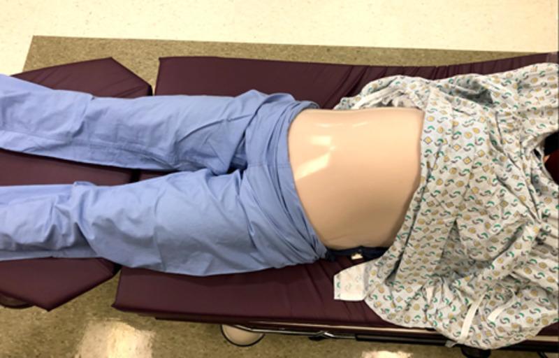
A clinical procedural image showing a low-fidelity medical simulation model of a pregnant patient in a supine position on a dark purple examination bed. The model is used for training in obstetrical maneuvers, specifically External Cephalic Version (ECV). The visual setup includes a lower body covered in light blue surgical scrubs and an upper torso draped in a patterned hospital gown. The central focus is a simulated gravid abdomen, constructed from skin-toned silicone layered over a pillowcase base. This abdominal component creates a smooth, rounded protuberance that replicates the anatomical appearance of a late-term pregnancy. The model is designed to house an internal 'uterus' mechanism—typically consisting of nested balloons and a fetal mannequin—to allow residents to practice palpation and the manual rotation of a fetus from a breech to a cephalic presentation. The image demonstrates the clinical application of simulation-based medical education (SBME) for improving procedural comfort and competency in obstetric care.
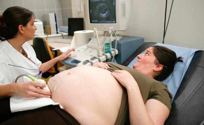
This clinical photograph illustrates a procedural setup for an obstetric ultrasound examination. The patient, a pregnant woman with a distended abdomen, is positioned supine on an examination table. A healthcare professional, positioned to the patient's right, is performing the scan using a curvilinear ultrasound transducer. A translucent coupling gel is visible on the lower abdominal region to facilitate acoustic transmission. The ultrasound machine's monitor is positioned at the patient's head level, displaying a real-time sonographic image of the fetus. The image demonstrates key aspects of maternal care and diagnostic imaging in obstetrics. Noteworthy anatomical features include the gravid uterus and striae gravidarum (stretch marks) on the abdominal skin. The clinical context relates to prenatal monitoring, fetal health assessment, and the study of maternal factors, such as obesity or insulin resistance, that may influence pregnancy and postpartum outcomes like lactation.
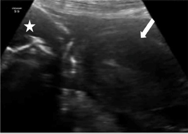
This diagnostic ultrasound image in the obstetric and gynecologic domain illustrates an abdominal pregnancy. The trans-abdominal scan reveals a fetus (marked with a white star) located outside the uterine cavity, positioned in close proximity to the maternal anterior abdominal wall. An empty, relatively amorphous uterus is identified separately (indicated by the white arrow). Key sonographic findings include the complete lack of myometrial tissue surrounding the fetus and the clear anatomical separation between the gestational sac and the uterine corpus. The fetus exhibits typical hyperechoic bony structures, while the adjacent uterine tissue presents a distinct, uniform echogenicity. This image serves as a clinical example of an extrauterine ectopic pregnancy, specifically an advanced abdominal pregnancy, emphasizing the diagnostic importance of identifying the relationship between the fetus and the empty uterus to prevent life-threatening maternal complications such as hemorrhage.
General and Obstetric Examination
PART 1: GENERAL EXAMINATION IN PREGNANCY
1. General Appearance
- Build, nutritional status, pallor, jaundice, cyanosis, edema
- Level of distress, anxiety, discomfort
- Signs of systemic disease (thyroid enlargement, facial puffiness suggesting pre-eclampsia)
2. Vital Signs
| Parameter | Clinical Relevance |
|---|---|
| Blood pressure | Hypertension (>140/90) - pre-eclampsia/gestational HTN; hypotension - aortocaval compression in supine position |
| Pulse rate | Tachycardia - anemia, infection, thyrotoxicosis, hemorrhage |
| Temperature | Pyrexia - TORCH infections, chorioamnionitis, UTI |
| Respiratory rate | Tachypnea - pulmonary embolism, severe anemia, cardiac disease |
| Weight | Excessive gain (>0.5 kg/week in 3rd trimester) - pre-eclampsia, polyhydramnios |
| Height | Short stature (<145 cm) - risk for cephalopelvic disproportion |
3. Systematic General Survey
- Pallor of conjunctivae - anemia
- Scleral icterus - obstetric cholestasis, HELLP
- Exophthalmos - Graves' disease
- Papilledema - severe pre-eclampsia
- Chloasma (melasma) - normal pregnancy pigmentation
- Moon face - Cushing's syndrome
- Facial edema - pre-eclampsia
- Thyroid enlargement - physiological or pathological goitre
- Lymphadenopathy - infection, malignancy
- Koilonychia - iron deficiency anemia
- Leukonychia - hypoalbuminemia
- Peripheral edema, especially pitting pedal edema
- Carpal tunnel signs - common in pregnancy
- Linea nigra, striae gravidarum - normal pregnancy changes
- Spider naevi - hepatic disease or normal pregnancy (estrogen effect)
- Jaundice
- Enlargement, darkening of areola and nipple, Montgomery's tubercles - normal changes
- Colostrum expression after 16 weeks
- Lumps, mastitis signs
PART 2: OBSTETRIC (ABDOMINAL) EXAMINATION
Sequence: Inspection → Palpation → Auscultation
A. INSPECTION
| Finding | Significance |
|---|---|
| Size of abdomen | Consistent with gestational age? |
| Shape | Longitudinal oval - normal; transverse - transverse lie |
| Umbilicus | Stretched/everted - common in pregnancy |
| Skin | Striae gravidarum, linea nigra, scars (previous LSCS) |
| Fetal movements | Visible rippling movements in thin women |
| Asymmetry | Uterine fibroids, fetal position anomalies |
B. PALPATION
1. Fundal Height Measurement
- Measured from the upper edge of the pubic symphysis to the fundus using a tape measure
- In centimeters, fundal height approximately equals gestational age in weeks (±2 cm) after 20 weeks
- Fundal height > dates: multiple pregnancy, polyhydramnios, LGA fetus, uterine fibroids, wrong dates
- Fundal height < dates: IUGR/SGA, oligohydramnios, transverse lie, wrong dates
2. Leopold's Maneuvers (Four Maneuvers)
| Maneuver | Action | Determines |
|---|---|---|
| 1st - Fundal Grip | Palpate the fundus with both hands | What is in the fundus? (Head = hard, round, ballotable; Breech = soft, irregular, not ballotable) |
| 2nd - Lateral/Umbilical Grip | Both hands on lateral sides of abdomen | Fetal back (smooth, firm, resistant) vs. limbs (irregular, small parts felt); determines position (LOA, ROA, etc.) |
| 3rd - Pawlik's Grip | Right hand grasps lower pole just above pubic symphysis | Presentation (head or breech) and whether it is engaged or free |
| 4th - Pelvic/Deep Pelvic Grip | Face patient's feet; both hands palpate lower uterine segment | Degree of engagement; descent of presenting part; direction of cephalic prominence |
Tip: If not confident of fetal position after Leopold's maneuvers, intrapartum ultrasound should confirm lie - Pfenninger and Fowler's Procedures for Primary Care, p. 7126.
3. Assessment of Lie and Presentation
-
Lie: Relationship of fetal long axis to maternal long axis
- Longitudinal (cephalic or breech) - normal
- Transverse - always abnormal at term
- Oblique - unstable, usually converts
-
Presentation: Part of fetus in lower uterine segment
- Cephalic (most common, ~96%)
- Breech (~4% at term): Frank breech, complete breech, footling/incomplete
- Shoulder/Transverse
-
Position: Relationship of denominator (occiput for vertex, sacrum for breech) to maternal pelvis
- LOA (Left Occiput Anterior) - most common
-
Engagement: Head engaged when biparietal diameter passes through pelvic inlet; in primigravida, occurs at ~36 weeks
C. AUSCULTATION
Fetal Heart Rate (FHR)
- Normal FHR: 110-160 bpm
- Heard best over the fetal back (for vertex: lower abdomen; for breech: above umbilicus)
- Tools: Pinard's stethoscope (from 20 weeks), handheld Doppler (from 12 weeks)
- Listen for 1 minute, count rate, note rhythm and character
- Bradycardia (<110 bpm): fetal distress, cord compression
- Tachycardia (>160 bpm): maternal fever, fetal infection, fetal anemia, prematurity
D. SPECIAL TESTS ON ABDOMINAL EXAMINATION
- Internal: finger in vagina - fetus floats away and returns (free head)
- External: tapping one flank while opposite hand feels fluid thrill - checks for ascites/polyhydramnios
- Positive in polyhydramnios; elicited by tapping one flank and feeling a transmitted impulse on the other, with the examiner's hand placed edge-on at the midline to block transmission through the abdominal wall
PART 3: VAGINAL (PELVIC) EXAMINATION IN OBSTETRICS
Indications:
- First antenatal visit (Pap smear, pelvic assessment)
- Assessment in labor (cervical dilatation, effacement, station, membranes)
- Suspected preterm labor
- Post-dates pregnancy
Findings to Assess (in labor - DECREM):
| Parameter | Description |
|---|---|
| Dilatation | 0-10 cm (fully dilated) |
| Effacement | Cervical length shortening (0-100%) |
| Consistency | Firm, medium, soft |
| RElation to pelvis | Position of cervix - posterior/mid/anterior |
| Engagement/station | -3 to +3 (ischial spines = 0) |
| Membranes | Intact or ruptured |
PART 4: CLINICAL SIGNS SPECIFIC TO PREGNANCY
| Sign | Description |
|---|---|
| Chadwick's sign | Bluish discoloration of vagina/cervix (6-8 weeks) |
| Hegar's sign | Softening of the isthmus (6-8 weeks) |
| Goodell's sign | Softening of the cervix |
| Osiander's sign | Pulsation felt in lateral vaginal fornices |
| Braxton-Hicks | Irregular, painless uterine contractions |
| Ballottement | Passive movement of fetus through fluid |
PART 5: SUMMARY - WHAT TO DOCUMENT AT EVERY ANTENATAL VISIT
- Gestational age (by LMP and/or ultrasound)
- Weight and BMI
- Blood pressure
- Urine dipstick (protein, glucose, nitrites)
- Symphysis-fundal height
- Fetal lie, presentation, position
- Engagement of presenting part
- Fetal heart rate
- Fetal movements (maternal perception and/or auscultation)
- Edema