
what is more likely to come for medical university exam for the topic cerebellum
cerebellum high yield topics medical university exam MBBS anatomy physiology
High-Yield Cerebellum Topics for Medical University Exams
1. FUNCTIONAL DIVISIONS (Most Frequently Tested)
| Division | Anatomical Region | Primary Input | Function |
|---|---|---|---|
| Vestibulocerebellum | Flocculonodular lobe | Vestibular (labyrinth) | Balance, eye movements |
| Spinocerebellum | Vermis + intermediate hemispheres | Spinal cord | Synergy/coordination of ongoing movement |
| Pontocerebellum (Cerebrocerebellum) | Lateral hemispheres | Cerebral cortex via pontine nuclei | Planning and initiation of movement |
Mnemonic: VeStibulocerebellum = Vestibular/balance; SPinocerebellum = SPindle/movement synergy; PonTOcerebellum = Planning/Timing.
2. THREE CEREBELLAR PEDUNCLES (Very High Yield)
| Peduncle | Direction | Key Contents |
|---|---|---|
| Superior | Mainly EFFERENT | Output to red nucleus and thalamus (dentato-rubro-thalamic tract) |
| Middle | Only AFFERENT | From contralateral pontine nuclei (corticopontocerebellar tract) |
| Inferior | Mixed | Afferent from spinal cord + brainstem; efferent to vestibular nuclei |
Exam pearl: The middle cerebellar peduncle is the largest and carries only afferents. The superior cerebellar peduncle is the main output route.
3. LAYERS OF THE CEREBELLAR CORTEX (Extremely High Yield)
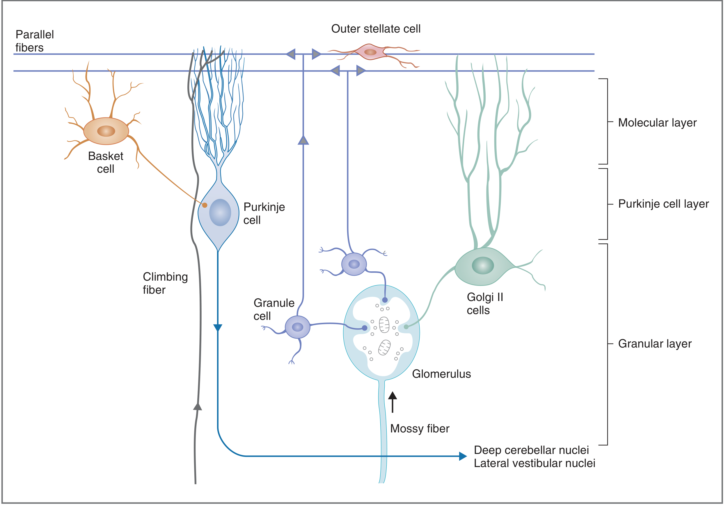
| Layer | Contents | Key Features |
|---|---|---|
| Granular (innermost) | Granule cells, Golgi II cells, glomeruli | Mossy fibers synapse here on granule cell dendrites in glomeruli |
| Purkinje cell (middle) | Purkinje cells only | One cell thick; output is ALWAYS inhibitory (GABA) |
| Molecular (outermost) | Basket cells, stellate cells, parallel fibers, Purkinje dendrites | Parallel fibers run here |
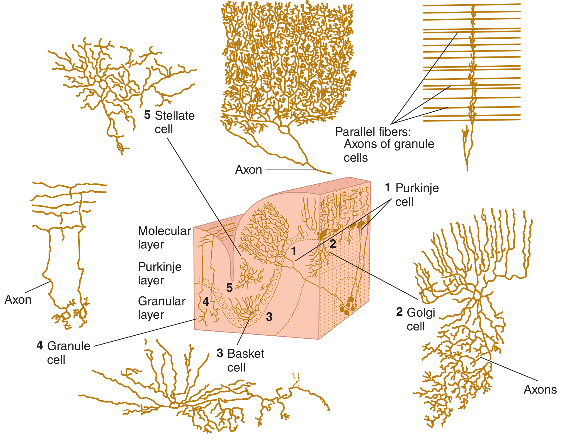
4. FIVE CELL TYPES - EXCITATORY vs INHIBITORY (Guaranteed MCQ)
| Cell | Location | Action |
|---|---|---|
| Granule cell | Granular layer | EXCITATORY (glutamate) - the ONLY excitatory interneuron |
| Purkinje cell | Purkinje layer | INHIBITORY (GABA) - sole OUTPUT of cerebellar cortex |
| Basket cell | Molecular layer | INHIBITORY (GABA) - inhibits Purkinje cells |
| Stellate cell | Molecular layer | INHIBITORY (GABA) - inhibits Purkinje cell dendrites |
| Golgi II cell | Granular layer | INHIBITORY (GABA) - inhibits granule cells (feedback) |
Exam pearl: Among all 5 cell types, only granule cells are excitatory. All others (Purkinje, basket, stellate, Golgi) are inhibitory. The Purkinje cell axon is the only output of the cerebellar cortex.
5. AFFERENT FIBER SYSTEMS (High Yield)
- Origin: Inferior olivary nucleus of medulla
- Project directly onto Purkinje cell dendrites
- Each Purkinje cell receives input from only ONE climbing fiber
- Produce complex spikes in Purkinje cells
- Role in motor learning
- Origin: Everything else (vestibulocerebellar, spinocerebellar, pontocerebellar)
- Synapse on granule cells in glomeruli
- Granule cells then send parallel fibers to Purkinje cells
- One Purkinje cell can receive up to 250,000 parallel fibers
- Produce simple spikes in Purkinje cells
6. DEEP CEREBELLAR NUCLEI (Medium-High Yield)
- Fastigial (most medial) - receives from vermis, projects to vestibular nuclei
- Globose - interpositus group
- Emboliform - interpositus group
- Dentate (most lateral) - receives from lateral hemispheres, projects via superior peduncle to red nucleus and thalamus (main output)
Key rule: Output of deep cerebellar nuclei to thalamus and brainstem is always EXCITATORY, despite receiving inhibitory Purkinje cell input.
7. SIGNS OF CEREBELLAR DISEASE (Very High Yield for Clinical Exams)
| Sign | Description |
|---|---|
| Dysmetria | Overshooting/undershooting a target |
| Intention tremor | Tremor that worsens as limb approaches target (vs. resting tremor in Parkinson's) |
| Dysdiadochokinesia | Inability to perform rapid alternating movements (e.g., pronation-supination) |
| Rebound phenomenon | Cannot arrest a movement; limb swings past when resistance is removed |
| Decomposition of movement | Complex movements are broken into segments, joint by joint |
| Nystagmus | Especially with flocculonodular lesions |
| Scanning (staccato) speech | Slurred, irregular speech rhythm |
| Truncal ataxia + wide-based gait | Vermis lesions |
8. LOCALIZATION OF CEREBELLAR LESIONS (Classic Exam Scenario)
- Flocculonodular lobe lesion (vestibulocerebellum): truncal ataxia, wide-based gait, nystagmus, vertigo, nausea/vomiting - NO limb ataxia
- Vermis lesion: truncal/gait ataxia predominantly
- Lateral hemisphere lesion: ipsilateral limb ataxia (appendicular ataxia), intention tremor, dysdiadochokinesia
- Cerebellar hemisphere in a child: think medulloblastoma (most common pediatric brain tumor)
9. CEREBELLAR AFFERENT TRACTS (Exam Table - Ganong's)
| Tract | Carries |
|---|---|
| Vestibulocerebellar | Vestibular impulses from labyrinths |
| Dorsal spinocerebellar | Proprioception from lower limbs (Golgi tendon organs, muscle spindles) |
| Ventral spinocerebellar | Proprioception from upper and lower limbs |
| Cuneocerebellar | Proprioception from upper limb and upper thorax |
| Pontocerebellar | Motor commands from cerebral cortex via pontine nuclei |
| Olivocerebellar | Whole-body proprioception via inferior olive - travels as climbing fibers |
10. MOTOR LEARNING ROLE (Short-Answer Favorite)
Quick Revision Summary (What to memorize first)
- Vestibulocerebellum = balance/eye movements | Spinocerebellum = coordination | Pontocerebellum = planning
- Three layers: Granular (inner) - Purkinje (middle) - Molecular (outer)
- Only granule cell is excitatory; Purkinje cell = sole inhibitory output (GABA)
- Climbing fibers = inferior olive → Purkinje directly (complex spikes, motor learning)
- Mossy fibers → granule cells → parallel fibers → Purkinje (simple spikes)
- Middle peduncle = only afferent, largest; Superior peduncle = main efferent
- Cerebellar signs are IPSILATERAL; intention tremor (not resting)
- Deep nuclei lateral to medial: Dentate, Emboliform, Globose, Fastigial
Generate a one-page study cheat sheet PDF for cerebellar anatomy
ls -la /tmp/workspace/ 2>/dev/null || mkdir -p /tmp/workspace && ls -la /tmp/workspace/
mkdir -p /tmp/workspace/cerebellum-cheatsheet
~/cerebellum-cheatsheet/generate_cheatsheet.py
from reportlab.lib.pagesizes import A4
from reportlab.lib import colors
from reportlab.lib.units import mm, cm
from reportlab.platypus import (
SimpleDocTemplate, Table, TableStyle, Paragraph, Spacer,
HRFlowable, KeepTogether
)
from reportlab.lib.styles import getSampleStyleSheet, ParagraphStyle
from reportlab.lib.enums import TA_CENTER, TA_LEFT, TA_JUSTIFY
from reportlab.platypus import FrameBreak
from reportlab.lib.colors import HexColor
import os
OUTPUT = "/tmp/workspace/cerebellum-cheatsheet/Cerebellum_Cheatsheet.pdf"
# ── Colour palette ────────────────────────────────────────────────────────────
C_NAVY = HexColor("#1B3A5C")
C_TEAL = HexColor("#1E7F8E")
C_AMBER = HexColor("#E8A020")
C_ROSE = HexColor("#C0392B")
C_GREEN = HexColor("#1E7F4E")
C_LGRAY = HexColor("#F2F5F8")
C_MGRAY = HexColor("#D6DCE4")
C_WHITE = colors.white
C_BLACK = colors.black
# ── Styles ────────────────────────────────────────────────────────────────────
styles = getSampleStyleSheet()
def S(name, **kw):
return ParagraphStyle(name, **kw)
title_style = S("TitleStyle",
fontSize=20, fontName="Helvetica-Bold",
textColor=C_WHITE, alignment=TA_CENTER,
spaceAfter=2)
subtitle_style = S("SubtitleStyle",
fontSize=9, fontName="Helvetica",
textColor=HexColor("#CCE4EE"), alignment=TA_CENTER,
spaceAfter=0)
sec_style = S("SecStyle",
fontSize=8.5, fontName="Helvetica-Bold",
textColor=C_WHITE, alignment=TA_LEFT,
leftPadding=4, spaceBefore=1, spaceAfter=1)
body_style = S("BodyStyle",
fontSize=7.2, fontName="Helvetica",
textColor=C_BLACK, leading=10,
leftPadding=0, spaceBefore=0)
body_bold = S("BodyBold",
fontSize=7.2, fontName="Helvetica-Bold",
textColor=C_NAVY, leading=10)
small_style = S("SmallStyle",
fontSize=6.5, fontName="Helvetica",
textColor=HexColor("#444444"), leading=9)
mnemonic_style = S("MnemonicStyle",
fontSize=7.4, fontName="Helvetica-BoldOblique",
textColor=C_ROSE, leading=10,
backColor=HexColor("#FFF4E5"),
borderPadding=(2,4,2,4))
pearl_style = S("PearlStyle",
fontSize=7.0, fontName="Helvetica-Oblique",
textColor=HexColor("#1B3A5C"), leading=10,
backColor=HexColor("#EAF4FF"),
borderPadding=(2,4,2,4))
# ── Helper: coloured section header ──────────────────────────────────────────
def sec_header(text, color=C_NAVY):
data = [[Paragraph(f"<b>{text}</b>", S("sh",
fontSize=8, fontName="Helvetica-Bold", textColor=C_WHITE,
alignment=TA_LEFT))]]
t = Table(data, colWidths=["100%"])
t.setStyle(TableStyle([
("BACKGROUND", (0,0), (-1,-1), color),
("TOPPADDING", (0,0), (-1,-1), 3),
("BOTTOMPADDING",(0,0), (-1,-1), 3),
("LEFTPADDING", (0,0), (-1,-1), 6),
]))
return t
def P(txt, style=body_style): return Paragraph(txt, style)
def SP(h=2): return Spacer(1, h)
# ── Build content ─────────────────────────────────────────────────────────────
story = []
# ─── TITLE BANNER ─────────────────────────────────────────────────────────────
banner_data = [[
Paragraph("CEREBELLUM", S("T2", fontSize=22, fontName="Helvetica-Bold",
textColor=C_WHITE, alignment=TA_CENTER)),
Paragraph("Anatomy & Physiology · Exam Cheat Sheet",
S("T3", fontSize=9, fontName="Helvetica", textColor=HexColor("#CCE4EE"),
alignment=TA_CENTER))
]]
banner = Table([[
Paragraph("🧠 CEREBELLUM — Anatomy & Physiology Cheat Sheet",
S("tb", fontSize=15, fontName="Helvetica-Bold",
textColor=C_WHITE, alignment=TA_CENTER))
]], colWidths=[190*mm])
banner.setStyle(TableStyle([
("BACKGROUND", (0,0), (-1,-1), C_NAVY),
("TOPPADDING", (0,0), (-1,-1), 8),
("BOTTOMPADDING",(0,0), (-1,-1), 8),
("LEFTPADDING", (0,0), (-1,-1), 6),
("ROUNDEDCORNERS", [4, 4, 4, 4]),
]))
story.append(banner)
story.append(SP(5))
# ─── ROW 1: Functional Divisions | Peduncles ──────────────────────────────────
# Left: Functional Divisions
div_header = sec_header("⚡ FUNCTIONAL DIVISIONS", C_TEAL)
div_data = [
[P("<b>Division</b>", body_bold), P("<b>Region</b>", body_bold), P("<b>Input</b>", body_bold), P("<b>Function</b>", body_bold)],
[P("Vestibulocerebellum", body_style), P("Flocculonodular lobe", small_style), P("Vestibular (labyrinth)", small_style), P("Balance, eye movements", small_style)],
[P("Spinocerebellum", body_style), P("Vermis + intermediate", small_style), P("Spinal cord", small_style), P("Synergy of ongoing movement", small_style)],
[P("Pontocerebellum", body_style), P("Lateral hemispheres", small_style), P("Cerebral cortex → pons", small_style), P("Planning & initiation", small_style)],
]
div_table = Table(div_data, colWidths=[28*mm, 30*mm, 34*mm, 36*mm])
div_table.setStyle(TableStyle([
("BACKGROUND", (0,0), (-1,0), C_MGRAY),
("BACKGROUND", (0,1), (-1,1), C_LGRAY),
("BACKGROUND", (0,2), (-1,2), C_WHITE),
("BACKGROUND", (0,3), (-1,3), C_LGRAY),
("GRID", (0,0), (-1,-1), 0.4, C_MGRAY),
("TOPPADDING", (0,0), (-1,-1), 2),
("BOTTOMPADDING",(0,0), (-1,-1), 2),
("LEFTPADDING", (0,0), (-1,-1), 3),
("VALIGN", (0,0), (-1,-1), "MIDDLE"),
]))
# Right: Peduncles
ped_header = sec_header("🔗 CEREBELLAR PEDUNCLES", C_ROSE)
ped_data = [
[P("<b>Peduncle</b>", body_bold), P("<b>Direction</b>", body_bold), P("<b>Key Contents</b>", body_bold)],
[P("Superior (SCP)", body_style), P("<font color='#C0392B'>Mainly EFFERENT</font>", small_style), P("Dentato-rubro-thalamic tract", small_style)],
[P("Middle (MCP)", body_style), P("<font color='#1E7F8E'>Only AFFERENT</font>", small_style), P("Corticopontocerebellar (LARGEST)", small_style)],
[P("Inferior (ICP)", body_style), P("Mixed", small_style), P("Spinocerebellar, vestibular", small_style)],
]
ped_table = Table(ped_data, colWidths=[26*mm, 24*mm, 40*mm])
ped_table.setStyle(TableStyle([
("BACKGROUND", (0,0), (-1,0), C_MGRAY),
("BACKGROUND", (0,1), (-1,1), C_LGRAY),
("BACKGROUND", (0,2), (-1,2), C_WHITE),
("BACKGROUND", (0,3), (-1,3), C_LGRAY),
("GRID", (0,0), (-1,-1), 0.4, C_MGRAY),
("TOPPADDING", (0,0), (-1,-1), 2),
("BOTTOMPADDING",(0,0), (-1,-1), 2),
("LEFTPADDING", (0,0), (-1,-1), 3),
("VALIGN", (0,0), (-1,-1), "MIDDLE"),
]))
row1 = Table([
[div_header, SP(3), ped_header],
[div_table, SP(3), ped_table],
], colWidths=[131*mm, 4*mm, 93*mm])
row1.setStyle(TableStyle([
("VALIGN", (0,0), (-1,-1), "TOP"),
("TOPPADDING", (0,0), (-1,-1), 0),
("BOTTOMPADDING",(0,0), (-1,-1), 0),
("LEFTPADDING", (0,0), (-1,-1), 0),
("RIGHTPADDING",(0,0), (-1,-1), 0),
]))
story.append(row1)
story.append(SP(5))
# ─── ROW 2: Cortical Layers | Cell Types ──────────────────────────────────────
layer_header = sec_header("🔬 CORTICAL LAYERS (outer → inner)", C_GREEN)
layer_data = [
[P("<b>Layer</b>", body_bold), P("<b>Contents</b>", body_bold), P("<b>Notes</b>", body_bold)],
[P("Molecular (outer)", body_style), P("Basket cells, stellate cells, parallel fibers, Purkinje dendrites", small_style), P("Parallel fibers run here", small_style)],
[P("Purkinje cell (middle)", body_style), P("Purkinje cells only", small_style), P("1 cell thick · output ALWAYS inhibitory (GABA)", small_style)],
[P("Granular (inner)", body_style), P("Granule cells, Golgi II cells, glomeruli", small_style), P("Mossy fibers synapse here; glomeruli = complex synapse clusters", small_style)],
]
layer_table = Table(layer_data, colWidths=[36*mm, 60*mm, 64*mm])
layer_table.setStyle(TableStyle([
("BACKGROUND", (0,0), (-1,0), C_MGRAY),
("BACKGROUND", (0,1), (-1,1), C_LGRAY),
("BACKGROUND", (0,2), (-1,2), C_WHITE),
("BACKGROUND", (0,3), (-1,3), C_LGRAY),
("GRID", (0,0), (-1,-1), 0.4, C_MGRAY),
("TOPPADDING", (0,0), (-1,-1), 2),
("BOTTOMPADDING",(0,0), (-1,-1), 2),
("LEFTPADDING", (0,0), (-1,-1), 3),
("VALIGN", (0,0), (-1,-1), "MIDDLE"),
]))
# Cell types
cell_header = sec_header("🧬 5 CELL TYPES — EXCITATORY vs INHIBITORY", C_AMBER)
cell_data = [
[P("<b>Cell</b>", body_bold), P("<b>Layer</b>", body_bold), P("<b>Type</b>", body_bold), P("<b>Key Role</b>", body_bold)],
[P("Granule", body_style), P("Granular", small_style), P("<font color='#1E7F4E'><b>EXCIT (Glu)</b></font>", small_style), P("→ parallel fibers → Purkinje", small_style)],
[P("Purkinje", body_style), P("Purkinje", small_style), P("<font color='#C0392B'><b>INHIB (GABA)</b></font>", small_style), P("SOLE OUTPUT of cortex", small_style)],
[P("Basket", body_style), P("Molecular", small_style), P("<font color='#C0392B'><b>INHIB (GABA)</b></font>", small_style), P("Inhibits Purkinje cell soma", small_style)],
[P("Stellate", body_style), P("Molecular", small_style), P("<font color='#C0392B'><b>INHIB (GABA)</b></font>", small_style), P("Inhibits Purkinje dendrites", small_style)],
[P("Golgi II", body_style), P("Granular", small_style), P("<font color='#C0392B'><b>INHIB (GABA)</b></font>", small_style), P("Feedback: inhibits granule cells", small_style)],
]
cell_table = Table(cell_data, colWidths=[22*mm, 22*mm, 30*mm, 44*mm])
cell_table.setStyle(TableStyle([
("BACKGROUND", (0,0), (-1,0), C_MGRAY),
("BACKGROUND", (0,1), (-1,1), HexColor("#E8F8EE")),
("BACKGROUND", (0,2), (-1,2), HexColor("#FFF0EE")),
("BACKGROUND", (0,3), (-1,3), HexColor("#FFF0EE")),
("BACKGROUND", (0,4), (-1,4), HexColor("#FFF0EE")),
("BACKGROUND", (0,5), (-1,5), HexColor("#FFF0EE")),
("GRID", (0,0), (-1,-1), 0.4, C_MGRAY),
("TOPPADDING", (0,0), (-1,-1), 2),
("BOTTOMPADDING",(0,0), (-1,-1), 2),
("LEFTPADDING", (0,0), (-1,-1), 3),
("VALIGN", (0,0), (-1,-1), "MIDDLE"),
]))
row2 = Table([
[layer_header, SP(3), cell_header],
[layer_table, SP(3), cell_table],
], colWidths=[163*mm, 4*mm, 61*mm])
row2.setStyle(TableStyle([
("VALIGN", (0,0), (-1,-1), "TOP"),
("TOPPADDING", (0,0), (-1,-1), 0),
("BOTTOMPADDING",(0,0), (-1,-1), 0),
("LEFTPADDING", (0,0), (-1,-1), 0),
("RIGHTPADDING",(0,0), (-1,-1), 0),
]))
story.append(row2)
story.append(SP(5))
# ─── ROW 3: Afferent Fibers | Deep Nuclei ────────────────────────────────────
fiber_header = sec_header("📥 AFFERENT FIBER SYSTEMS", C_TEAL)
fiber_data = [
[P("<b>Climbing Fibers</b>", body_bold), P("<b>Mossy Fibers</b>", body_bold)],
[
P("Origin: <b>Inferior olivary nucleus</b> (medulla)<br/>"
"→ Synapse directly on Purkinje cell dendrites<br/>"
"→ <b>1 climbing fiber per Purkinje cell</b><br/>"
"→ Produce <b>complex spikes</b><br/>"
"→ Role in <b>motor learning</b><br/>"
"→ Activity ↑ when learning a NEW movement", small_style),
P("Origin: ALL others (spinocerebellar, vestibulocerebellar, pontocerebellar)<br/>"
"→ Synapse on <b>granule cells</b> in glomeruli<br/>"
"→ Granule cells → <b>parallel fibers</b> → Purkinje<br/>"
"→ 1 Purkinje cell receives up to <b>250,000 parallel fibers</b><br/>"
"→ Produce <b>simple spikes</b><br/>"
"→ Majority of cerebellar input", small_style),
],
]
fiber_table = Table(fiber_data, colWidths=[87*mm, 88*mm])
fiber_table.setStyle(TableStyle([
("BACKGROUND", (0,0), (-1,0), C_MGRAY),
("BACKGROUND", (0,1), (0,1), HexColor("#EAF4FF")),
("BACKGROUND", (1,1), (1,1), HexColor("#F0FBF5")),
("GRID", (0,0), (-1,-1), 0.4, C_MGRAY),
("TOPPADDING", (0,0), (-1,-1), 3),
("BOTTOMPADDING",(0,0), (-1,-1), 3),
("LEFTPADDING", (0,0), (-1,-1), 4),
("VALIGN", (0,0), (-1,-1), "TOP"),
]))
# Deep nuclei
nuc_header = sec_header("🏛 DEEP CEREBELLAR NUCLEI (lateral → medial)", C_ROSE)
nuc_data = [
[P("<b>Nucleus</b>", body_bold), P("<b>Receives from</b>", body_bold), P("<b>Projects to</b>", body_bold)],
[P("<b>Dentate</b> (most lateral)", small_style), P("Lateral hemispheres", small_style), P("Red nucleus + thalamus via SCP", small_style)],
[P("Emboliform", small_style), P("Intermed. hemisphere", small_style), P("Red nucleus", small_style)],
[P("Globose", small_style), P("Intermed. hemisphere", small_style), P("Red nucleus", small_style)],
[P("<b>Fastigial</b> (most medial)", small_style), P("Vermis", small_style), P("Vestibular nuclei via ICP", small_style)],
]
nuc_table = Table(nuc_data, colWidths=[44*mm, 40*mm, 41*mm])
nuc_table.setStyle(TableStyle([
("BACKGROUND", (0,0), (-1,0), C_MGRAY),
("BACKGROUND", (0,1), (-1,1), HexColor("#FFF0EE")),
("BACKGROUND", (0,2), (-1,2), C_LGRAY),
("BACKGROUND", (0,3), (-1,3), C_LGRAY),
("BACKGROUND", (0,4), (-1,4), HexColor("#FFF0EE")),
("GRID", (0,0), (-1,-1), 0.4, C_MGRAY),
("TOPPADDING", (0,0), (-1,-1), 2),
("BOTTOMPADDING",(0,0), (-1,-1), 2),
("LEFTPADDING", (0,0), (-1,-1), 3),
("VALIGN", (0,0), (-1,-1), "MIDDLE"),
]))
row3_top = Table([
[fiber_header, SP(3), nuc_header],
[fiber_table, SP(3), nuc_table],
], colWidths=[178*mm, 4*mm, 128*mm])
row3_top.setStyle(TableStyle([
("VALIGN", (0,0), (-1,-1), "TOP"),
("TOPPADDING", (0,0), (-1,-1), 0),
("BOTTOMPADDING",(0,0), (-1,-1), 0),
("LEFTPADDING", (0,0), (-1,-1), 0),
("RIGHTPADDING", (0,0), (-1,-1), 0),
]))
story.append(row3_top)
story.append(SP(5))
# ─── ROW 4: Clinical Signs | Lesion Localization | Mnemonics ─────────────────
signs_header = sec_header("⚠ SIGNS OF CEREBELLAR DISEASE", C_ROSE)
signs_data = [
[P("<b>Sign</b>", body_bold), P("<b>Description</b>", body_bold)],
[P("Ataxia", small_style), P("<b>IPSILATERAL</b> to lesion; wide-based unsteady gait", small_style)],
[P("Dysmetria", small_style), P("Overshooting/undershooting target", small_style)],
[P("Intention tremor", small_style), P("Worsens near target (vs resting tremor in Parkinson's)", small_style)],
[P("Dysdiadochokinesia", small_style), P("Cannot do rapid alternating movements", small_style)],
[P("Rebound phenomenon", small_style), P("Cannot arrest movement when resistance removed", small_style)],
[P("Decomposition", small_style), P("Breaks multi-joint movement into separate joints", small_style)],
[P("Nystagmus", small_style), P("Especially flocculonodular lesion", small_style)],
[P("Scanning speech", small_style), P("Slurred, staccato, irregular rhythm", small_style)],
]
signs_table = Table(signs_data, colWidths=[34*mm, 70*mm])
signs_table.setStyle(TableStyle([
("BACKGROUND", (0,0), (-1,0), C_MGRAY),
*[("BACKGROUND", (0,i), (-1,i), C_LGRAY if i%2==0 else C_WHITE) for i in range(1,9)],
("GRID", (0,0), (-1,-1), 0.4, C_MGRAY),
("TOPPADDING", (0,0), (-1,-1), 2),
("BOTTOMPADDING",(0,0), (-1,-1), 2),
("LEFTPADDING", (0,0), (-1,-1), 3),
("VALIGN", (0,0), (-1,-1), "MIDDLE"),
]))
# Localization
loc_header = sec_header("📍 LESION LOCALIZATION", C_GREEN)
loc_data = [
[P("<b>Lesion Site</b>", body_bold), P("<b>Features</b>", body_bold)],
[P("Flocculonodular lobe", small_style), P("Truncal ataxia + nystagmus + vertigo/N&V · NO limb ataxia", small_style)],
[P("Vermis", small_style), P("Gait/truncal ataxia (proximal muscles)", small_style)],
[P("Lateral hemisphere", small_style), P("Ipsilateral limb ataxia, intention tremor, dysdiadochokinesia", small_style)],
[P("Child + lateral", small_style), P("Think Medulloblastoma (most common pediatric brain tumour)", small_style)],
]
loc_table = Table(loc_data, colWidths=[34*mm, 72*mm])
loc_table.setStyle(TableStyle([
("BACKGROUND", (0,0), (-1,0), C_MGRAY),
("BACKGROUND", (0,1), (-1,1), C_LGRAY),
("BACKGROUND", (0,2), (-1,2), C_WHITE),
("BACKGROUND", (0,3), (-1,3), C_LGRAY),
("BACKGROUND", (0,4), (-1,4), HexColor("#FFF0EE")),
("GRID", (0,0), (-1,-1), 0.4, C_MGRAY),
("TOPPADDING", (0,0), (-1,-1), 2),
("BOTTOMPADDING",(0,0), (-1,-1), 2),
("LEFTPADDING", (0,0), (-1,-1), 3),
("VALIGN", (0,0), (-1,-1), "MIDDLE"),
]))
# Mnemonics box
mn_header = sec_header("💡 MNEMONICS & PEARLS", C_AMBER)
pearls = [
("Only EXCITATORY cell:", "Granule cell (glutamate). All others = GABA inhibitory."),
("Purkinje cell output:", "SOLE cortical output · ALWAYS GABA inhibitory → deep nuclei"),
("Deep nuclei output:", "ALWAYS EXCITATORY (to thalamus/brainstem) despite inhibitory Purkinje input"),
("MCP = largest peduncle:", "Contains ONLY afferents (pontocerebellar)"),
("Climbing fibers:", "1 per Purkinje · from inferior olive · motor learning · complex spikes"),
("Deep nuclei (lat→med):", "Don't Eat Greasy Food (Dentate · Emboliform · Globose · Fastigial)"),
("Divisions mnemonic:", "VeStibulo = Vestibular/balance | SPino = Synergy | PonTo = Planning/Timing"),
("Cerebellar vs Cortical:", "Cerebellar signs IPSILATERAL · Cortical signs CONTRALATERAL"),
]
mn_content = ""
for k, v in pearls:
mn_content += f"<b>• {k}</b> {v}<br/>"
mn_para = Paragraph(mn_content, S("mns",
fontSize=6.8, fontName="Helvetica", leading=10,
textColor=C_BLACK))
mn_cell = Table([[mn_para]], colWidths=[90*mm])
mn_cell.setStyle(TableStyle([
("BACKGROUND", (0,0), (-1,-1), HexColor("#FFFBF0")),
("TOPPADDING", (0,0), (-1,-1), 4),
("BOTTOMPADDING",(0,0), (-1,-1), 4),
("LEFTPADDING", (0,0), (-1,-1), 5),
("RIGHTPADDING", (0,0), (-1,-1), 5),
("BOX", (0,0), (-1,-1), 0.5, C_AMBER),
]))
# Assemble right column
right_col = Table([
[loc_header],
[loc_table],
[SP(4)],
[mn_header],
[mn_cell],
], colWidths=[109*mm])
right_col.setStyle(TableStyle([
("TOPPADDING", (0,0), (-1,-1), 0),
("BOTTOMPADDING",(0,0), (-1,-1), 0),
("LEFTPADDING", (0,0), (-1,-1), 0),
("RIGHTPADDING", (0,0), (-1,-1), 0),
]))
row4 = Table([
[signs_header, SP(3), right_col],
[signs_table, SP(3), ""],
], colWidths=[107*mm, 4*mm, 117*mm])
row4.setStyle(TableStyle([
("VALIGN", (0,0), (-1,-1), "TOP"),
("TOPPADDING", (0,0), (-1,-1), 0),
("BOTTOMPADDING",(0,0), (-1,-1), 0),
("LEFTPADDING", (0,0), (-1,-1), 0),
("RIGHTPADDING", (0,0), (-1,-1), 0),
("SPAN", (2,0), (2,1)),
]))
story.append(row4)
story.append(SP(5))
# ─── FOOTER ──────────────────────────────────────────────────────────────────
footer_data = [[
Paragraph(
"Sources: Costanzo Physiology 7e · Ganong's Review of Medical Physiology 26e · Neuroanatomy through Clinical Cases 3e | © Orris Medical Study Aid",
S("ft", fontSize=5.8, fontName="Helvetica-Oblique",
textColor=HexColor("#888888"), alignment=TA_CENTER))
]]
footer = Table(footer_data, colWidths=[190*mm])
footer.setStyle(TableStyle([
("TOPPADDING", (0,0), (-1,-1), 3),
("BOTTOMPADDING",(0,0), (-1,-1), 3),
("LINEABOVE", (0,0), (-1,0), 0.5, C_MGRAY),
]))
story.append(footer)
# ─── Build PDF ────────────────────────────────────────────────────────────────
doc = SimpleDocTemplate(
OUTPUT,
pagesize=A4,
leftMargin=10*mm, rightMargin=10*mm,
topMargin=10*mm, bottomMargin=8*mm,
)
doc.build(story)
print(f"PDF saved to: {OUTPUT}")
cd /tmp/workspace/cerebellum-cheatsheet && python generate_cheatsheet.py 2>&1
ls -lh /tmp/workspace/cerebellum-cheatsheet/Cerebellum_Cheatsheet.pdf
python3 -c " from pypdf import PdfReader r = PdfReader('/tmp/workspace/cerebellum-cheatsheet/Cerebellum_Cheatsheet.pdf') print(f'Pages: {len(r.pages)}') print(f'Page size: {r.pages[0].mediabox}') text = r.pages[0].extract_text() print(f'Text length: {len(text)} chars') print('First 300 chars:', text[:300]) "
Cerebellum Cheatsheet
PDF Document · PDF
| Section | Content |
|---|---|
| Functional Divisions | Vestibulocerebellum / Spinocerebellum / Pontocerebellum table |
| Cerebellar Peduncles | Superior / Middle / Inferior with direction + key contents |
| Cortical Layers | Molecular / Purkinje / Granular with contents and notes |
| 5 Cell Types | Excitatory vs Inhibitory colour-coded table |
| Afferent Fibers | Climbing vs Mossy fibers side-by-side comparison |
| Deep Nuclei | Dentate, Emboliform, Globose, Fastigial (lateral → medial) |
| Clinical Signs | All 8 signs of cerebellar disease |
| Lesion Localization | Flocculonodular / Vermis / Lateral hemisphere + medulloblastoma |
| Mnemonics & Pearls | 8 high-yield exam pearls including "Don't Eat Greasy Food" |