
A 7-year-old boy presented to the Paediatric OPD with red sores covered with honey-coloured crusts over the face and limbs. His mother gave history of such recurrent episodes. Wound swab was taken and sent to Microbiology laboratory for Gram staining and culture and sensitivity. Gram staining showed Gram positive cocci arranged in clusters and the culture grew a bacterial agent that is coagulase test positive and showed growth on mannitol salt agar. a) What is the probable diagnosis? b) Identify the causative pathogen. c) Give a short description of its virulence factors and toxins produced by this pathogen. d) Describe the laboratory diagnosis of this infection. e) What is MRSA and write a note on its clinical significanc
Staphylococcus aureus gram stain cocci clusters impetigo honey crust
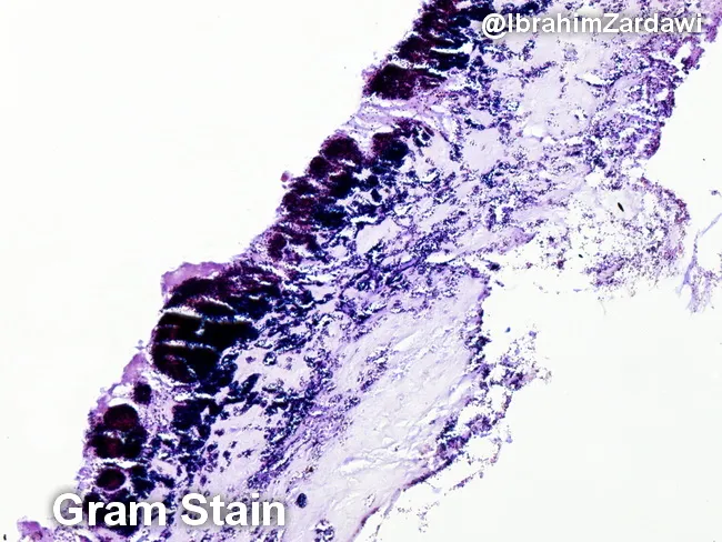
This histology image is a light-micrograph of a Gram-stained valvular vegetation from a case of infective endocarditis. The specimen is cardiac valvular tissue; the vegetation comprises a fibrin-rich, platelet-laden thrombus with embedded bacterial colonies along the endocardial surface. Gram staining highlights Gram-positive cocci as purple to deep purple clusters within the thrombus, indicating organisms such as Streptococcus spp., Staphylococcus spp., or Enterococcus. The bacterial colonies are adherent to the valvar surface within a dense inflammatory milieu, with neutrophilic infiltrate and tissue necrosis limited to microdomains. The histologic architecture demonstrates vegetations typical of endocarditis: a friable, topographically irregular lesion on the valve with microabscess-like foci and bacterial encasement within fibrin. The presence of Gram-positive bacteria on histology supports a bacterial endocarditis diagnosis and guides antimicrobial therapy pending culture confirmation. Clinically, such histologic confirmation is critical for distinguishing infectious from non-infectious valve pathology, for clarifying the etiologic category of pathogens (oral flora-related viridans Streptococcus, Staphylococcus aureus, enterococci), and for correlating with blood culture, echocardiographic findings, and patient risk factors. This image is valuable for educational purposes, illustrating Gram stain morphology in endocardial vegetations and reinforcing the need for targeted antibiotics and possible surgical consultation.
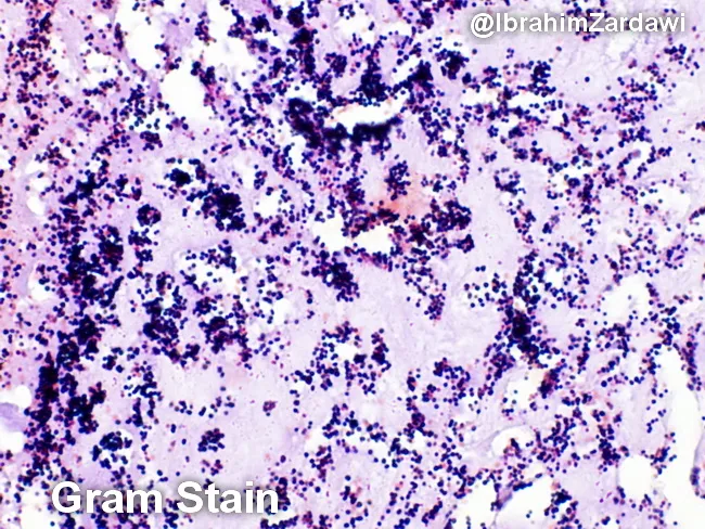
Imaging modality: Light microscopy; Gram staining of a histologic section of a cardiac valve vegetation from suspected infective endocarditis. The specimen shows a dense, friable vegetative mass within valve tissue characterized by a fibrinous, platelet-rich matrix in which numerous Gram-positive cocci are adherent. On the Gram stain, the cocci appear as purple spherical cells arranged in clusters and sometimes in short chains, consistent with Gram-positive organisms. The background demonstrates inflammatory cells and debris typical of endocardial infection. The histologic architecture indicates vegetative lesions with microbial colonization and a biofilm component. These findings correlate with infective endocarditis due to cocci such as Staphylococcus aureus or viridans-group streptococci in clinical practice, though Gram stain alone cannot specify species. The image is a micrograph provided with permission by Dr. Ibrahim Zardawi; image shows classic histopathology of bacterial endocarditis. The diagnostic significance lies in confirming bacterial endocarditis and guiding immediate management including empiric antibiotic therapy targeting Gram-positive cocci while awaiting culture results. This image is valuable for pathology education, microbiology correlation, and endocarditis research. Potential clinical use cases include training in Gram stain interpretation, distinguishing bacterial endocarditis from sterile vegetations, and supporting diagnosis when blood cultures are pending.
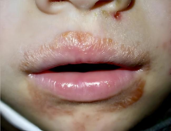
This clinical photograph displays a perioral skin condition in a pediatric patient, characteristic of impetigo. The image focuses on the lower face, specifically the lips and surrounding cutaneous areas. Visible are multiple well-demarcated, brownish to honey-colored crusts (meliceric crusts) distributed along the vermillion border of both the upper and lower lips. The crusting is most prominent at the superior aspect of the upper lip and the inferior aspect of the lower lip. Additional focal lesions, including a small erythematous papule with overlying yellow crust, are located near the philtrum and the base of the nose. The underlying skin and the lips themselves appear mildly inflamed and dry. This presentation is typical for non-bullous impetigo, a superficial bacterial skin infection often caused by Staphylococcus aureus or Streptococcus pyogenes. The visual features demonstrate the classic diagnostic sign of dried exudate that forms thick, adhered crusts in the perioral region.
Clinical Case: Honey-Crusted Lesions in a 7-Year-Old Boy
a) Probable Diagnosis
b) Causative Pathogen
- Gram stain: Gram-positive cocci in clusters (characteristic of staphylococci)
- Coagulase test positive - the defining test that distinguishes S. aureus from coagulase-negative staphylococci
- Growth on mannitol salt agar (MSA) with fermentation of mannitol (turns the medium yellow due to acid production) - a selective and differential medium for S. aureus
c) Virulence Factors and Toxins of S. aureus
Structural / Surface Virulence Factors
| Component | Function |
|---|---|
| Peptidoglycan (thick cell wall) | Resists desiccation; survives on dry surfaces for weeks |
| Protein A | Binds the Fc region of IgG, blocking opsonisation and phagocytosis |
| Capsule / slime layer | Inhibits phagocytosis; promotes biofilm formation on catheters/prostheses |
| Fibronectin-binding proteins (FnBPs) | Adhesins - mediate attachment to host tissues and extracellular matrix |
| Clumping factor (ClfA, ClfB) | Binds fibrinogen; responsible for clumping on glass slide (bound coagulase) |
| Panton-Valentine Leukocidin (PVL) | Pore-forming toxin destroying neutrophils and macrophages; strongly associated with community-MRSA skin infections and necrotising pneumonia |
Secreted Toxins
| Toxin | Disease / Mechanism |
|---|---|
| Coagulase (free) | Converts fibrinogen to fibrin, creating a fibrin "cloak" around the organism - evades phagocytosis |
| Exfoliative toxins A & B (ET-A, ET-B) | Serine proteases that cleave desmoglein-1 in the skin; cause Staphylococcal Scalded Skin Syndrome (SSSS) in neonates and infants; responsible for blister formation in bullous impetigo |
| Enterotoxins A-E, G-J (heat-stable) | Superantigens; cause staphylococcal food poisoning (rapid onset vomiting within 1-6 hours) |
| Toxic Shock Syndrome Toxin-1 (TSST-1) | Superantigen - triggers massive cytokine release; causes Toxic Shock Syndrome (TSS) with fever, hypotension, diffuse erythematous rash, multi-organ failure |
| Hyaluronidase | Degrades hyaluronic acid in connective tissue, facilitating spread |
| Fibrinolysin (Staphylokinase) | Dissolves fibrin clots; aids dissemination |
| Lipase / Phospholipase | Breaks down lipids in skin; aids in cutaneous infection |
| Leucocidin (alpha-toxin, beta-toxin, delta-toxin) | Cytotoxins that lyse erythrocytes, leukocytes, platelets; contribute to tissue necrosis |
| Nuclease (DNase) | Thermostable - used in lab identification |
d) Laboratory Diagnosis
Specimen Collection
- Wound swab from beneath the crust (remove crust first, swab the serous fluid from the base of the lesion for best yield)
1. Microscopy (Gram Stain)
- Smear prepared and heat-fixed
- Stained with crystal violet (primary stain), iodine (mordant), decolorised with acetone-alcohol, counterstained with safranin
- Result: Gram-positive cocci (purple) arranged in clusters (grape-like bunches)
- Gram stain is useful for pyogenic infections (as in this case) but NOT for blood infections or purely toxin-mediated diseases
2. Culture
| Medium | Purpose | Appearance of S. aureus |
|---|---|---|
| Blood agar (BAP) | Non-selective primary medium | Golden/yellow colonies; beta-hemolysis (clear zone) |
| Mannitol Salt Agar (MSA) | Selective (7.5% NaCl suppresses other organisms) + differential (mannitol fermentation) | Yellow colonies with yellow halo (mannitol fermented, acid turns phenol red indicator yellow) |
| Chromogenic agar | Selective and differential; used for MRSA screening | Mauve/pink colonies |
| Nutrient/MacConkey agar | Growth possible; not differential for staph | Grows on nutrient agar |
- Incubation: 37°C for 18-24 hours
- S. aureus grows rapidly on non-selective media
- Colonies appear golden-yellow (due to carotenoid pigment, hence "aureus") with a surrounding zone of beta-hemolysis on blood agar
3. Identification Tests
| Test | Result in S. aureus | Significance |
|---|---|---|
| Catalase test | Positive (bubbles with H₂O₂) | Distinguishes staphylococci from streptococci (strep = catalase negative) |
| Coagulase test (tube/slide) | Positive | KEY test - distinguishes S. aureus from coagulase-negative staphylococci (CoNS) |
| Mannitol fermentation | Positive (acid) | Confirmed on MSA |
| DNase test | Positive (clears DNA in agar) | Useful confirmatory test |
| Protein A test | Positive | Latex agglutination |
| MALDI-TOF mass spectrometry | Species confirmation | Gold standard in modern labs |
| Molecular (PCR/NAAT) | Detects nuc gene (S. aureus specific) or mecA gene (MRSA) | Used for screening and MRSA identification |
4. Antibiotic Susceptibility Testing (AST / C&S)
- Kirby-Bauer disk diffusion or MIC by broth microdilution
- Important drugs to test: penicillin, oxacillin/cefoxitin, clindamycin, TMP-SMX, vancomycin
- Cefoxitin 30μg disk used to detect MRSA (zone of inhibition <22 mm = resistant)
- D-test performed if erythromycin-resistant but clindamycin-sensitive (to detect inducible clindamycin resistance via erm gene)
e) MRSA - Definition, Mechanism, and Clinical Significance
What is MRSA?
Mechanism of Resistance
- Normal beta-lactam antibiotics kill bacteria by binding Penicillin Binding Proteins (PBPs) and inhibiting peptidoglycan cross-linking, causing cell wall lysis
- MRSA carries the mecA gene (on a mobile genetic element called SCCmec) which encodes a modified binding protein: PBP2a (PBP2')
- PBP2a has very low affinity for beta-lactam antibiotics - they cannot bind to it, so cell wall synthesis continues unimpeded even in the presence of these antibiotics
- Detection: latex agglutination for PBP2a, PCR for mecA, growth on chromogenic media, or cefoxitin disk susceptibility testing (Quick Compendium of Clinical Pathology 5e)
Types of MRSA
| Type | Abbreviation | Features |
|---|---|---|
| Hospital-acquired MRSA | HA-MRSA | Multi-drug resistant; affects immunocompromised/post-surgical patients; causes line infections, pneumonia, wound infections |
| Community-acquired MRSA | CA-MRSA | Often carries PVL gene; causes skin/soft tissue infections in healthy individuals; first reported 1993 |
| Livestock-associated MRSA | LA-MRSA | Seen in farmers/veterinary workers |
Clinical Significance of MRSA
-
Epidemiology: MRSA is now the most common cause of community-acquired skin and soft tissue infections in many settings. In major cities, MRSA is the most common pathogen in ED patients presenting with skin infections (Rosen's Emergency Medicine).
-
Clinical presentations of HA-MRSA:
- Surgical site infections (post-operative wounds)
- Catheter-related bloodstream infections (CRBSI)
- Ventilator-associated pneumonia (VAP) and necrotising pneumonia
- Infective endocarditis (especially right-sided in IV drug users)
- Osteomyelitis and septic arthritis
- Meningitis (post-neurosurgical)
-
Clinical presentations of CA-MRSA:
- Skin abscesses, furuncles, carbuncles (often with central necrosis, frequently mistaken for spider bites)
- Impetigo (as in this case)
- Necrotising fasciitis (rare but life-threatening)
- Recurrences are common; household and close-contact spread well-recognised
-
Treatment challenges:
- Resistant to all beta-lactams (penicillins, cephalosporins, carbapenems)
- For skin infections (non-invasive): oral TMP-SMX, doxycycline/minocycline, clindamycin (after D-test confirmation), linezolid
- For invasive/systemic MRSA: Vancomycin is the drug of choice (IV); alternatives include daptomycin, linezolid, tigecycline, telavancin
- In severely ill patients, carbapenems (meropenem) combined with vancomycin offer synergy
- Cephalosporins and macrolides are typically ineffective against MRSA
- Fluoroquinolones are avoided (resistance develops rapidly) (Rosen's Emergency Medicine)
-
Emerging resistances:
- VISA (Vancomycin-Intermediate S. aureus) and VRSA (Vancomycin-Resistant S. aureus) - rare but of grave concern
- Inducible clindamycin resistance - mediated by erm genes; detected by the D-test
-
Infection control:
- Contact precautions (gown + gloves) for colonised/infected patients
- Nasal decolonisation with mupirocin for carriers
- Chlorhexidine body washes
- Regular hand hygiene and cleaning of shared equipment
- Screening of high-risk patients on admission (PCR/NAAT swabs from nares, axilla, groin)
Summary Table for This Case
| Feature | Finding |
|---|---|
| Diagnosis | Non-bullous (crusted) Impetigo |
| Causative organism | Staphylococcus aureus |
| Gram stain | Gram-positive cocci in clusters |
| Culture media | Blood agar (golden colonies, beta-haemolysis), Mannitol Salt Agar (yellow colonies) |
| Key identification test | Coagulase positive |
| Key virulence factors | Exfoliative toxins, protein A, fibronectin-binding proteins, PVL, TSST-1 |
| MRSA resistance gene | mecA (encodes PBP2a) |
| MRSA treatment | Vancomycin (IV) / TMP-SMX, clindamycin, doxycycline (oral) |
56-year-old male working in a sheep farm developed a vesicular lesion / blister on his left forearm. The lesion is painless but itchy. There was no history of fever, myalgia and loss of appetite. The vesicle fluid is aspirated and sent to Microbiology laboratory for Gram staining and culture. Gram staining showed Spore forming Gram positive bacilli. a) What is the clinical condition? b) What are its various clinical presentations? c) Write a short description of its laboratory diagnosis. d) Write a note on Zoonosis and list any five Zoonotic diseases and its causative age
cutaneous anthrax eschar black necrotic lesion forearm
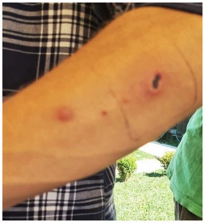
This clinical photograph shows the cutaneous manifestation of anthrax on a human forearm. The primary lesion is a classic necrotic eschar, characterized by a dark, coal-black central area of tissue necrosis and ulceration. This central eschar is surrounded by a raised, erythematous ring and a halo of inflammatory edema. Proximal to the main lesion, there are smaller secondary findings, including an erythematous papule and a tiny macule, which may represent satellite lesions or evolving stages of infection. The skin around the primary eschar has been marked with a pen, likely to monitor the progression of peripheral edema or cellulitis. The image demonstrates the typical appearance of Bacillus anthracis infection in the skin, highlighting the progression from a pruritic papule to a characteristic painless ulcer with a black necrotic center, a key diagnostic feature for clinical identification in infectious disease and dermatology.
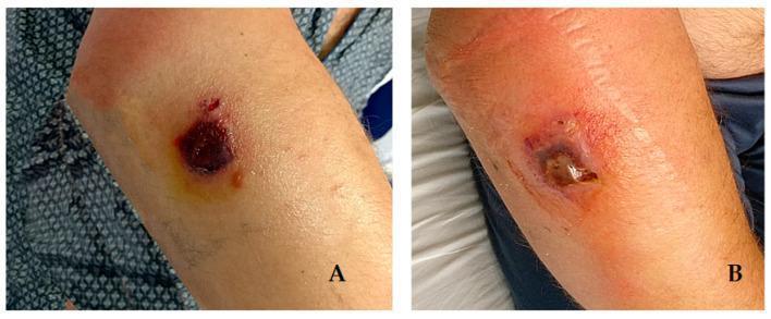
Two-panel clinical photograph illustrating the progression of a cutaneous anthrax lesion on a patient's right forearm (Case 2). Panel A (8 days post-exposure) shows a characteristic black, necrotic eschar, approximately one centimeter in diameter, with a distinct depressed center. It is surrounded by a yellowish serous ring and mild erythema. Panel B (20 days post-exposure) demonstrates the evolution of the lesion, showing a more defined ulcerated area with a central eschar containing clear serous fluid. There is significantly increased perilesional erythema and soft tissue edema, extending across the forearm. This progression illustrates the transition from a painless itchy papule to a malignant pustule with associated systemic inflammatory response, typical of infection with Bacillus anthracis. The image serves as a clinical reference for infectious disease staging and dermatological manifestations of occupational zoonotic exposures.
Clinical Case: Painless Vesicular Lesion in a Sheep Farm Worker
a) Clinical Condition
- Occupation: sheep farm worker (primary animal reservoir for Bacillus anthracis)
- Painless (a hallmark feature), itchy vesicular lesion on the forearm (exposed area)
- No systemic symptoms (fever, myalgia) - consistent with localised cutaneous form
- Gram stain: spore-forming Gram-positive bacilli = Bacillus anthracis
b) Clinical Presentations of Anthrax
1. Cutaneous Anthrax (~95% of cases)
| Stage | Time | Appearance |
|---|---|---|
| Papule | Day 1-3 post-inoculation | Purpuric macule/papule resembling insect bite; pruritic |
| Vesicle | Within 48 hours | 1-3 mm vesicle forms with surrounding non-pitting edema |
| Ulcer + Satellite vesicles | Day 3-5 | Central vesicle ulcerates; small ring of vesicles ("pearly wreath") forms around it |
| Black Eschar | Day 5-7 | Painless, depressed, coal-black necrotic eschar with intense surrounding erythema and edema |
| Resolution | Week 1-2 | Eschar dries, loosens, and sloughs; heals without permanent scar |
- Painlessness of the lesion despite necrosis (diagnostically important - helps distinguish from brown recluse spider bite which is painful)
- Marked surrounding non-pitting edema (may be massive, especially on face/neck)
- Regional lymphangitis and lymphadenopathy (tender, enlarged, may suppurate)
- Without treatment, up to 20% mortality (from septicaemia); near-zero mortality with antibiotics
- Note: antibiotic therapy does not alter the progression of the local lesion (toxin-mediated), but prevents systemic dissemination (Dermatology 5e)
2. Inhalational Anthrax ("Woolsorter's Disease") - Most lethal form
- Caused by inhalation of spores (aerosols from infected hides/wool, or bioterrorism)
- Spores are phagocytosed by alveolar macrophages, transported to mediastinal lymph nodes where they germinate
- Biphasic illness:
- Phase 1 (2-10 days): Flu-like illness - malaise, fever, non-productive cough; may mimic influenza
- Phase 2 (abrupt deterioration within 24-48 hours): Overwhelming sepsis, shock, haemorrhagic mediastinitis, respiratory distress, stridor
- CXR/CT: widened mediastinum, hilar adenopathy, pleural effusions (haemorrhagic)
- Mortality: ~50% even with intensive treatment; without treatment near 100%
3. Gastrointestinal Anthrax
- From ingestion of undercooked contaminated meat
- Two sub-forms:
- Oropharyngeal: Sore throat, neck swelling (cervical lymphadenitis), tonsil involvement, fever, dysphagia, respiratory distress
- Intestinal: Nausea, vomiting, fever → severe abdominal pain, haematemesis, ascites, bloody diarrhoea, acute abdomen (mesenteric lymphadenitis)
- Incubation: 2-5 days
- Mortality: approximately 50% (Rosen's Emergency Medicine)
4. Injection Anthrax
- Complication of intravenous drug use (primarily heroin)
- More than 50 cases in the UK and Germany
- Presents with fever and soft-tissue swelling/infection of an extremity
- Can cause deep tissue infection and septicaemia
c) Laboratory Diagnosis of Anthrax
Specimen Collection
- Vesicle fluid aspirate (as in this case) - collected before antibiotic therapy if possible
- Swab from base of ulcer/beneath eschar for cutaneous anthrax
- Blood cultures (bacteraemia)
- Sputum/BAL for inhalational anthrax
- Biopsy (punch biopsy) for histology, immunohistochemistry, and PCR
Biosafety note: Suspected anthrax specimens must be handled with BSL-3 precautions. Laboratory must be notified in advance.
1. Direct Microscopy (Gram Stain)
- Smear prepared from vesicle fluid
- Result: Large, square-ended, Gram-positive bacilli (rod-shaped), occurring singly or in pairs in clinical material; may form long bamboo-like chains on artificial media
- Spores visible - located centrally or sub-terminally, do not distend the cell
- Organisms are encapsulated (capsule visible with M'Fadyean's methylene blue stain - shows blue rods within pink/purple capsular "halo")
- Note: On Gram stain, the organisms are numerous and easily seen in lesion material (Andrews' Diseases of the Skin)
2. Culture
| Medium | Appearance | Notes |
|---|---|---|
| Blood agar (BAP) | Large (2-5 mm), grey-white, opaque, irregular "medusa head" or "comet tail" colonies; non-haemolytic | Non-haemolysis distinguishes it from B. cereus (which is beta-haemolytic) |
| Nutrient agar | Rough, dull, flat colonies with irregular edges | Spore formation enhanced |
| PLET agar (Polymyxin-Lysozyme-EDTA-Thallous acetate) | Selective medium for B. anthracis | Ground glass appearance |
- Incubation: 37°C aerobic, 18-24 hours
- On prolonged incubation or growth at <37°C, spores form
- B. anthracis colonies show a characteristic "beaten-egg-white" consistency and when teased with a loop, strands stand up and fall back ("stringy" or "tenacious" consistency - negative gamma phage test is key)
3. Identification Tests
| Test | Result in B. anthracis | Significance |
|---|---|---|
| Gram stain | Large Gram-positive rods, central/subterminal spores | Morphological ID |
| Capsule staining (M'Fadyean's polychrome methylene blue) | Positive - blue bacilli with pink capsule halo | Specific for B. anthracis |
| Motility | Non-motile | Distinguishes from other Bacillus spp. which are motile |
| Haemolysis | Non-haemolytic | B. cereus = beta-haemolytic |
| Catalase | Positive | All Bacillus spp. are catalase-positive |
| String of pearls test (penicillin agar) | Positive - chains of spherical protoplasts like a string of pearls | Highly characteristic for B. anthracis |
| Gamma phage lysis | Susceptible (lysed by gamma phage) | Specific confirmatory test |
| Gelatin hydrolysis | Positive (inverted pine-tree pattern) | Biochemical test |
| Bicarbonate agar (CO₂) | Forms mucoid colonies (capsule production) | Virulence testing |
4. Molecular and Advanced Tests
- PCR - detects specific genes: pagA (protective antigen), cya (edema factor), lef (lethal factor), capA/B/C (capsule genes)
- Immunohistochemistry (IHC) on biopsy - detects B. anthracis antigens in tissue
- ELISA / Western blot - serology for anti-protective antigen (PA) antibodies; become positive from day 10 onwards, peak at day 40
- MALDI-TOF mass spectrometry - rapid species identification
5. Antibiotic Susceptibility
- Sensitive to: ciprofloxacin, doxycycline, penicillin (natural strains), clindamycin, amoxicillin
- First-line treatment: ciprofloxacin or doxycycline (for 7-10 days for cutaneous; 60 days if inhalational co-exposure)
d) Zoonosis - Definition and Five Examples
Definition
- Direct contact with animals, their secretions, excretions, or tissues (e.g., anthrax, brucellosis, rabies)
- Indirect contact via contaminated environment, soil, water, food (e.g., leptospirosis)
- Vector-borne transmission via arthropods (e.g., ticks, mosquitoes, fleas) - these are also called arboviruses when transmitted by arthropod vectors
- Inhalation of infected aerosols or spores (e.g., anthrax, Q fever, hantavirus)
- Ingestion of contaminated food/water (e.g., salmonellosis, brucellosis)
Five Important Zoonotic Diseases and Their Causative Agents
| # | Disease | Causative Agent | Animal Reservoir | Transmission to Humans |
|---|---|---|---|---|
| 1 | Anthrax | Bacillus anthracis (spore-forming Gram-positive bacillus) | Sheep, cattle, horses, goats | Contact with infected animals/hides/wool; inhalation of spores; ingestion |
| 2 | Rabies | Rabies virus (Rhabdoviridae, Lyssavirus) | Dogs, bats, foxes, jackals, raccoons | Bite/scratch from infected animal; saliva contact with mucous membranes |
| 3 | Brucellosis | Brucella spp. (B. melitensis - goats; B. abortus - cattle; B. suis - pigs) | Cattle, goats, sheep, pigs, dogs | Contact with infected animals (aborted foetuses, placenta); ingestion of unpasteurised milk/cheese; inhalation |
| 4 | Plague | Yersinia pestis (Gram-negative bacillus) | Rodents (rats, ground squirrels) | Bite of infected fleas (Xenopsylla cheopis); direct contact with infected tissue; inhalation (pneumonic plague) |
| 5 | Leptospirosis ("Weil's disease") | Leptospira interrogans (spirochaete) | Rats, cattle, dogs, pigs | Contact with water/soil contaminated with infected animal urine; through skin abrasions or mucous membranes |
- Q fever - Coxiella burnetii; sheep/cattle; inhalation of birth products
- Salmonellosis - Salmonella spp.; poultry, reptiles; ingestion
- Toxoplasmosis - Toxoplasma gondii; cats; ingestion of oocysts/undercooked meat
- Cat scratch disease - Bartonella henselae; cats; scratch/bite
Summary for This Case
| Feature | Finding |
|---|---|
| Clinical condition | Cutaneous Anthrax |
| Causative organism | Bacillus anthracis |
| Gram stain | Large, Gram-positive, spore-forming bacilli |
| Key identifying features | Non-motile, non-haemolytic, coagulase-negative, capsulated; sensitive to gamma phage; "string of pearls" test positive |
| Confirmatory test | Gamma phage lysis, PCR, capsule staining (M'Fadyean's) |
| Treatment | Ciprofloxacin or Doxycycline for 7-10 days |
| Disease category | Zoonosis (animal source: sheep) |