Define circulatory system from G.A text book
Reading File
Finding Sources
Finding Sources
Finding Sources
Finding Sources
Finding Sources
Reading File
Reading File
Loading Image
Loading Image
I now have rich content and diagrams from both the General Anatomy (THIEME Atlas of Anatomy - G.A) and the Color Atlas of Human Anatomy Vol. 2. Here is a complete answer:
The Circulatory System
Sources: General Anatomy and Musculoskeletal System - THIEME Atlas of Anatomy (G.A); Color Atlas of Human Anatomy, Vol. 2 - Internal Organs
Definition
The circulatory system is a closed system of tubes (blood vessels) in which blood circulates continuously, driven by the heart acting as a central pump. The heart is divided into a right half and a left half, each consisting of an atrium and a ventricle. Regardless of oxygen content, vessels carrying blood away from the heart are called arteries, and vessels carrying blood toward the heart are called veins.
"Circulation of blood occurs in a closed system of tubes consisting of blood vessels, with the heart serving as the central pump."
- Color Atlas of Human Anatomy, Vol. 2 (Internal Organs)
Organization
Human postnatal circulation can be schematically illustrated as a figure-of-eight, with the heart at its intersection acting as both a suction and pressure pump. Blood is driven through the circulation by arterial blood pressure, defined as:
Arterial blood pressure = Cardiac output × Peripheral resistance
Two Main Circuits
1. Pulmonary Circulation (Lung Circuit)
- Deoxygenated blood from systemic circulation flows: right atrium → right ventricle → pulmonary trunk → right and left pulmonary arteries → lung capillaries
- At the alveoli, blood is oxygenated and CO₂ is released
- Oxygenated blood returns via pulmonary veins → left atrium
2. Systemic Circulation (Body Circuit)
- Oxygenated blood flows: left atrium → left ventricle → aorta → individual organ circuits
- Large arteries branch repeatedly into arterioles and then capillaries, where exchange of gases and metabolic products occurs
- Deoxygenated blood is collected in venules → veins → inferior vena cava (from legs and lower trunk) and superior vena cava (from head, arms, and upper trunk) → right atrium
3. Portal Circulation (Special Sub-circuit)
Venous blood from unpaired abdominal organs (stomach, intestine, pancreas, spleen) travels via the portal vein to a capillary bed in the liver before returning to the inferior vena cava via the hepatic veins.
Functional Pressure Systems
Blood is transported along a pressure gradient between two systems:
| System | Pressure | Function | Components |
|---|---|---|---|
| High-pressure system | ~100 mmHg average | Supply function | Arteries, arterioles |
| Low-pressure system | < 20 mmHg | Reservoir function | Veins, lungs |
- Elastic-type arteries (near the heart, e.g., aorta) expand during systole and recoil during diastole, converting intermittent pumping into continuous flow
- Muscular-type arteries (distant from heart) can vasodilate and vasoconstrict to regulate local blood flow and vascular resistance
- Veins are called "capacitance vessels" - they accommodate approximately 80% of total blood volume and serve as a blood reservoir
Lymphatic System (Associated Circuit)
The lymphatic system acts within the circulatory system to return excess tissue fluid (lymph) back to the venous system. Unlike blood vessels, lymphatic vessels begin as blind-ended capillaries and drain into the thoracic duct and the right lymphatic duct, which empty into the superior vena cava. Lymph nodes are biological filters interspersed along lymph vessels.
Diagrams
Fig. A - Basic functional diagram (High- vs. Low-pressure systems):
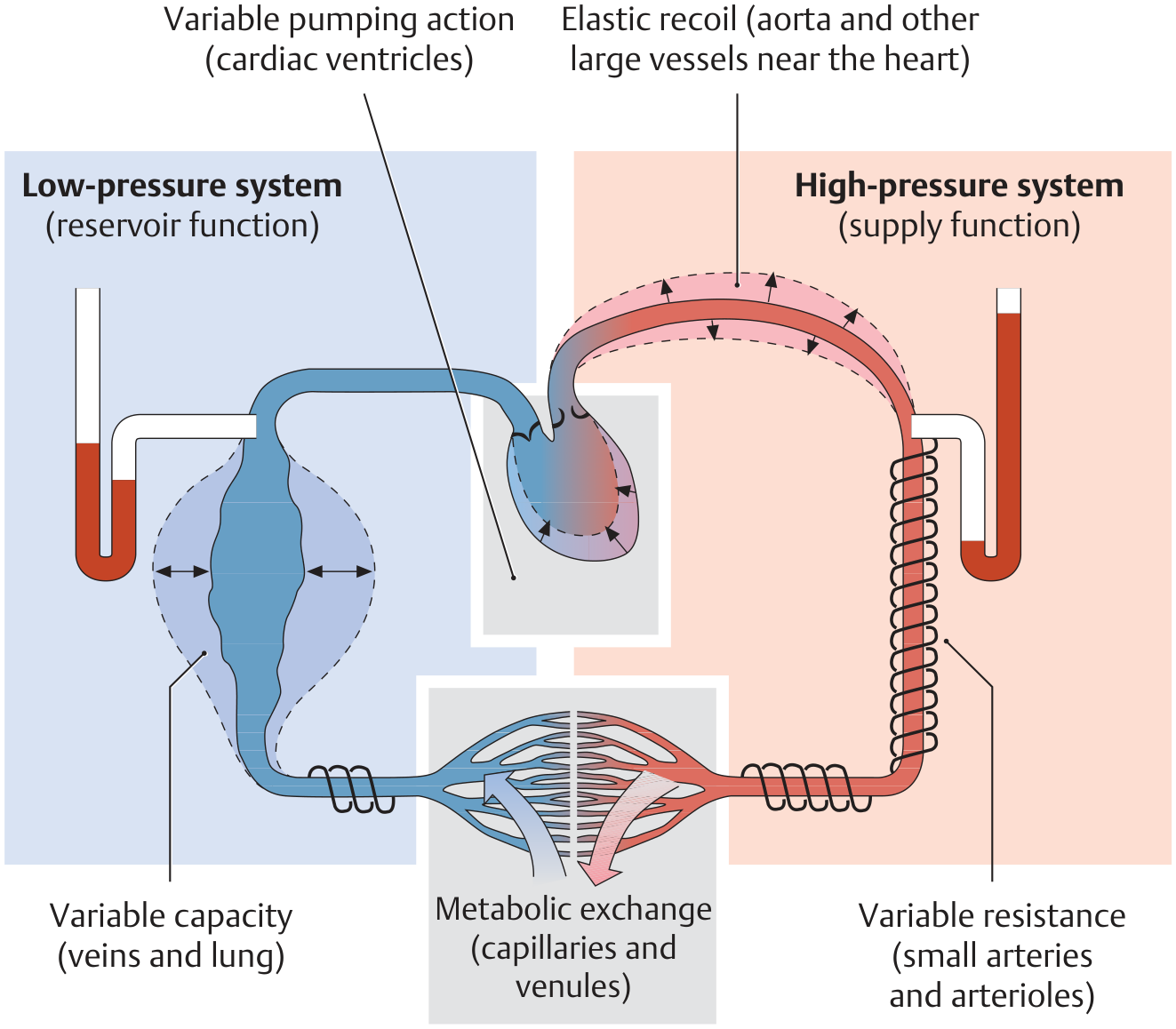
Fig. B - Schematic overview of the full circulatory system (figure-of-eight model):
Sources:
- General Anatomy and Musculoskeletal System, THIEME Atlas of Anatomy (G.A), Section 6.1 - Circulatory System and Lymphatic Vessels
- Color Atlas of Human Anatomy, Vol. 2 - Internal Organs, p. 30-31
Define circulatory system from General Anatomy text book
Finding Sources
Reading File
I now have the complete, verbatim text directly from the General Anatomy and Musculoskeletal System - THIEME Atlas of Anatomy. Here is the full definition:
Circulatory System
General Anatomy and Musculoskeletal System - THIEME Atlas of Anatomy
Chapter 6.1 - Overview of the Human Cardiovascular System
Definition
"Special circulatory organs are needed to transport and distribute the blood, ensuring that it is made accessible to all the cells in the body. These organs consist of the heart and vascular system (blood vessels and lymphatics)."
The circulatory system is a closed vascular system composed of:
- The heart - the central pump that maintains blood flow
- Blood vessels - arteries, capillaries, and veins
- The lymphatic system - running parallel to the venous system
Key rule on vessel naming (regardless of oxygen content):
- All vessels leading away from the heart = Arteries
- All vessels leading toward the heart = Veins
The exchange of gases, nutrients, and waste products takes place in the capillary region.
Two Main Functional Circuits
1. Pulmonary Circulation
Deoxygenated venous blood from the upper and lower body is returned through the superior and inferior vena cava → right atrium → right ventricle → pulmonary arteries → lungs, where it is oxygenated.
2. Systemic Circulation
Oxygen-enriched blood from the lungs returns through the pulmonary veins → left atrium → left ventricle → aorta → body tissues.
A special subdivision is the portal circulation: venous blood from the unpaired abdominal organs (stomach, bowel, pancreas, and spleen) is carried by the portal vein to the capillary bed of the liver before returning to the inferior vena cava via the hepatic veins. This ensures nutrient-rich blood from digestive organs undergoes filtering and metabolic processing in the liver first.
Functional Pressure Gradient
Blood moves through the circulatory system along a pressure gradient between two functional systems:
| System | Average Pressure | Role |
|---|---|---|
| Arterial high-pressure system | ~100 mmHg | Supply / distribution |
| Venous low-pressure system | < 20 mmHg | Reservoir / return |
The two systems meet at the capillary region, where metabolic exchange occurs.
- Elastic-type arteries (near heart, e.g., aorta) - expand during systole and recoil during diastole, converting the heart's intermittent pumping into continuous blood flow
- Muscular-type arteries (distant from heart) - actively vasodilate and vasoconstrict to regulate local blood flow and vascular resistance
- Veins (capacitance vessels) - accommodate approximately 80% of total blood volume, serving as a blood reservoir
Lymphatic System
The lymphatic system runs parallel to the venous system. It originates with blind-ended vessels in the capillary region, collects extracellular fluid deposited there, and returns it to the venous blood through lymphatic vessels. Lymph nodes are interspersed along these pathways to filter the lymph.
Diagrams
Fig. A - Basic functional diagram (High- vs. Low-pressure systems):
Fig. B - Schematic overview of systemic, pulmonary, and portal circulations:
Source: General Anatomy and Musculoskeletal System, THIEME Atlas of Anatomy - Section 6.1, Overview of the Human Cardiovascular System
It's too short
Finding Sources
Reading File
Reading File
Reading File
Reading File
Reading File
Reading File
Reading File
Reading File
Loading Image
Now I have comprehensive content covering all aspects of the circulatory system from the General Anatomy textbook. Here is the full, detailed answer:
The Circulatory System
General Anatomy and Musculoskeletal System - THIEME Atlas of Anatomy
Sections 6.1, 6.2, and 6.3
1. Definition and Overview
Special circulatory organs are needed to transport and distribute blood, ensuring that it is made accessible to all cells in the body. These organs consist of:
- The heart - the central pump
- The vascular system - blood vessels (arteries, capillaries, veins)
- The lymphatic system - running parallel to the venous system
The system of blood vessels consists of arteries, capillaries, and veins:
- Arteries carry blood from the heart and distribute it throughout the body
- Veins return blood to the heart
- Exchange of gases, nutrients, and waste products occurs in the capillary region
Important rule: All blood vessels leading away from the heart are called arteries, and all vessels leading toward the heart are called veins - regardless of their oxygen content. (For example, the umbilical vein carries oxygen-rich blood, yet is still called a vein because it leads toward the heart.)
Blood flow in this closed vascular system is maintained by the pumping action of the heart.
2. The Two Main Functional Circuits
Functionally, the circulatory system is divided into two main circuits:
A. Pulmonary Circulation
Deoxygenated venous blood from the upper and lower body regions is returned through the superior and inferior vena cava → right atrium → right ventricle → pulmonary arteries → lungs, where it receives oxygen.
B. Systemic Circulation
Oxygen-enriched blood from the lungs returns through the pulmonary veins → left atrium → left ventricle → aorta → body tissues.
A special part of the systemic circuit is the portal circulation, which includes two successive capillary beds. Before venous blood returns to the inferior vena cava from the capillary beds of the unpaired abdominal organs (stomach, bowel, pancreas, and spleen), it is carried by the portal vein to the capillary bed of the liver. This ensures that nutrient-rich blood from the digestive organs undergoes numerous filtering and metabolic processes in the liver before being returned to the inferior vena cava via the hepatic veins.
Schematically, postnatal human circulation can be illustrated as a figure-of-eight, with the heart at its intersection:
3. Functional Pressure Systems
Blood is transported through the circulatory system along a pressure gradient between two functional systems:
| System | Pressure | Role | Components |
|---|---|---|---|
| High-pressure system | ~100 mmHg average | Supply function | Arteries, arterioles |
| Low-pressure system | < 20 mmHg | Reservoir function | Veins, lungs |
The two systems meet in the capillary region of the terminal vascular bed, where metabolic exchange takes place.
- Elastic-type arteries (near the heart, e.g., the aorta): temporarily expand to accommodate ejected blood during systole; during diastole the vessel lumen undergoes elastic recoil, transforming the heart's intermittent pumping into continuous blood flow
- Muscular-type arteries (distant from the heart): can actively expand (vasodilation) and contract (vasoconstriction), providing effective control of vascular resistance and regulation of local blood flow
- Veins (capacitance vessels): accommodate approximately 80% of total blood volume and serve as a blood reservoir
4. Structure of Blood Vessels (Section 6.2)
The wall of every blood vessel is built from three fundamental layers:
Layer 1 - Tunica Intima (innermost)
- A layer of spindle-shaped endothelial cells aligned along the vessel axis, resting on a basement membrane and thin subendothelial connective tissue
- In muscular-type arteries, separated from the media by an internal elastic membrane
- Function: Exchange of gases, fluids, and substances through the vessel wall
Layer 2 - Tunica Media (middle)
- An approximately circular arrangement of smooth muscle cells, elastic and collagenous fibers, and proteoglycans
- Muscular-type arteries may have an external elastic membrane separating the media from the adventitia
- Function: Regulates blood flow (by contraction/relaxation of smooth muscle)
Layer 3 - Tunica Adventitia (outermost)
- Composed of longitudinally aligned connective tissue elements; veins may additionally contain smooth muscle here
- Contains autonomic nerves to the vessel wall muscle and, in larger vessels, vasa vasorum (vessels that supply the outer third of the vessel wall)
- Function: Integrates the blood vessel into its surrounding tissue
Note: While arteries have thick, densely packed smooth muscle layers in the media, veins contain far more connective tissue (collagenous and elastic fibers), giving them a looser structure. Veins also lack a conspicuous internal elastic membrane.
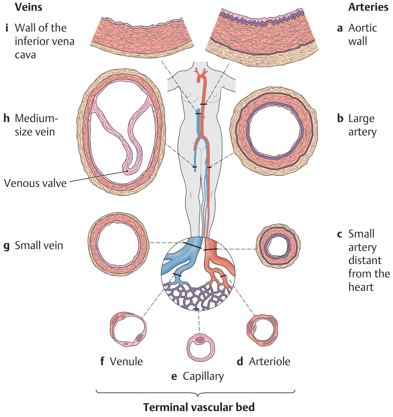
5. The Terminal Vascular Bed (Microcirculation - Section 6.3)
The terminal vascular bed is the site of microcirculation - where gases, fluids, and substances are actually exchanged between blood and body cells. It consists of:
- Afferent arterial limb - precapillary arterioles
- Capillary bed - the exchange zone
- Efferent venous limb - postcapillary venules
The Capillaries
The smallest vessels, capillaries (5-15 μm in diameter), consist only of an endothelial layer and a basal lamina, to which pericytes may be attached. Due to the extensive branching of vessels in the capillary bed:
- Total vascular cross-section increases approximately 800-fold compared to the aorta
- Blood flow velocity drops from 50 cm/s in the aorta to only 0.05 cm/s in capillaries
- With an average capillary length of 0.5 mm, approximately 1 second is available for metabolic exchange
Precapillary Sphincters
Precapillary sphincters (circular muscle cells) are located at the junction of metarterioles and capillaries and regulate blood flow within the capillary network. When they contract, branching capillaries are closed - under resting conditions, only about 25-35% of all capillaries are perfused. Arterioles and venules may also be interconnected by arteriovenous anastomoses (shunts).
Fluid Exchange in Capillaries (Starling Forces)
Fluid exchange between capillaries and surrounding tissue (interstitium) is regulated by a changing pressure gradient between:
- Hydrostatic pressure (blood pressure in capillaries) - pushes fluid out
- Colloid osmotic pressure (~25 mmHg) - pulls fluid in
At the arterial end: hydrostatic pressure = 35 mmHg > colloid osmotic pressure (25 mmHg) → fluid is filtered out into surrounding tissue.
At the venous end: hydrostatic pressure falls to ~15 mmHg < colloid osmotic pressure (25 mmHg) → fluid is reabsorbed back into the capillary.
Any excess fluid that is not reabsorbed is collected by the lymphatic capillaries and returned to the venous system.
Types of Capillary Endothelium
| Type | Structure | Location |
|---|---|---|
| Type I | Closed, no fenestrations, continuous basal lamina | Nervous system |
| Type II | Closed, with pinocytotic activity | Cardiac and skeletal muscle |
| Type III | Fenestrated by a diaphragm | Gastrointestinal tract |
| Type IV | Intercellular gaps, no continuous basal lamina | Liver |
6. Arterial and Venous Pressure in Different Positions
Changing from a recumbent to a standing position radically alters pressure relationships in the circulatory system. Hydrostatic effects cause:
- Pressure to rise sharply in the lower parts of the body
- Pressure to decrease in the upper body
- Approximately 500 mL of blood to shift into the lower limb veins
This rise in venous pressure greatly increases transmural pressure in the lower limb veins, while pressure in the head and neck veins may fall so low that veins collapse. The "hydrostatic indifference level" (just below the diaphragm) is the point where pressure remains unchanged.
7. Lymphatic System
The lymphatic system runs parallel to the venous system:
- Originates with blind-ended vessels in the capillary region
- Collects extracellular fluid deposited in the interstitium
- Returns it to the venous blood through progressively larger lymphatic vessels
- Lymph nodes are interspersed along these pathways to biologically filter the lymph
8. Early Embryonic Circulatory System (Section 1.6)
The cardiovascular system of a 3 to 4-week-old human embryo consists of a well-functioning two-chambered heart and three distinct circulatory systems:
- An intraembryonic systemic circulation (ventral and dorsal aorta, branchial arch and aortic arches, anterior and posterior cardinal veins)
- An extraembryonic vitelline circulation (omphalomesenteric arteries and veins)
- A placental circulation (umbilical arteries and veins)
The vascular pathways show a largely symmetrical arrangement at this stage. With further development, the original symmetry is lost in favor of a left-sided preponderance (e.g., the left fourth aortic arch becomes the definitive aortic arch).
Source: General Anatomy and Musculoskeletal System, THIEME Atlas of Anatomy - Sections 1.6, 6.1, 6.2, and 6.3
Nervous System From Gray's Deeper
Your credits are exhausted. Your daily credits reset tomorrow, or you can purchase a top-up for more.
This is a shared conversation. Sign in to Orris to start your own chat.