
https://youtu.be/FMttHoW2QiE?si=_nTSUZUhM_EXQPQG
analyse_media
This is a Dr. Najeeb Lectures video on Cell Membrane Transport (Cell Biology). Here's a full breakdown:
Cell Membrane Transport | Dr. Najeeb Lectures
Channel: Dr. Najeeb Lectures (2.45M subscribers) | Published: March 18, 2021 | Runtime: ~1h 25min
What the Lecture Covers
The video is a comprehensive whiteboard-style medical lecture on how substances cross the plasma membrane. Dr. Najeeb draws hand-colored diagrams in real time covering:
1. Passive Transport (no ATP required)
- Simple Diffusion - solutes move down their concentration gradient (high → low). Covered at 02:24.
- Facilitated Diffusion - passive movement aided by membrane channel or carrier proteins (e.g., glucose channels, sodium channels, ligand-gated channels). Covered at 16:17 onward.
- Voltage-gated sodium channels (19:55)
- Ligand-operated/gated channels (27:08)
- Transporter/carrier proteins (32:27)
2. Active Transport (ATP required)
- Primary Active Transport - uses ATP directly. Examples: Na⁺/K⁺ ATPase pump, Ca²⁺ transporters (39:16 onward).
- Secondary Active Transport - exploits electrochemical gradients established by primary pumps. Examples: sodium-glucose co-transporter (SGLT), counter-transport. Covered at 42:46.
3. Vesicular Transport (Bulk Transport)
- Endocytosis (bringing material IN):
- Pinocytosis - cell "drinking" (1:14:55)
- Phagocytosis - cell "eating," e.g., macrophages (1:16:40)
- Receptor-mediated endocytosis - e.g., LDL uptake in liver cells (1:17:53)
- Exocytosis - expelling material OUT of the cell (1:23:25)
Key Concepts at a Glance
| Mechanism | Energy | Direction | Example |
|---|---|---|---|
| Simple Diffusion | None | High → Low | O₂, CO₂, lipids |
| Facilitated Diffusion | None | High → Low | Glucose (GLUT), ions via channels |
| Primary Active Transport | ATP | Against gradient | Na⁺/K⁺ pump |
| Secondary Active Transport | Gradient (indirect ATP) | Against gradient | SGLT1/2 (Na⁺-glucose co-transport) |
| Endocytosis | ATP | Into cell | Phagocytosis, pinocytosis |
| Exocytosis | ATP | Out of cell | Neurotransmitter release |
Teaching Style
Dr. Najeeb uses a large whiteboard with multi-colored hand-drawn diagrams - lipid bilayers, ion gradients, receptor proteins, and vesicles are all illustrated live. This is his signature style, widely regarded as excellent for visual learners in medical education.
Full access to 800+ exclusive videos is available at DrNajeebLectures.com for a one-time fee.
Make notes from the above video for my exams
Reading File
Finding Sources
Finding Sources
Reading File
Reading File
Finding Sources
Finding Sources
Finding Sources
Reading File
Reading File
Reading File
Finding Sources
Reading File
Finding Sources
Finding Sources
Reading File
Loading Image
Loading Image
Here are your complete exam notes on Cell Membrane Transport, built from the Dr. Najeeb lecture and backed by Guyton & Hall, Histology (Ross), and other authoritative textbooks.
Cell Membrane Transport - Exam Notes
Based on Dr. Najeeb Lectures | Sources: Guyton & Hall, Ross Histology, Goodman & Gilman
1. THE CELL MEMBRANE - Overview
- The plasma membrane is a phospholipid bilayer that acts as a selective barrier between the intracellular and extracellular environments.
- It regulates homeostasis: allows nutrients in, keeps wastes out, and blocks harmful substances.
- Key rule: The lipid bilayer is a natural barrier to most polar/charged molecules - they need help to get through.
2. OVERVIEW OF TRANSPORT TYPES
| Type | Energy? | Direction | Protein needed? |
|---|---|---|---|
| Simple Diffusion | No | High → Low | No |
| Facilitated Diffusion | No | High → Low | Yes (channel or carrier) |
| Primary Active Transport | Yes (ATP directly) | Against gradient | Yes (pump) |
| Secondary Active Transport | Yes (indirect, gradient-driven) | Against gradient | Yes (cotransporter) |
| Vesicular Transport | Yes (ATP) | Bulk, both ways | No - uses vesicle budding |
3. PASSIVE TRANSPORT (No ATP)
3a. Simple Diffusion
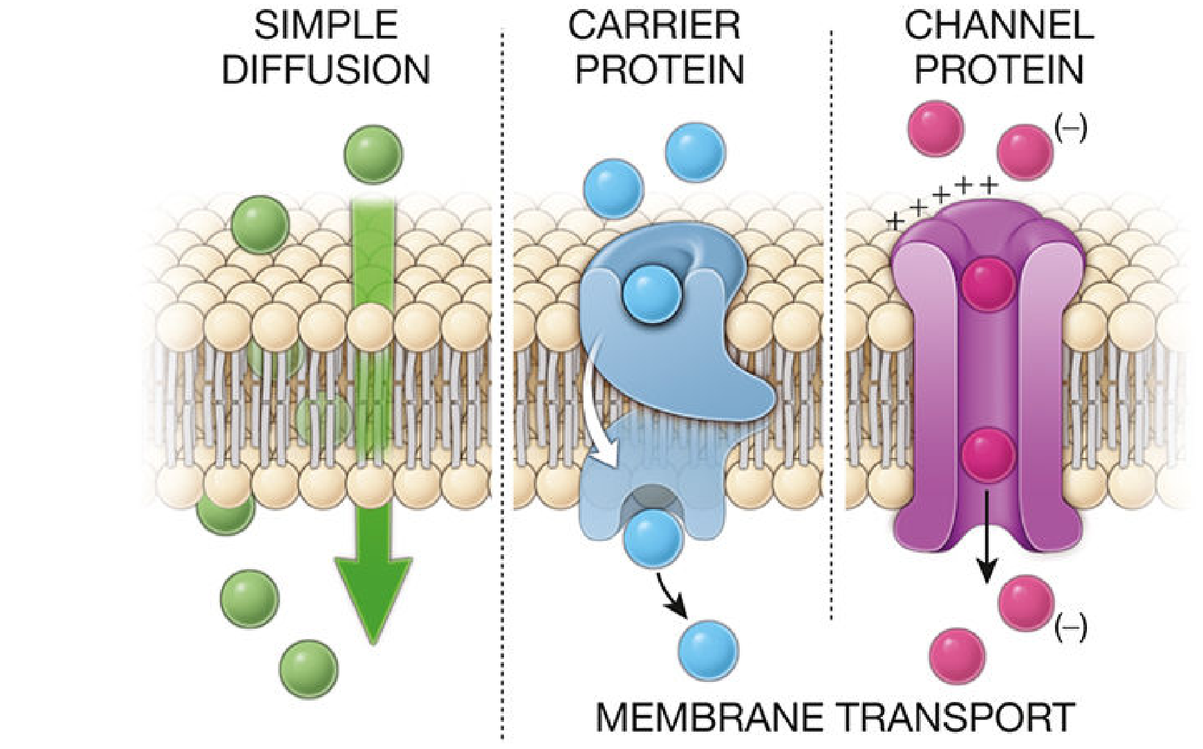
- Movement of molecules down their concentration gradient (high → low) with no energy and no protein needed.
- Driven purely by random kinetic motion (Brownian motion).
What can cross by simple diffusion?
- Small, lipid-soluble molecules: O₂, CO₂, N₂, alcohols, steroid hormones, fat-soluble vitamins (A, D, E, K)
- O₂ and CO₂ are the classic examples - they dissolve directly in the lipid bilayer.
Rate of diffusion is proportional to:
- Concentration difference across membrane: Net diffusion ∝ (C_outside - C_inside)
- Lipid solubility of the substance
- Size and number of membrane pores
- Temperature
Key fact: Simple diffusion shows a linear relationship - the higher the concentration gradient, the faster the diffusion, with no maximum rate (unlike facilitated diffusion). (Guyton & Hall, p.65)
3b. Facilitated Diffusion
Still passive (no ATP), still moves down the concentration gradient, but requires a membrane protein.
Two types of proteins are involved:
i. Channel Proteins (Ion Channels)
- Form hydrophilic pores/tunnels through the membrane.
- Contain a pore domain (ion-selectivity filter) - regulates which ions pass.
- Transport is fast (ions flow freely when open).
Types of ion channels (by gating mechanism):
| Channel Type | Opens in Response To | Example |
|---|---|---|
| Voltage-gated | Change in membrane potential | Na⁺ channels in neurons (action potential) |
| Ligand-gated | Binding of a chemical (ligand) | Acetylcholine receptor at NMJ |
| Mechanically-gated | Physical stretch/pressure | Channels in inner ear hair cells |
- Aquaporins - special channel proteins for water. At least 13 types in mammals. Aquaporin-2 density is regulated by ADH (antidiuretic hormone). Water diffuses through them in single file, so fast that the red blood cell membrane transports 100x its own volume per second. (Guyton & Hall, p.65)
ii. Carrier Proteins
- Bind a specific molecule, then undergo conformational change to shuttle it across.
- Highly selective - usually one type of molecule per carrier.
- Show a maximum transport rate (Vmax) - at high concentrations, carriers become saturated and rate plateaus. This is the key difference from simple diffusion.
- Examples: GLUT transporters (glucose into cells), amino acid carriers.
Glucose transport (GLUT family):
- GLUT transporters are carrier proteins - not Na⁺-dependent - facilitate diffusion of glucose into most cells.
- In the gut and kidney, glucose is absorbed via SGLT (active transport - see below).
Facilitated diffusion vs. simple diffusion (important exam point):
- Simple diffusion: rate keeps increasing linearly with concentration.
- Facilitated diffusion: rate plateaus at Vmax (carrier saturation).
4. ACTIVE TRANSPORT (Requires Energy)
Moves substances against their concentration/electrochemical gradient. Requires ATP.
4a. Primary Active Transport
- Uses ATP directly to drive the pump.
- Most important example: Na⁺/K⁺ ATPase pump (sodium-potassium pump).
Na⁺/K⁺ ATPase - The Master Pump
- Location: Basolateral membrane of most cells.
- Action: Pumps 3 Na⁺ OUT and 2 K⁺ IN per cycle, using 1 ATP.
- Net result: Creates low intracellular Na⁺ (12 mEq/L inside vs. 140 mEq/L outside) and a negative intracellular charge (-70 mV).
- This creates the electrochemical gradient exploited by secondary active transport.
Other primary active transporters:
- Ca²⁺-ATPase (SERCA pump): pumps Ca²⁺ into the ER or out of the cell. Keeps cytoplasmic Ca²⁺ very low (~10⁻⁷ mol/L).
- H⁺/K⁺ ATPase: in stomach parietal cells - secretes gastric acid. Blocked by PPIs (omeprazole, pantoprazole).
4b. Secondary Active Transport
- Does not use ATP directly.
- Uses the electrochemical gradient of Na⁺ (created by the Na⁺/K⁺ pump) to drive a second substance against its gradient.
- So it indirectly depends on ATP (because the pump that made the gradient used ATP).
Two subtypes:
| Subtype | Direction of co-transported substance | Example |
|---|---|---|
| Co-transport (Symport) | Na⁺ and cargo move in the same direction | SGLT1/2 (Na⁺-glucose), Na⁺-amino acid cotransporters |
| Counter-transport (Antiport) | Na⁺ and cargo move in opposite directions | Na⁺/H⁺ exchanger |
SGLT (Sodium-Glucose Co-Transporter) - High Yield!
- SGLT1 - in small intestine (absorbs glucose from gut lumen) and kidney proximal tubule (late segment).
- SGLT2 - in kidney proximal tubule (early segment; reabsorbs ~90% of filtered glucose).
- Mechanism: Na⁺ flows down its gradient INTO the cell, dragging glucose against its gradient into the cell simultaneously.
- Clinical relevance: SGLT2 inhibitors (gliflozins - dapagliflozin, empagliflozin) block this transporter → glucosuria → lower blood glucose in Type 2 DM. Also have cardioprotective and renoprotective effects.
(Guyton & Hall, p.68; Brenner & Rector's The Kidney)
5. VESICULAR TRANSPORT (Bulk Transport)
Used for large molecules or bulk quantities of material that cannot cross through protein channels. Requires energy (ATP).
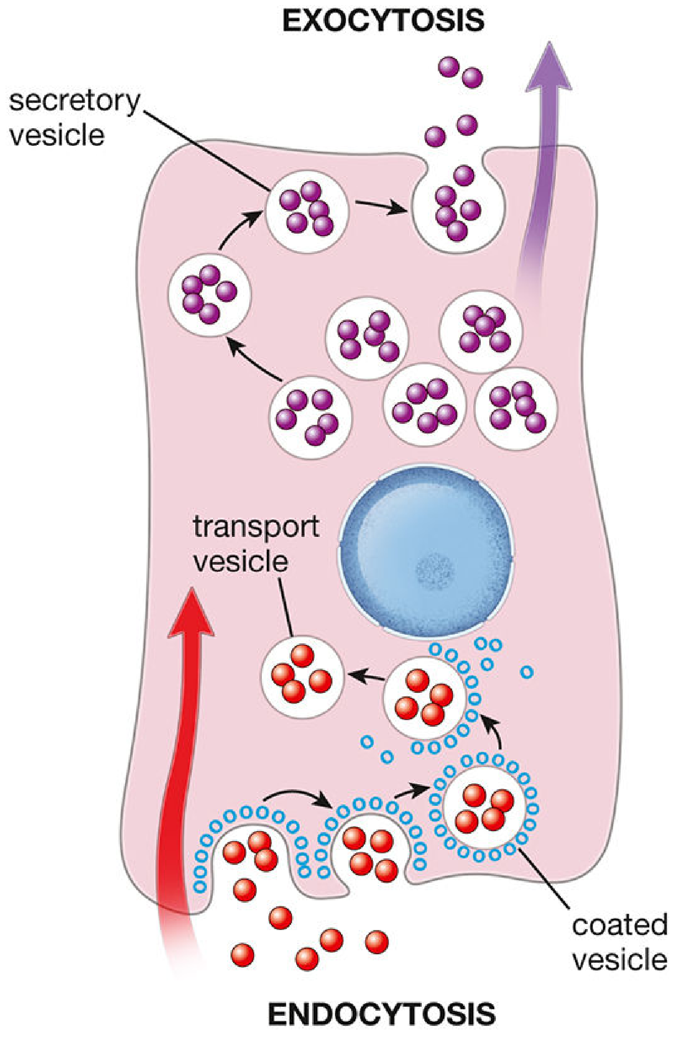
Key principle: Exocytosis and endocytosis are coupled - if exocytosis is blocked (e.g., by tetanus or botulinum toxin), endocytosis is also blocked. SNARE proteins mediate both. (Ross Histology, p.145)
5a. Endocytosis (INTO the cell)
General term for vesicular transport bringing material inside the cell.
Three Major Types:
1. Pinocytosis ("cell drinking")
-
Micropinocytosis - nonspecific ingestion of extracellular fluid and small proteins via vesicles < 150 nm.
- Constitutive process (happens continuously).
- Uses caveolin and flotillin proteins in lipid rafts.
- Clathrin-independent and actin-independent.
- Especially prominent in: endothelial cells of blood vessels, smooth muscle cells.
-
Macropinocytosis - non-specific uptake of large volumes of extracellular fluid.
- Actin-dependent - actin cytoskeleton rearranges to form membrane ruffles.
- Vesicles (macropinosomes) are large - enter cytoplasm and fuse with lysosomes.
2. Phagocytosis ("cell eating")
- Ingestion of large particles - bacteria, dead cells, debris.
- Performed by specialized cells: macrophages, neutrophils, dendritic cells.
- Requires actin cytoskeleton remodeling.
- Particles are engulfed into a phagosome, which fuses with a lysosome → phagolysosome → enzymatic degradation.
- This is an immune defense mechanism.
3. Receptor-Mediated Endocytosis (Clathrin-Dependent)
- Most specific form of endocytosis.
- Cell surface receptors bind specific ligands (e.g., LDL, transferrin, insulin, hormones).
- The receptor-ligand complex clusters at clathrin-coated pits on the plasma membrane.
- The pit invaginates and pinches off as a clathrin-coated vesicle.
- Inside the cell: vesicle loses its clathrin coat → fuses with early endosome.
- The ligand is degraded in lysosomes; the receptor is often recycled back to the surface.
- Classic example: LDL receptor uptake in liver cells (LDL carries cholesterol). Mutation in LDL receptor → familial hypercholesterolemia.
5b. Exocytosis (OUT of the cell)
- Secretory vesicles (from Golgi apparatus) move to the plasma membrane and fuse with it, releasing cargo into the extracellular space.
- Examples: neurotransmitter release from synaptic vesicles, hormone secretion, mucus release.
- After exocytosis, vesicular membrane is retrieved by endocytosis (prevents cell from swelling).
6. QUICK SUMMARY TABLE - High Yield for Exams
| Mechanism | Protein Needed | Energy | Examples |
|---|---|---|---|
| Simple diffusion | No | No | O₂, CO₂, steroids, ethanol |
| Facilitated diffusion (channel) | Yes - channel | No | Na⁺, K⁺, Cl⁻ via ion channels; H₂O via aquaporins |
| Facilitated diffusion (carrier) | Yes - carrier | No | GLUT1-5 (glucose), amino acids |
| Primary active transport | Yes - pump | Yes (ATP) | Na⁺/K⁺ ATPase, Ca²⁺ pump, H⁺/K⁺ ATPase |
| Secondary active transport | Yes - cotransporter | Yes (indirect) | SGLT1/2 (Na-glucose), Na⁺/H⁺ exchanger |
| Pinocytosis | No | Yes | Endothelial cells absorbing fluid |
| Phagocytosis | No | Yes | Macrophages eating bacteria |
| Receptor-mediated endocytosis | Yes - receptor | Yes | LDL uptake, transferrin, insulin |
| Exocytosis | No | Yes | Neurotransmitter release, hormone secretion |
7. HIGH-YIELD EXAM POINTS
- Simple vs. Facilitated diffusion: Simple = linear rate increase; Facilitated = Vmax saturation.
- Na⁺/K⁺ ATPase: Pumps 3 Na⁺ OUT, 2 K⁺ IN per ATP. Creates -70 mV inside cell.
- SGLT2 inhibitors (e.g., dapagliflozin, empagliflozin) - used in T2DM. Cause glucosuria.
- Aquaporins - channel proteins for water only. At least 13 types. AQP2 regulated by ADH.
- Clathrin - coats endocytic vesicles in receptor-mediated endocytosis.
- Caveolin/Flotillin - proteins in micropinocytosis (clathrin-independent).
- Exo- and endo-cytosis are coupled: tetanus/botulinum toxin blocks both via SNARE proteins.
- Lipid-soluble substances cross freely through the bilayer (no help needed).
- Carrier proteins show Vmax (saturation kinetics); channels do not.
- Secondary active transport does not use ATP directly - it uses the Na⁺ gradient built by the Na⁺/K⁺ pump.
Sources: Guyton and Hall Textbook of Medical Physiology (14e); Histology: A Text and Atlas with Correlated Cell and Molecular Biology (Ross, 8e); Goodman & Gilman's The Pharmacological Basis of Therapeutics; Brenner & Rector's The Kidney
This is a shared conversation. Sign in to Orris to start your own chat.