
Psoriasis causes and treatment and management
psoriasis treatment
psoriasis causes treatment management guidelines 2026
plaque psoriasis skin lesion clinical photo
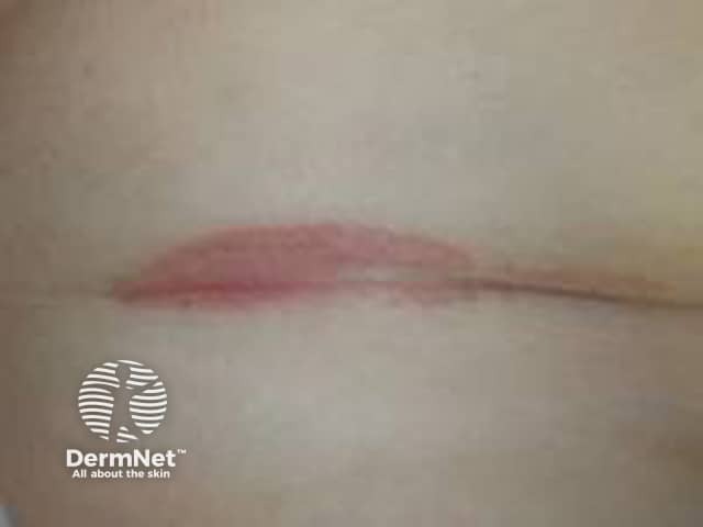
Clinical photography of the antecubital fossa region shows a single erythematous plaque within a flexural skin fold. The lesion is well-demarcated, with a smooth pink-to-erythematous surface and minimal scales evident, reflecting inverse/ flexural psoriasis features in moist environments where scaling is less conspicuous. The plaque lies centrally within the fold, with intact surrounding skin and no obvious crusting or excoriation. Lighting emphasizes a uniform coloration without effusion or ulceration. The regional skin may appear slightly edematous or glistening from moisture, a common appearance in intertriginous psoriasis. In clinical practice, such lesions are often symmetric or multipart and may co-occur with plaques on extensor surfaces; history of chronic plaque psoriasis or family history supports the diagnosis. Differential considerations include irritant dermatitis, candidal intertrigo, or seborrheic dermatitis; biopsy would reveal epidermal hyperplasia with parakeratosis and neutrophilic microabscesses typical of plaque psoriasis, though histology is not demonstrated here. The image is intended for dermatology education, aiding recognition of flexural psoriasis, differentiation from dermatitis, and guiding topical therapy, including high-potency corticosteroids or vitamin D analogs with occlusion, while avoiding excessive moisture. This photo supports image-based teaching, clinical pattern recognition, and correlation with patient history in practice and research.
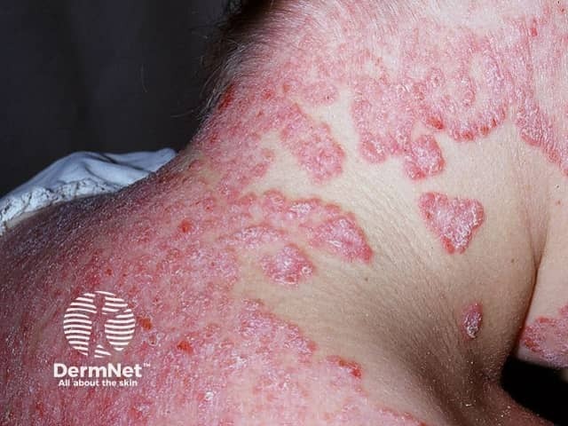
Clinical photography of facial skin demonstrates classic psoriatic plaques involving the malar region and surrounding cheeks. The lesions are erythematous to violaceous, well-demarcated, and markedly scaly with silvery-white scale, consistent with plaque-type psoriasis (psoriasis vulgaris). Involved areas show surface coarseness, thickening (acanthosis) and mild induration, while adjacent skin remains relatively spared. Perilesional inflammation creates a ring of erythema, and some plaques exhibit superficial fissuring. The distribution is facial rather than scalp, trunk, or limbs, suggesting involvement limited to the head-and-face region in this image. No vesiculation or purulence is evident. The appearance may mimic seborrheic dermatitis, eczema, or contact dermatitis; however, the well-circumscribed plaques with thick scale and the typical red-silver presentation strongly favor psoriasis. Skin photo shows high-contrast textural differences between lesional and non-lesional skin. Clinically, facial psoriasis can be challenging due to cosmetic and functional concerns, including itching and social impact. This image supports differential diagnosis discussions in dermatology education and serves as a reference for recognizing facial plaque psoriasis, assessing severity, and guiding treatment planning (topical steroids, vitamin D analogs, or phototherapy). It is useful for teaching photodocumentation, lesion morphology, and pattern recognition in psoriasis spectrum. This image thus enhances reasoning for diagnosis, prognosis, and management planning.
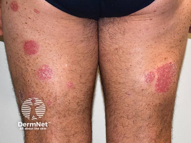
Clinical photography of bilateral lower legs demonstrates features typical of plaque psoriasis. Modality: Clinical photograph; technique: Standard color digital image captured in visible light without dermoscopic magnification or contact; staining and contrast agents not used. Anatomical localization: skin of the legs, pretibial and extensor regions, bilateral (anterior aspects). Visual impression: well-circumscribed, erythematous plaques with diffuse or loosely adherent silvery scales; plaques are raised with well-defined borders and variable size, some larger patches measuring several centimeters in diameter. The distribution is symmetric and predominantly on extensor surfaces, consistent with plaque psoriasis vulgaris; surrounding skin may show mild xerosis or epidermal scaling. Pathology cannot be confirmed from a photo alone, but clinical correlation with history and examination would favor psoriasis; differential diagnoses include eczema/atopic dermatitis, tinea corporis, pityriasis rosea, and nummular dermatitis. Clinical significance: this image aids diagnosis, patient education, and monitoring of treatment response over time; it supports documentation for dermatology records and can be used in educational or research contexts to illustrate chronic plaque psoriasis phenotype. Potential clinical use cases: baseline assessment, response evaluation to topical therapies, phototherapy planning, and AI-driven lesion recognition datasets. Ethical note: ensure patient consent for image use in training datasets.
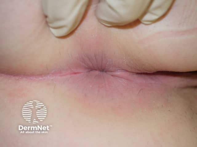
Clinical photograph of external genital skin showing an erythematous, well-demarcated plaque in an intertriginous fold. The lesion is pink to red, with a smooth surface and minimal peripheral scaling within moist skin areas; a slight fissure is seen along the crease. The surrounding skin is mildly inflamed but otherwise unremarkable. This appearance is characteristic of genital psoriasis, most compatible with inverse psoriasis in moist genital folds where scaling is less prominent than on extensor surfaces. Differential diagnoses include candidal balanitis or vulvitis, irritant contact dermatitis, lichen sclerosus, eczema, and less commonly erosive lesions. Clinical context such as past or concurrent psoriasis elsewhere, absence of itch or burning, and response to topical steroids can help confirmation. Histopathology would typically show epidermal hyperplasia with parakeratosis, regular elongation of rete ridges, and a dermal perivascular lymphocytic infiltrate if biopsy were performed; in practice, biopsy is not required for typical genital psoriasis. The diagnostic significance lies in recognizing genito-pubic psoriasis to guide therapy and avoid misdiagnosis as infection. This image is useful for education on genital psoriasis recognition, differential diagnosis, and management considerations including topical corticosteroids, vitamin D analogs, and calcineurin inhibitors, with referrals for systemic therapy if extensive disease. Clinical photo for reference.
Psoriasis: Causes, Treatment, and Management
What it is
Causes / Pathogenesis
- Strong heritable component: if both parents have psoriasis, a child's risk is up to 50%; with one affected parent, about 16% (Textbook of Family Medicine, p. 928).
- The PSORS1 locus and multiple genome-wide association study (GWAS) loci contribute to risk, though pinning down the exact causal genes within these regions remains difficult (Dermatology 2-Volume Set 5e).
- Psoriasis involves both the innate and adaptive immune system plus abnormal keratinocyte proliferation. Dendritic cells activate T cells, particularly Th17 cells, which are central to disease (Goldman-Cecil Medicine; Fitzpatrick's Dermatology).
- Key cytokine axis: dendritic cells produce IL-23 (which drives Th17 development) and TNF-α. Th17 cells then produce IL-17A, which acts directly on keratinocytes to drive the hyperproliferative, inflamed epidermis characteristic of psoriatic plaques. This IL-23/Th17/IL-17 axis is now the primary target of modern biologic therapy (Fitzpatrick's Dermatology, p. 227).
- Histologically: markedly thickened epidermis with rapid keratinocyte turnover, abnormal differentiation, neutrophilic/lymphocytic infiltrates, and prominent dilated capillaries reaching close to the skin surface.
- Infections - classically streptococcal pharyngitis, which frequently precedes guttate psoriasis in children (Textbook of Family Medicine, p. 928)
- Skin trauma (Koebner phenomenon)
- Withdrawal of systemic corticosteroids - can provoke pustular psoriasis
- Psychological/emotional stress, alcohol, smoking
- Certain drugs (lithium, beta-blockers, antimalarials)
- Cold, dry weather
Clinical Types
Treatment and Management
- Topical corticosteroids - mainstay for limited disease, often combined with vitamin D analogues for added efficacy
- Vitamin D3 analogues (calcipotriene/calcipotriol, calcitriol, tacalcitol) - normalize keratinocyte proliferation/differentiation
- Topical retinoids (tazarotene), anthralin, tar preparations
- Topical calcineurin inhibitors for sensitive areas (face, flexures, genitals)
- Phototherapy: narrowband UVB is highly cost-effective for widespread disease; combining topical agents (e.g., calcipotriol) with UVB improves and speeds clearance
- Conventional systemics:
- Methotrexate - the systemic agent against which others are benchmarked
- Cyclosporine - rapid onset but not suitable for long-term use due to toxicity
- Oral retinoids (acitretin)
- Biologic agents (targeted immunomodulators) - produce the most dramatic responses in appropriate candidates:
- TNF-α inhibitors: infliximab, adalimumab, etanercept
- IL-17A antagonists (e.g., secukinumab, ixekizumab, bimekizumab) - among the most effective agents available, given IL-17A's central pathogenic role
- IL-23 inhibitors (e.g., guselkumab, risankizumab) - target the upstream driver of the Th17 pathway
- Newer oral IL-23 receptor antagonists (e.g., icotrokinra) have recently expanded oral options for moderate-to-severe disease
- Guttate psoriasis is often self-limited (resolves in 6-12 weeks) and may not need treatment in mild cases; widespread cases benefit from UVB plus topical therapy.
- Antistreptococcal treatment (antibiotics) for strep-triggered guttate psoriasis has been studied but evidence for benefit remains limited (recent Cochrane/systematic review data).
- Psoriatic arthritis - joint symptoms warrant rheumatology referral
- Cardiovascular risk - blood pressure, lipids, fasting glucose at least annually
- Metabolic syndrome, obesity, insulin resistance
- Depression/anxiety and quality-of-life impact
- Smoking and alcohol counseling
- Screening for lymphoma and skin cancer risk in patients on immunosuppressants or phototherapy