
Achalasia cardia to gry full marks in mbbs physiology university exams
achalasia bird's beak barium swallow esophagus
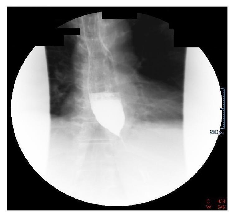
A diagnostic fluoroscopic image of a barium swallow (esophagram) demonstrating classic radiologic features of achalasia or pseudoachalasia. The image shows a markedly dilated thoracic esophagus filled with radiopaque barium contrast. There is a characteristic abrupt, symmetrical tapering of the distal esophagus at the gastroesophageal junction, creating the diagnostic 'bird's beak' appearance. Significant barium column stasis is visible proximal to the lower esophageal sphincter, indicating functional or mechanical obstruction. Minimal contrast is seen passing into the stomach, which is partially visualized in the lower quadrant. This finding is highly suggestive of impaired lower esophageal sphincter relaxation or extrinsic compression, correlating clinically with symptoms such as progressive dysphagia and weight loss.
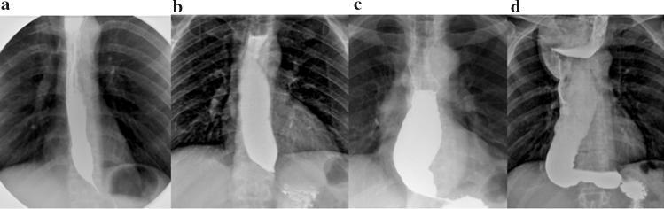
A series of four diagnostic X-ray radiography images (a-d) showing a barium swallow study illustrating the progressive stages of achalasia. All images demonstrate a 'bird’s beak' appearance at the gastroesophageal junction (EGJ) due to distal esophageal narrowing and impaired relaxation. Stage I (a) shows a straight esophagus with a maximum diameter ≤4 cm. Stage II (b) exhibits moderate dilation between 4–6 cm while maintaining a straight vertical axis. Stage III (c) demonstrates significant dilation ≥6 cm but remains relatively straight. Stage IV (d) represents end-stage disease with marked dilation (≥6 cm) and a characteristic 'sigmoid' or S-shaped configuration, indicating advanced esophageal tortuosity and loss of longitudinal muscle tone. The contrast distribution shows increasing proximal pooling and stasis as the disease progresses from Stage I to Stage IV, illustrating the severity of functional obstruction and esophageal decompensation.
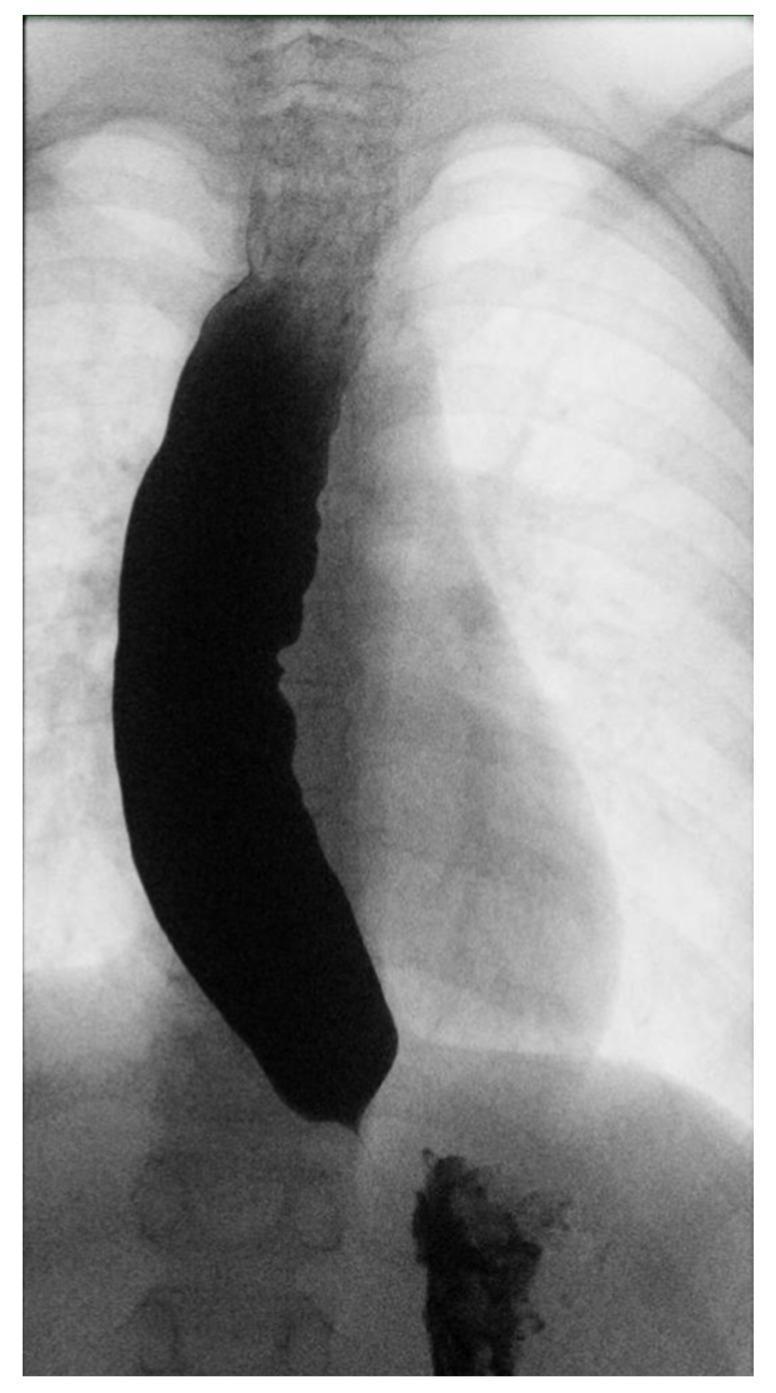
Diagnostic barium swallow radiograph (esophagogram) of the chest and upper abdomen demonstrating classic radiographic features of achalasia. The primary finding is a markedly dilated and tortuous esophagus, which is opacified by a thick column of barium contrast. The esophageal body shows significant proximal expansion with irregular, undulating contours. Distally, the esophagus tapers sharply at the gastroesophageal junction, creating a pathognomonic 'bird's beak' appearance, indicative of a failure of the lower esophageal sphincter (LES) to relax. There is significant stasis of contrast material within the esophageal lumen, with only a minimal, trickle amount of barium observed passing into the stomach. The image illustrates impaired esophageal motility and delayed emptying, which are key clinical hallmarks of esophageal achalasia. This imaging is essential for students and clinicians studying swallowing disorders and motility pathologies in gastroenterology.
Achalasia Cardia - Complete MBBS Physiology Answer (Full Marks)
DEFINITION
- Failure of relaxation of the Lower Esophageal Sphincter (LES) during swallowing
- Absence of peristalsis (aperistalsis) in the smooth muscle esophagus
- Harrison's Principles of Internal Medicine 22E, p. 738
NORMAL PHYSIOLOGY OF SWALLOWING (Background context - earns marks)
- The LES is normally maintained in a state of tonic contraction (resting pressure 15-30 mmHg)
- With each swallow, the inhibitory neurons (nitrergic/VIPergic) of the myenteric (Auerbach) plexus release Nitric Oxide (NO) and VIP → cause LES relaxation
- Simultaneously, a peristaltic wave propagates down the esophagus from pharynx to LES
- This coordinated relaxation allows the bolus to pass into the stomach
ETIOLOGY & PATHOPHYSIOLOGY (The key physiology section)
Primary Cause: Degeneration of Myenteric (Auerbach) Plexus
| Neuron Type | Mediator | Normal Function | Effect of Loss |
|---|---|---|---|
| Inhibitory neurons | NO + VIP | Deglutitive LES relaxation, peristalsis sequencing | LES fails to relax; aperistalsis |
| Excitatory neurons | ACh (cholinergic) | LES tone, esophageal contraction | Partially preserved → unopposed tonic contraction of LES |
- Elevated resting LES pressure
- No LES relaxation on swallowing
- No propagating peristalsis
Cause of Ganglion Cell Degeneration
- Autoimmune process in genetically susceptible individuals
- Triggered by latent HSV-1 (Herpes Simplex Virus-1) infection
- Myenteric plexus is infiltrated by cytotoxic T lymphocytes
- Antibodies against myenteric neurons detectable in serum
- Associated with specific HLA alleles
Why CCK Paradoxically Increases LES Pressure in Achalasia
- Normally: CCK stimulates inhibitory neurons → LES relaxation
- In achalasia: inhibitory neurons are absent → CCK acts unopposed on smooth muscle → LES pressure paradoxically rises (diagnostic clue)
TYPES OF ACHALASIA (Chicago Classification on HRM)
| Type | Manometric Feature | Prognosis |
|---|---|---|
| Type I (Classic) | Aperistalsis + minimal pressurization | Moderate response to treatment |
| Type II | Aperistalsis + panesophageal pressurization | Best response (~100% to pneumatic dilation) |
| Type III (Spastic) | Premature/spastic contractions + failed LES relaxation | Poorest response to dilation; better with myotomy |
CLINICAL FEATURES
- Progressive dysphagia - both solids AND liquids (unlike mechanical obstruction which is solids first)
- Regurgitation of undigested, nonacid, nonbilious food (retained in esophagus for hours/days)
- Chest pain - squeezing, retrosternal; present in ~2/3 early in course; resembles angina
- Weight loss
- Heartburn (paradoxical - due to bacterial fermentation of retained food, NOT true reflux)
- Aspiration pneumonia/lung abscess - in advanced disease (10% of cases)
- Halitosis and hiccups
INVESTIGATIONS
1. Barium Swallow (Best initial test)
- "Bird's beak" / "Rat tail" appearance - dilated esophagus tapering symmetrically at LES
- Loss of primary peristalsis in distal 2/3 (smooth muscle segment)
- Air-fluid level in esophagus
- Progressive esophageal dilatation → sigmoid deformity in advanced disease
2. Esophageal Manometry (Gold Standard - Confirms diagnosis)
- Impaired/absent LES relaxation on swallowing (integrated relaxation pressure > 15 mmHg)
- Aperistalsis in smooth muscle esophagus
- Elevated resting LES pressure (often > 35 mmHg)
- High-resolution manometry (HRM) allows subtyping (Type I, II, III)
3. Endoscopy (OGD - Mandatory to exclude pseudoachalasia)
- Dilated esophagus, retained food
- LES closed but passable with gentle pressure (unlike malignancy)
- Exclude carcinoma of gastric cardia mimicking achalasia ("pseudoachalasia" = up to 5% of suspected cases)
DIFFERENTIAL DIAGNOSIS
| Condition | Distinguishing Feature |
|---|---|
| Pseudoachalasia (carcinoma gastric cardia) | Age >60, abrupt onset <1 year, marked weight loss; CT/EUS needed |
| Chagas disease | Trypanosoma cruzi; endemic in South America; identical manometry |
| Distal Esophageal Spasm (DES) | Intermittent dysphagia; LES relaxes normally; "corkscrew" esophagus |
| Opioid-induced dysmotility | Drug history; resolves on stopping opioids |
TREATMENT
1. Pharmacological (Temporizing measures - not very effective)
- Calcium channel blockers (nifedipine) - relax smooth muscle
- Nitrates (isosorbide dinitrate) - NO donor → LES relaxation
- Botulinum toxin injection - blocks ACh release from excitatory neurons at LES; effective in elderly/poor surgical candidates; effect lasts 6-12 months
2. Pneumatic (Balloon) Dilation
- Forceful dilation of LES disrupts circular muscle fibers
- 90% effective, especially Type II achalasia (100% efficacy)
- Risk: esophageal perforation (~1%)
3. Surgical: Heller's Cardiomyotomy
- Laparoscopic division of LES muscle fibers (myotomy)
- ~90% efficacy; combined with partial fundoplication (Dor or Toupet) to prevent GERD
- Preferred for Type III achalasia
4. POEM (Per-Oral Endoscopic Myotomy)
- Endoscopic myotomy via submucosal tunnel; latest modality
- Success rates >90%; increasingly preferred
COMPLICATIONS
- Aspiration pneumonia / lung abscess
- Esophageal carcinoma - both squamous cell carcinoma AND adenocarcinoma risk significantly elevated in achalasia patients (long-term)
- Megaesophagus / sigmoid esophagus (end-stage)
- Nutritional deficiency / weight loss
HIGH-YIELD EXAM POINTS (Rapid Recall)
| Point | Fact |
|---|---|
| Defect | Loss of inhibitory neurons (NO/VIP) in Auerbach plexus |
| LES in achalasia | Fails to relax (high resting pressure + impaired deglutitive relaxation) |
| Barium sign | "Bird's beak" / "Rat tail" appearance |
| Gold standard investigation | Esophageal manometry |
| CCK effect | Paradoxically increases LES pressure (inhibitory neurons absent) |
| Dysphagia type | Both solids AND liquids from the start |
| Regurgitation character | Non-acid, non-bilious, undigested food |
| Gold standard treatment | Heller's myotomy (laparoscopic) |
| Most common complication | Aspiration pneumonia |
| Most dangerous late complication | Esophageal carcinoma |
| Pseudoachalasia caused by | Carcinoma of gastric cardia |
MEMORY AIDS
-
ACHALASIA = Auerbach plexus damage + Complete aperistalsis + High LES pressure + Aspiration risk + LES fails to relax + Autoimmune mechanism + Solid+liquid dysphagia + Impaired inhibitory (NO/VIP) neurons + Autonomic neuropathy
-
Bird's beak on barium = the narrowed LES looks like a bird's beak in the dilated esophagus