
Medial wall of middle ear diagram easy
medial wall middle ear tympanic cavity diagram anatomy labeled
Medial Wall of the Middle Ear - Easy Diagram & Notes

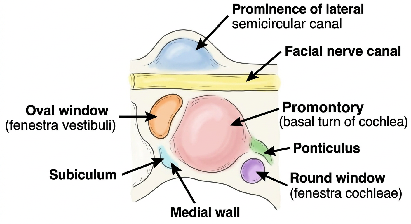
Structures on the Medial Wall (from top to bottom)
SUPERIOR
┌─────────────────────────────────────────┐
│  Prominence of LATERAL SEMICIRCULAR     │  ← bulge above facial canal
│             CANAL                       │
├─────────────────────────────────────────┤
│  FACIAL NERVE CANAL (Canal of Fallopius)│  ← runs horizontally here
├─────────────────────────────────────────┤
│  OVAL WINDOW (Fenestra Vestibuli)       │  ← kidney-shaped, ~3.25×1.75 mm
│  (stapes footplate sits here)           │    connects to vestibule
├─────────────────────────────────────────┤
│  PROMONTORY (central bulge)             │  ← basal turn of cochlea beneath
│  (tympanic plexus on its surface)       │    most prominent landmark
├─────────────────────────────────────────┤
│  ROUND WINDOW (Fenestra Cochleae)       │  ← below & behind oval window
│  (closed by secondary tympanic membrane)│    connects to scala tympani
└─────────────────────────────────────────┘
INFERIOR
5 Key Structures - Quick Memory Aid
| Structure | What it is | Clinical Point |
|---|---|---|
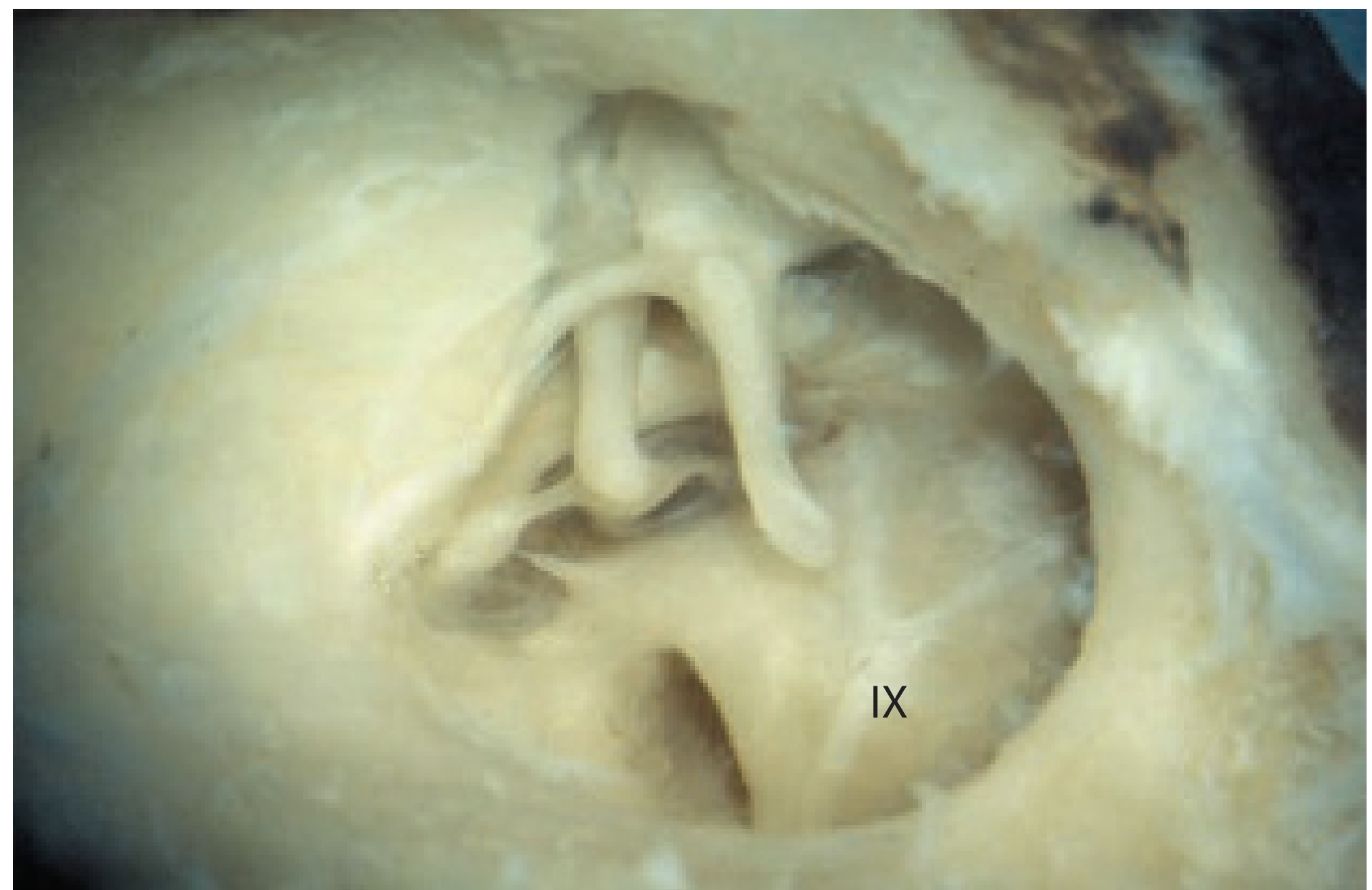
| Promontory | Bony bulge from basal cochlear turn | Tympanic plexus (CN IX) runs over it |
| Oval Window | Opening to vestibule; kidney-shaped | Stapes footplate here; otosclerosis fixes it |
| Round Window | Opening to scala tympani | Covered by secondary tympanic membrane |
| Facial Nerve Canal | Horizontal segment of CN VII | Most common site of facial nerve injury in ear surgery |
| Lateral SCC prominence | Bulge of horizontal semicircular canal | Landmark in mastoid surgery |
Two Important Bony Ridges (between oval and round windows)
- Ponticulus - ridge from promontory to the pyramid (posterior wall), above the round window niche
- Subiculum - ridge separating the oval window from the round window niche
Cadaveric View (Scott-Brown's ENT textbook, Fig 46.7)
Easy Mnemonic to Remember Structures (Top to Bottom)
- Lateral semicircular canal prominence
- Facial nerve canal (horizontal segment)
- Oval window
- Promontory
- Round window
More easy
Simple clean labeled anatomical diagram of the medial wall of the middle ear (labyrinthine wall), showing all key structures clearly labeled with arrows: Prominence of lateral semicircular canal (at top), Facial nerve canal (horizontal segment, below semicircular canal), Oval window (fenestra vestibuli, kidney-shaped opening, middle area), Promontory (large central rounded bulge, basal turn of cochlea), Round window (fenestra cochleae, below and behind oval window), Ponticulus (bony ridge above round window), Subiculum (bony ridge below oval window). White background, simple illustration style like a medical textbook diagram, bold clear labels, color-coded structures, easy to understand for students
Medial Wall = "The Inner Wall" (faces the inner ear)
        ┌──────────────────────────────┐
  TOP   │  🔵 Lateral SCC prominence   │  ← bump from semicircular canal
        │                              │
        │  🟡 Facial nerve canal       │  ← CN VII runs across here
        │                              │
        │  ⬜ OVAL WINDOW              │  ← stapes sits here
        │                              │
 MIDDLE │  🔴 PROMONTORY (big bump)    │  ← most important landmark
        │                              │
        │  ⬜ ROUND WINDOW             │  ← has a membrane covering it
  BOT   └──────────────────────────────┘
Super Simple Summary
| # | Structure | Remember it as |
|---|---|---|
| 1 | Lateral SCC prominence | "Bump at the top" |
| 2 | Facial nerve canal | "CN7 highway" |
| 3 | Oval window | "Where stapes fits" |
| 4 | Promontory | "Big central hill" = cochlea underneath |
| 5 | Round window | "Closed hole below" |
One-Line Mnemonic
"Lazy Foxes Often Play Roundly" Lateral SCC → Facial nerve → Oval window → Promontory → Round window
Course of tensor tympani muscle in ear
tensor tympani muscle origin course insertion function anatomy middle ear
Simple clear anatomical diagram showing the complete course of the tensor tympani muscle in the middle ear, step by step labeled: 1) Origin - from cartilaginous part of Eustachian tube, greater wing of sphenoid, and walls of its own bony canal (semicanal), 2) The muscle runs posteriorly inside the semicanal (bony canal above Eustachian tube), 3) Enters tympanic cavity, lies on medial wall just below facial nerve canal, becomes a slender tendon, 4) Tendon hooks around the processus cochleariformis (a small bony pulley/hook), turning 90 degrees laterally, 5) Inserts into the medial aspect of the handle (manubrium) of the malleus. Show the Eustachian tube below, facial nerve canal above, processus cochleariformis as a small hook/pulley, and the malleus with tensor tympani tendon attached. White background, simple medical textbook style, bold clear arrows and labels, easy for students
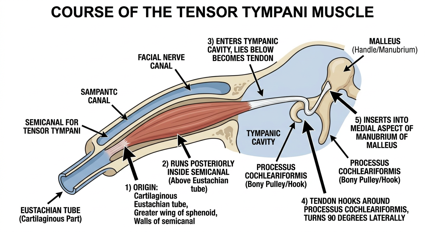
Course of Tensor Tympani Muscle - Step by Step
🔴 ORIGIN (3 sources)
- Cartilaginous part of Eustachian tube
- Greater wing of sphenoid
- Walls of its own bony canal (semicanal for tensor tympani)
➡️ COURSE
ORIGIN                    COURSE                        INSERTION
──────                    ──────                        ─────────
Cartilage of         Runs POSTERIORLY         Hooks around
Eustachian tube  →   inside semicanal    →    PROCESSUS         →   Handle of
Greater wing         (above ET, in            COCHLEARIFORMIS       MALLEUS
of sphenoid          petrous temporal         (turns 90° LATERAL)   (medial aspect,
Walls of             bone)                                          upper part)
semicanal
The KEY Step - Processus Cochleariformis
- This is a small bony pulley/hook at the anterior end of the medial wall
- The tendon of tensor tympani bends 90 degrees around it
- Then travels laterally to reach the malleus handle
- Think of it like a rope over a pulley
Easy Summary Table
| Feature | Detail |
|---|---|
| Type | Long, slender muscle |
| Location | Semicanal - above Eustachian tube |
| Origin | Cartilage of ET + sphenoid + bony canal walls |
| Direction | Runs posteriorly → turns 90° laterally |
| Pulley | Processus cochleariformis |
| Insertion | Medial aspect, upper handle of malleus |
| Nerve supply | Medial pterygoid nerve (branch of V3 - mandibular) |
| Blood supply | Superior tympanic branch of middle meningeal artery |
| Action | Pulls malleus medially → tenses tympanic membrane → dampens loud sounds |
One-Line Memory Trick
"Tensor Tympani - Eustachian tube travels Posteriorly, Pulley (processus cochleariformis), then Pulls malleus medially" = E → P → P → P
Clinical Point
- Supplied by V3 (trigeminal) - NOT facial nerve (facial nerve supplies stapedius)
- Contraction tenses the eardrum - protective reflex against loud sounds
- Paralysis = hypersensitivity to sound (hyperacusis can occur in combination with stapedius paralysis in Bell's palsy)