
Mucosal Melanoma
Reading File
Now let me search the medical library and PubMed in parallel for mucosal melanoma.
Finding Sources
Finding Sources
Searching PubMed
"mucosal melanoma"[MeSH Terms] AND treatment
Excellent results. Now let me read the most comprehensive sources - Fitzpatrick's (dedicated section), KJ Lee's detailed section, and Scott-Brown's in parallel.
Reading File
Reading File
Reading File
Searching PubMed
"mucosal melanoma" AND immunotherapy
Good content gathered. Let me now fetch the Sabiston surgical content and the Fitzpatrick's image, and also get more content from the Scott-Brown's microscopic section.
Reading File
Reading File
Loading Image
I now have comprehensive information from multiple authoritative textbooks. Let me compile the full response.
Mucosal Melanoma
Overview
Mucosal melanoma is a rare and aggressive subtype of melanoma arising from melanocytes within mucosal epithelium. It represents only ~1.3% of all melanomas (some sources cite 0.5-3%) but carries a distinctly worse prognosis than cutaneous melanoma due to occult location and delayed diagnosis.
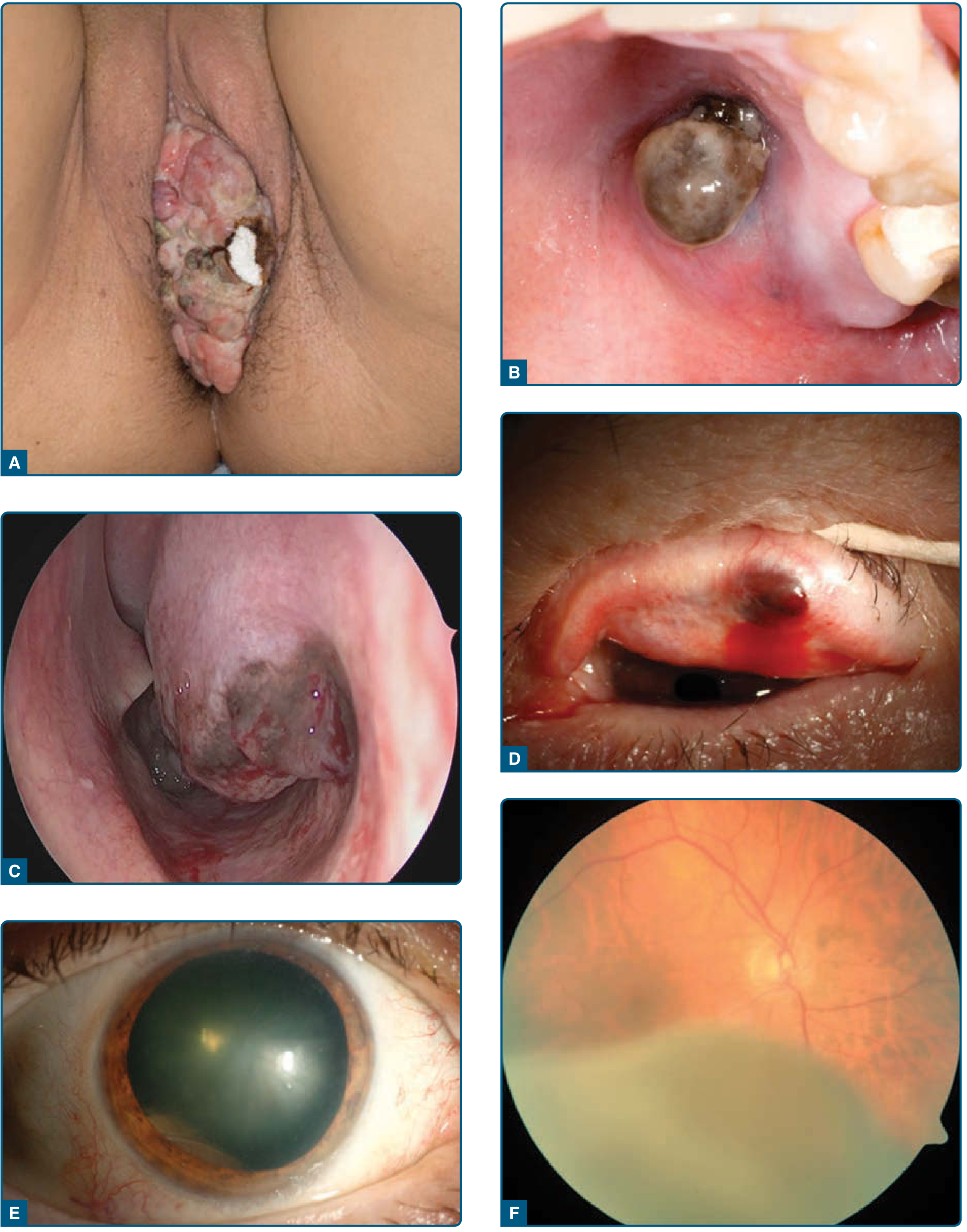
Figure: Mucosal melanomas at various sites - A. Introitus vaginae; B. Gingiva; C. Intranasal; D. Conjunctiva; E,F. Uveal melanoma (Fitzpatrick's Dermatology)
Sites of Origin
The most common anatomical sites are (in approximate frequency):
Head and Neck (~55% of all mucosal melanomas):
- Nasal cavity (most common H&N site)
- Paranasal sinuses
- Oral cavity (palate most often)
- Oropharynx / nasopharynx
In H&N, prognosis by site: Nasal (best) > Oral cavity > Paranasal sinuses (worst) - K.J. Lee's Essential Otolaryngology
Female genital tract (~50% of mucosal melanomas in women):
- Vulva and vagina
Anorectal: ~16.5% of all mucosal melanomas
Other: Urethral mucosa, conjunctiva
Epidemiology
- Sex: More frequent in women overall, largely due to vulvovaginal primaries
- Head and neck mucosal melanoma: More common in men, but inconsistently reported; wide age range (20-80 years)
- Accounts for ~10% of all head and neck melanomas
- Disease course is often indolent with vague early symptoms, so patients frequently present at an advanced stage
Clinical Features
- Lesions begin with a radial growth phase - macular pigmentation - before becoming tumorous
- Usually pigmented (light tan to black, depending on melanin production)
- Amelanotic variants do occur and are more easily missed
- Signs of bleeding are common at presentation
- Depth of invasion does NOT predict prognosis (unlike cutaneous melanoma) - K.J. Lee's
Histopathology
Macroscopic: Pigmented; color ranges from light tan to jet black. Amelanotic examples occur.
Microscopic:
- Composed of epithelioid and/or spindle cells
- Recognized variants: plasmacytoid, rhabdoid, small cell, giant cell, balloon cell, neurotropic, desmoplastic
- Cells are markedly pleomorphic, may contain pigment
- Conspicuous nucleoli and intranuclear inclusions are typical
- Adjacent inflammatory infiltrate and necrosis may be present
- Melanocytic atypia or melanoma in situ may be seen in background mucosa
Immunohistochemistry:
| Marker | Result |
|---|---|
| S-100 protein | Positive (nuclear + cytoplasmic) |
| HMB-45 | Positive |
| Melan-A | Positive |
| SOX10 | Positive |
| Epithelial markers (cytokeratin) | Negative (focal positivity in up to 10%) |
| Desmoplastic variant | May be negative for ALL melanocytic markers |
- Scott-Brown's Otorhinolaryngology
Molecular Biology
Mucosal melanoma has a distinct molecular profile from cutaneous melanoma:
| Mutation | Frequency | Notes |
|---|---|---|
| BRAF V600E | < 10% | Much lower than cutaneous (~50%). Higher in vulvovaginal origin (~26%) |
| NRAS / KRAS (RAS alterations) | 5-30% | Activates MAPK pathway similar to BRAF |
| NF1 | Frequent | |
| c-KIT (receptor tyrosine kinase) | 5-20% | Higher in vulvovaginal/anorectal; targetable with multi-kinase inhibitors (e.g., imatinib) |
Key point: KIT mutation status has NOT been shown to associate with survival outcomes despite being targetable.
Because BRAF mutations are rare, vemurafenib (BRAF inhibitor) is unlikely to benefit most mucosal melanoma patients - Scott-Brown's
Staging
Mucosal melanoma uses a distinct AJCC staging system (not the same T1-T4 system as cutaneous melanoma). There is no Stage I or II - all localized mucosal melanomas start at:
- Stage III - localized disease (T3)
- Stage IVA - deep invasion into adjacent bone, nerve, etc. (T4a)
- Stage IVB - very advanced local / nodal disease (T4b/N1)
- Stage IVC - distant metastases (M1)
The AJCC 8th edition updated staging criteria based on the poor prognosis even of "localized" disease, with 5-year survival rates reported at ~38% per some studies, and as low as ~15% for oral mucosal melanoma (median survival < 2 years).
Management
Surgery
- Surgical resection to negative margins is the primary treatment modality
- Wide local excision is the goal
- Extensive radical resections (e.g., abdominoperineal resection, pelvic exenteration) do not improve overall survival, though may be needed for local disease control - Sabiston
- Sentinel lymph node (SLN) biopsy: Role not well established. For anal melanoma, a negative inguinal SLN can spare that region from radiation fields
Lymph Node Management (Head & Neck)
- Therapeutic neck dissection for clinically positive nodes: Yes
- Elective neck dissection: NOT recommended - K.J. Lee's
Radiation Therapy
- High-dose radiation (>54 Gy, standard fractionation) can improve locoregional control
- Used as adjuvant therapy post-surgery
Systemic Therapy
- Immunotherapy (checkpoint inhibitors): Response rate is lower than in cutaneous melanoma but still superior to cytotoxic chemotherapy - Sabiston
- c-KIT inhibitors (e.g., imatinib): For KIT-mutant tumors, though survival benefit unclear
- BRAF/MEK inhibitors: Limited role given low BRAF mutation frequency
Prognosis
- Overall poor - delayed detection due to occult sites
- 5-year overall survival: ~15-38% depending on site and series
- Oral mucosal melanoma: median survival < 2 years, 5-year OS ~15%
- Most patients with head and neck mucosal melanoma succumb to distant metastasis despite locoregional control
- Worst prognosis among paranasal sinus cancers (compared to minor salivary gland tumors, low-grade sarcomas) - K.J. Lee's
Key Distinguishing Points vs. Cutaneous Melanoma
| Feature | Mucosal Melanoma | Cutaneous Melanoma |
|---|---|---|
| Frequency | ~1.3% of all melanomas | ~98% |
| BRAF V600E | < 10% | ~50% |
| KIT mutations | 5-20% | Rare |
| Breslow depth | Not a prognostic factor | Key staging parameter |
| Staging | Starts at Stage III | Starts at Stage I |
| Prognosis | Much worse | Better (if caught early) |
| Response to immunotherapy | Lower | Higher |
Sources:
- Fitzpatrick's Dermatology, Ch. 116 (Mucosal Melanoma)
- K.J. Lee's Essential Otolaryngology, Head & Neck Melanoma chapter
- Scott-Brown's Otorhinolaryngology Head & Neck Surgery, Ch. 26
- Sabiston Textbook of Surgery, 21e (Mucosal Melanoma section)
- Cummings Otolaryngology Head and Neck Surgery
Mucosal melanoma anal canal
Mucosal melanoma of the anal canal is a rare but highly aggressive malignancy, accounting for about 1-2% of all anal cancers and roughly 16.5% of all mucosal melanomas. It carries one of the worst prognoses of any GI malignancy.
Searching sources...
Finding Sources
Finding Sources
Searching PubMed
"anorectal melanoma"[All Fields] AND treatment
Excellent - multiple high-quality sources identified. Let me read the key textbook sections and the most recent systematic review in parallel.
Reading File
Reading File
Reading File
Reading Article
PMID: 39361336
Now let me get the Mulholland and Sleisenger sections plus the 2023 J Surg Oncol review abstract for the most complete picture.
Reading File
Reading Article
PMID: 37395165
Reading Article
PMID: 41809872
I now have comprehensive, current data from multiple authoritative textbooks and recent literature. Here is the full review:
Mucosal Melanoma of the Anal Canal
Definition and Epidemiology
Anorectal melanoma (ARM) is a rare, aggressive primary malignancy arising from melanocytes in the mucosa of the anal canal and distal rectum.
| Statistic | Figure |
|---|---|
| % of all anorectal malignancies | < 1-2% |
| % of all anal canal tumors | ~0.5% |
| Rank among all melanomas | 3rd most common (after cutaneous & ocular) |
| Most common primary GI melanoma site | Yes - anal canal |
| % of all mucosal melanomas | ~16.5% |
| Median age at diagnosis | 66 years |
| Sex | Slight female predominance (51% F : 49% M) |
| Median time to diagnosis | ~4 months (from symptom onset) |
ARM accounts for less than 2% of all melanomas and 37% of mucosal melanomas that originate in the GI tract, alongside esophageal primaries. - Paolino et al., Melanoma Res 2024
Anatomy and Origin
- Tumors arise from neural crest-derived melanocytes within the transitional and squamous epithelium of the anal canal and anoderm
- Can affect any part of the anal canal and distal rectum; most are found within 6 cm of the anal verge
- Spread is submucosal into the rectum without early invasion of adjacent organs
- Lymphatic drainage follows two routes:
- Mesenteric/perirectal nodes (superior drainage)
- Inguinal nodes (inferior drainage)
- Lymph node metastasis to mesenteric nodes present in up to 32% at diagnosis
- Distant metastasis present in 13% at diagnosis
Clinical Presentation
Patients typically present with common anorectal complaints, leading to frequent misdiagnosis:
Symptoms (from systematic review, n=166):
| Symptom | Frequency |
|---|---|
| Rectal bleeding | 84.9% |
| Pain | 68.7% |
| Change in bowel habits | 28.5% |
| Tenesmus | 16.9% |
| Mucous discharge | 6% |
Classic pitfall: The lesion is frequently mistaken for a thrombosed hemorrhoid and first diagnosed only after an inadvertent "hemorrhoidectomy" is performed. - Fischer's Mastery of Surgery
- Clinical presentation is nodular in 98.2% of cases
- Amelanotic in 20-29% of cases - making visual diagnosis unreliable
- Symptoms of pain, pruritus, and bleeding mimic benign anorectal disease - Mulholland & Greenfield's Surgery
Histopathology and IHC
Macroscopic: Usually pigmented polypoid/nodular mass; 20-30% amelanotic and may resemble benign polyps
Histology: Epithelioid and/or spindle cells with marked pleomorphism, prominent nucleoli, intranuclear inclusions
Immunohistochemistry (positive):
- S-100 protein
- HMB-45 (Human Melanoma Black-45)
- Melan-A
- SOX10
Molecular Biology
Unlike cutaneous melanoma, anorectal melanoma has a distinct and unfavorable molecular landscape:
| Mutation | Notes |
|---|---|
| BRAF V600E | Rare (< 10%) - BRAF inhibitors (vemurafenib, dabrafenib) have limited role |
| c-KIT | 5-20%; higher frequency in anorectal and vulvovaginal sites; targetable with imatinib |
| NRAS/KRAS | 5-30%; activates MAPK pathway |
| Low tumor mutational burden | Contributes to reduced immunotherapy response vs. cutaneous |
| PD-L1 | Expression variable; used to guide checkpoint inhibitor selection |
Molecular testing for PD-L1, BRAF, and c-KIT mutations should be performed on all tumors to guide systemic therapy. - Fischer's Mastery of Surgery
Workup and Staging
Recommended Work-up
- MRI of pelvis - assess local extent, sphincter involvement, nodal disease
- PET-CT scan - distant metastasis
- Colonoscopy and anorectal examination - characterize local tumor
- Complete cutaneous and ophthalmologic examination - rule out cutaneous primary with GI metastasis
- CT chest/abdomen
- Molecular tumor analysis (BRAF, KIT, PD-L1)
AJCC Staging
ARM uses the mucosal melanoma-specific AJCC staging (no Stage I/II):
- Stage III: Localized disease (T3N0M0)
- Stage IVA: Deep invasion of adjacent structures (T4a)
- Stage IVB: Extensive local/nodal disease (T4b/N1)
- Stage IVC: Distant metastases (M1)
Treatment
Surgery - The Cornerstone
The central debate has been between two operations:
| Feature | Wide Local Excision (WLE) | Abdominoperineal Resection (APR) |
|---|---|---|
| Local control | Lower | Higher R0 rate |
| Overall survival | No significant difference | No significant difference |
| Quality of life | Better (sphincter preservation) | Worse (permanent colostomy) |
| Morbidity | Lower | Higher |
| Preferred? | Yes, when margins achievable | For large/sphincter-involving tumors |
Current consensus: Multiple large case series show no survival advantage for APR over WLE. WLE is the preferred approach when negative margins can be achieved. APR is reserved for larger lesions with sphincter involvement or when WLE cannot achieve clear margins. - Sabiston, Fischer's, Mulholland
Practical approach (Fischer's Mastery):
"Our practice is to locally excise small lesions with adjuvant radiation. For larger lesions, neoadjuvant radiation is used first - if the tumor shrinks, local excision; if it remains large with no distant disease, a circumferential operation (possibly APR) is performed."
Lymph Node Management
- No role for prophylactic/elective inguinal lymph node dissection
- Lymphadenectomy reserved for radiographically or clinically apparent nodal disease
- For anal melanoma: a negative SLN biopsy in the superficial inguinal region can omit that region from radiation fields
Radiation Therapy
- Adjuvant radiation can improve locoregional control
- High dose (>54 Gy) used for head/neck mucosal melanoma; same principles apply
- Benefit is reduced in advanced disease
- Neoadjuvant radiation used in larger lesions to downstage before surgery
Systemic Therapy
Chemotherapy: Limited role - melanomas of the anal canal respond poorly to conventional cytotoxic chemotherapy. - Mulholland & Greenfield's
Immunotherapy (Checkpoint Inhibitors):
- Response rates are lower than for cutaneous melanoma due to lower tumor mutational burden
- Still superior to cytotoxic chemotherapy regimens
- Nivolumab + ipilimumab (CheckMate 067): Significant improvement in long-term OS in advanced cutaneous melanoma; being evaluated in mucosal subtypes
- Early studies suggest improved short-term OS for anorectal melanoma with immunotherapy - Sabiston
- The optimal regimen continues to evolve
Targeted Therapy:
- c-KIT inhibitors (imatinib): For KIT-mutant tumors; modest responses reported; survival benefit not definitively established
- BRAF inhibitors: Very limited role given low BRAF mutation frequency
- Case reports exist for both; larger prospective data are lacking
Emerging approaches (2026): Cancer vaccines, immunotherapy combinations, and personalized medicine approaches are under investigation. - De Nardi et al., World J Gastroenterol 2026
Prognosis
| Metric | Value |
|---|---|
| 5-year overall survival | 14-31% (most series ~20%) |
| Median overall survival | ~11 months (systematic review, 2024) |
| Anal melanoma (vs. rectal) | Better OS than rectal or anorectal junction melanoma |
| Rectal melanoma median OS | ~7 months |
The prognosis is extremely poor regardless of surgical approach. Most patients succumb to distant metastatic disease rather than local failure. The occult location, late presentation, distinct molecular profile, and lower immunotherapy response all contribute to the dismal outcomes.
Key Points Summary
- Rare (< 2% of anorectal cancers) but the most common primary GI melanoma
- Frequently misdiagnosed as thrombosed hemorrhoid - high index of suspicion needed
- 20-29% amelanotic - may mimic polyps or SCC
- BRAF mutations rare (< 10%) - targeted therapy with BRAF inhibitors has limited role
- c-KIT mutations (5-20%) - relevant therapeutic target
- WLE = APR in survival - prefer sphincter-saving WLE when margins achievable
- No elective inguinal node dissection
- Immunotherapy response lower than cutaneous melanoma but still preferred over chemotherapy
- Median OS only ~11 months; 5-year survival 14-31%
- Multidisciplinary tumor board involvement is mandatory
Sources:
- Fischer's Mastery of Surgery, 8th ed., Ch. 137 (Anal Melanoma)
- Sabiston Textbook of Surgery, 21e (Anal Adenocarcinoma and Anal Melanoma)
- Mulholland & Greenfield's Surgery, 7e (Melanoma of the Anal Canal)
- Tintinalli's Emergency Medicine (Anorectal Tumors)
- Paolino G et al. Anorectal melanoma: systematic review. Melanoma Res 2024 - PMID 39361336
- Fastner S et al. Anorectal melanoma. J Surg Oncol 2023 - PMID 37395165
- De Nardi P et al. Primary melanoma of the GI tract. World J Gastroenterol 2026 - PMID 41809872
This is a shared conversation. Sign in to Orris to start your own chat.