


I'm planning to read cleft lip and palate from mccarthy. Kindly summaries and simplify this pages in an note form. Make sure you don't miss any vital points which are important to know and study. Explain me with relevant pictures given in the book.
📚 McCarthy Chapter 46 - Embryology of the Head and Neck
Study Notes (Pages 2451-2460)
🔷 OVERVIEW & INTRODUCTION
- Understanding of normal and abnormal craniofacial development has increased dramatically
- Pathogenesis of common surgical malformations clarified by correlating:
- Experimentally induced malformations
- Those caused by identical teratogens or genetic defects
- Technical advances have enabled study at the molecular level
- New information necessitates reassessment of pathogenesis of some human malformations
- Chapter follows progressive stages: normal development → abnormal alterations at each stage
Key Point: Embryogenesis of cleft lip and palate is covered in detail in Chapter 48 - this chapter provides the foundational embryology
🔷 NORMAL DEVELOPMENT: AN OVERVIEW (Fig. 46-1)
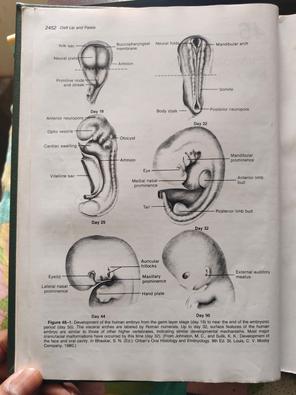
Developmental Timeline (Figure 46-1)
| Day | Key Structures Visible |
|---|---|
| Day 19 | Neural plate, yolk sac, primitive node and streak, buccopharyngeal membrane, amnion |
| Day 22 | Neural folds, mandibular arch (arch I), somites, posterior neuropore, body stalk |
| Day 25 | Anterior neuropore, optic vesicle, cardiac swelling, otocyst, visceral arches I-III |
| Day 32 | Eye, medial nasal prominence, mandibular prominence, limb buds, arches I-IV |
| Day 44 | Eyelid, lateral nasal prominence, auricular hillocks, maxillary prominence, hand plate |
| Day 50 | External auditory meatus visible |
Critical Exam Point: Most major craniofacial malformations have already occurred by Day 32. Up to Day 32, human surface features resemble other higher vertebrates - indicating similar developmental mechanisms.
🔷 EARLY DEVELOPMENT: GASTRULATION & GERM LAYERS (Fig. 46-2)
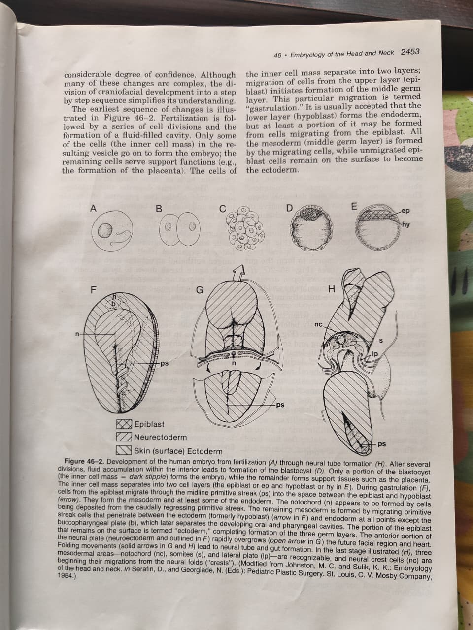
Sequence (A through H):
- Fertilization (A) → cell divisions → fluid-filled cavity = blastocyst (D)
- Only the inner cell mass (dark stipple) forms the embryo; remaining cells form support tissues (e.g., placenta)
- Inner cell mass separates into 2 layers:
- Epiblast (ep) = upper layer
- Hypoblast (hy) = lower layer → forms endoderm
- Gastrulation (F): Epiblast cells migrate through the midline primitive streak (ps) into the space between epiblast and hypoblast → forms mesoderm (the middle germ layer)
- Unmigrated epiblast cells on the surface → become ectoderm
The Three Germ Layers:
| Layer | Origin | Derivatives |
|---|---|---|
| Ectoderm | Remaining epiblast | Skin, nervous system, neural crest |
| Mesoderm | Migrated epiblast cells | Muscle, bone, connective tissue |
| Endoderm | Hypoblast + some migrating cells | Gut lining, pharynx |
Notochord:
- Formed from cells deposited by the caudally regressing primitive streak
- Later mesoderm is formed by migrating primitive streak cells
- Buccopharyngeal plate (b) = separates developing oral and pharyngeal cavities
- At stage H: Three mesodermal areas recognizable: notochord (nc), somites (s), lateral plate (lp)
- Neural crest cells (nc) begin migrations from the neural folds ("crests") at this stage
🔷 EPITHELIA vs. MESENCHYME (Fig. 46-3)
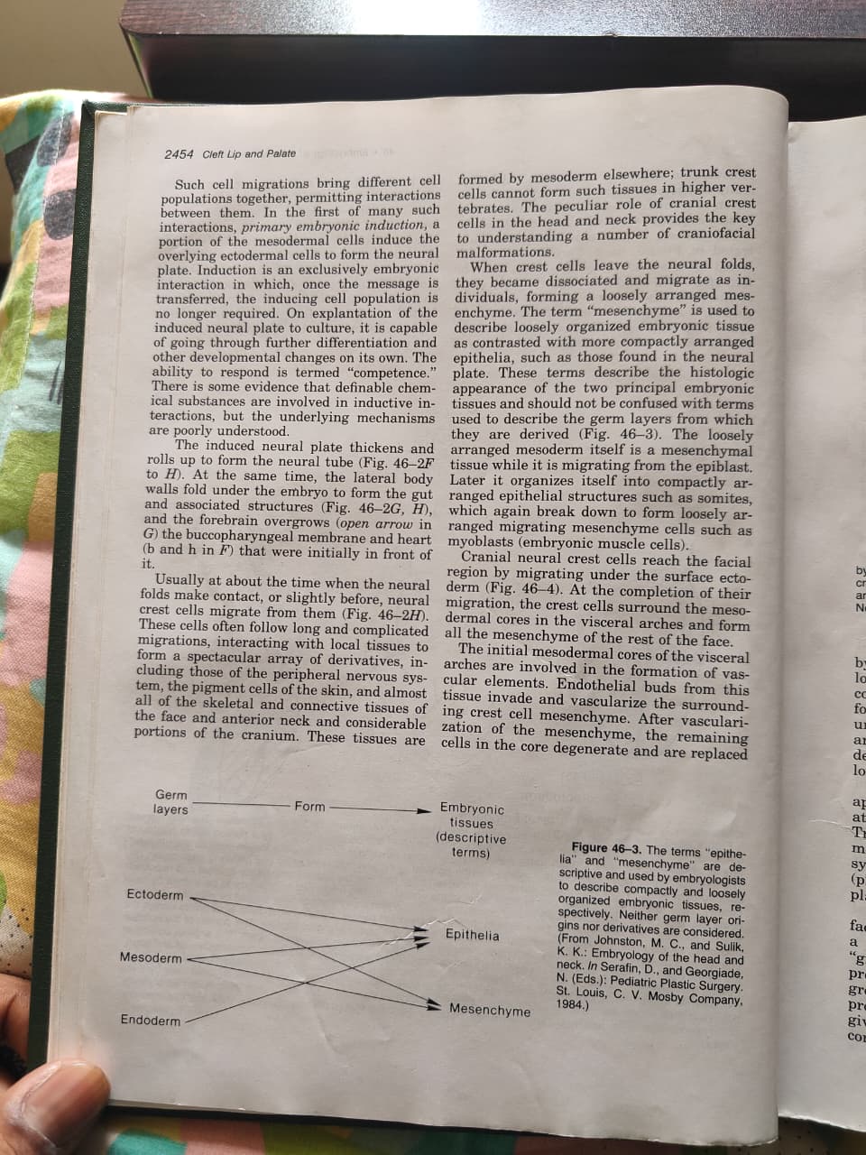
Key Distinction:
- Epithelium = compactly arranged embryonic tissue (e.g., neural plate)
- Mesenchyme = loosely organized embryonic tissue (migrating cells)
- These are descriptive terms (histological appearance) - NOT the same as germ layer origins!
Figure 46-3 Diagram - Important Concept:
- All three germ layers (ectoderm, mesoderm, endoderm) can give rise to BOTH epithelia and mesenchyme
- Loosely arranged mesoderm is mesenchymal WHILE migrating from epiblast
- Later it organizes into compactly arranged epithelial structures (e.g., somites)
- Which again break down to form loosely migrating myoblasts (embryonic muscle cells)
🔷 PRIMARY EMBRYONIC INDUCTION
- Cell migrations bring different populations together → enabling interactions
- First key interaction = Primary Embryonic Induction:
- Mesodermal cells induce overlying ectodermal cells to form the neural plate
- Induction = exclusively embryonic; once message is transferred, the inducing cell population is no longer required
- Explanted induced neural plate can continue differentiation on its own
- Ability to respond = "competence"
- Chemical substances are likely involved but mechanisms poorly understood
Neural Tube Formation:
- Induced neural plate thickens → rolls up → neural tube (Fig. 46-2F to H)
- Simultaneously: lateral body walls fold under → form gut and associated structures
- Forebrain overgrows the buccopharyngeal membrane and heart
🔷 NEURAL CREST (Fig. 46-4)
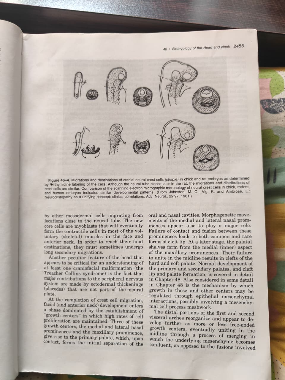
Neural Crest Cells - The Most Important Concept in Craniofacial Embryology:
- Neural crest cells migrate from the neural folds just before/at time of neural fold contact
- Follow long, complicated migration paths
- Interact with local tissues to form a spectacular array of derivatives
Derivatives of Neural Crest Cells:
| System | Derivative |
|---|---|
| Peripheral nervous system | Ganglia, nerves |
| Pigment cells | Melanocytes of skin |
| Skeletal/connective tissue | Almost ALL skeletal and connective tissue of face and anterior neck, considerable portions of cranium |
| Meningeal tissue | Cranial meninges |
KEY EXAM FACT: In the head/neck, skeletal and connective tissues are formed by neural crest (NOT by mesoderm as in the trunk). This is unique to the head and anterior neck. Trunk crest cells CANNOT form such tissues in higher vertebrates.
Cranial vs. Trunk Crest:
- Cranial crest = forms bone, cartilage, connective tissue of face
- Trunk crest = cannot form these skeletal/connective tissues in higher vertebrates
- The peculiar role of cranial crest cells provides the key to understanding craniofacial malformations
After Crest Cell Migration:
- Crest cells surround the mesodermal cores in visceral arches
- Form the mesenchyme of the rest of the face
- Initial mesodermal cores → involved in formation of vascular elements
- Endothelial buds invade and vascularize the crest cell mesenchyme
- After vascularization, remaining mesodermal cells in core degenerate
- Replaced by other mesodermal cells migrating from near the neural tube
- New core cells = myoblasts → form contractile cells of voluntary (skeletal) muscles of face and anterior neck
Ectodermal Placodes:
- Important for Treacher Collins syndrome understanding
- Major contributions to peripheral nervous system are made by ectodermal thickenings (placodes) that are NOT part of the neural plate
- Olfactory placodes - some evidence they are derived from neural folds
- Otic placodes - develop in close relation to neural plate and tube
- Ganglionic placodes - form at some distance from neural plate and tube → contribute neurons to cranial sensory ganglia
🔷 CLEFT LIP/PALATE - BRIEF OVERVIEW (covered in detail in Ch. 48)
Primary Palate Formation:
- After crest cell migration, facial development enters a phase dominated by "growth centers" with high rates of cell proliferation
- Three growth centers give rise to the primary palate:
- Medial nasal prominence (x2)
- Maxillary prominence
- Upon contact, these form the initial separation of the oral and nasal cavities
- Morphogenetic movements of medial and lateral nasal prominences also play a major role
- Failure of contact and fusion between prominences → cleft lip (both common and rare forms)
Secondary Palate Formation:
- Palatal shelves form from the medial (inner) aspect of the maxillary prominences
- Their failure to unite in the midline → clefts of hard and soft palate
Merging vs. Fusion:
- 1st and 2nd visceral arch distal portions reorganize → unite in midline through a process of merging (underlying mesenchyme becomes confluent)
- This differs from fusions involved in primary and secondary palate formation (which require breakdown of contracting epithelia)
🔷 GASTRULATION & ORGANIZATIONAL PLANS OF ECTODERM AND MESODERM
Mesoderm's "Blueprint" Function:
- Much of the "blueprint" of the head is determined during or immediately after gastrulation
- Structural relationships of the upper face depend on organizational plans of the mesoderm and anterior neural plate
- Mesoderm first induces overlying ectoderm to differentiate into neural tissues
- Induced neural plates have considerable ability to "self-organize" even with artificial inducers
- The specificity for derivatives such as eyes and olfactory placodes is in the competent ectoderm
- Quantity and positioning of derivatives is erratic without mesoderm (Holtfreter and Hamburger, 1955)
Mesodermal Organization:
- Has two major components:
- Notochord - organizing function
- Paraxial mesoderm - somites
- A third component: lateral plate - mostly angiogenic (blood vessels) in the head
- A cranial extension from the notochord = prechordal plate (sometimes called "prechordal plate")
- Never forms notochord but has similar organizing abilities
- Both notochord and prechordal plate initially attached to endoderm
- Attachment is lost when paraxial mesoderm becomes confluent across midline
Exogastrulation Experiments (Holtfreter, 1934):
- High salt concentrations caused gastrulating mesodermal cells to balloon outward (exogastrulas)
- Instead of going interior, mesoderm connected to ectoderm by only a stalk
- Mass of meso-/endodermal cells could form notochord
- Incomplete (partial) gastrulation → cycloptic eyes (Holtfreter and Hamburger, 1955)
🔷 ORGANIZATIONAL PLAN (Fig. 46-5)
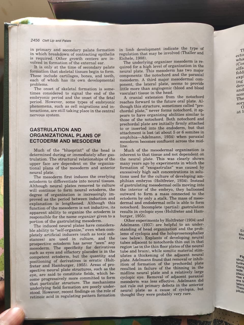
Figure 46-5 Shows:
- A & B: Pre-gastrulation layout - buccal plate (bp), anterior pituitary anlage (p), eye field (ef), prechordal plate (pp), notochord (n), neural crest (nc), olfactory placode (op)
- C: Neural tube segmentation into neuromeres (numbered 1-9, with cranial nerves V, VII, IX, X labeled)
- D: Post-tubulation cross-section showing relationship of placode and ganglion to neural tube
Neural Tube Segmentation:
- Neural tube is segmented into neuromeres (C)
- Ganglionic placode cells add neuroblasts to developing cranial sensory ganglia
- Roman numerals in C = cranial nerves (V, VII, IX, X)
- Contact occurs between visceral endoderm and ectoderm at locations of all ganglionic placodes EXCEPT the trigeminal
- Segmentation in mesoderm → well-defined somites (s) in postotic region + poorly defined somitomeres (se) in preotic region
- Somites are further divided into: dermatomes (d), myotomes (m), sclerotomes (s)
- Somitomeres in visceral arch region do NOT have dermatomes
- Lateral plate (in post-tubulation section) becomes segmented to form cores in each visceral arch
- These cores become surrounded by neural crest cells
Occipital Somites:
- Organization forms pattern similar to cervical and trunk regions
- Dermatome → dermis of skin over somite
- Myotome → contractile cells of voluntary (skeletal) muscle
- Sclerotome → skeletal and connective tissues, including most of cranial base and vault
Olfactory Placodes:
- Controversial whether derived from neural folds
- Otic placodes also develop in close relation to neural plate and tube
- Organization of mesoderm is at least partly dependent on neural plate and tube
- Combination of neural tube and notochord in culture → formation of cartilage
- Presence of neural tube alone → formation of muscle
- Lateral plate forms most skeletal/connective tissues in trunk (including skin dermis) BUT in head and anterior neck, lateral plate forms only the cores of the visceral arches (predominantly endothelial cells for blood vessel linings)
🔷 HOLOPROSENCEPHALIES & MIDLINE DEFECTS (Fig. 46-6 and 46-7)
Medial Anterior Neural Plate Deficiency:
- Variable degrees of deficiency → series of malformations termed "holoprosencephalies"
- Name: partial or complete failure of anterior neural tube to form cerebral hemispheres with ventricles → only ONE (holo) forebrain (prosencephaly) cavity in severe cases
- Spectrum from mild to severe (cyclopia)
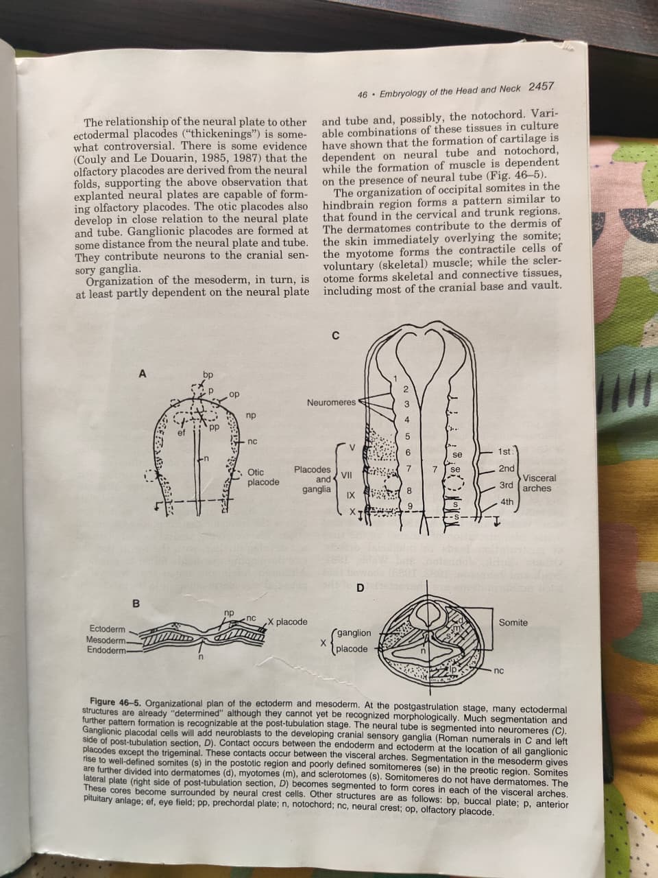
Fetal Alcohol Syndrome (FAS) (Figure 46-6):
- One of the mildest forms of holoprosencephaly
- Webster, Walsh, and Lipson (1980): acute ethanol administration at time of gastrulation → midfacial defects
- Sulik, Johnston, and Webb (1981): malformations characteristic of FAS
- Others (Sulik and Johnston, 1982, 1983) confirmed this
FAS Facial Features (Figure 46-6A, B - children; C - FAS mouse embryo; D - control mouse):
- Narrow forehead
- Short palpebral fissures
- Small nose
- Small midface
- Long upper lip with deficient philtrum
Mechanism of FAS:
- Midline deficiency of the anterior neural plate → more closely approximated olfactory placodes (Fig. 46-7)
- → Small medial nasal prominences
- → Comparable midline defects in developing brain (Sulik, Lauder, and Dehart, 1984)
- Contact of the placode has been shown to lead to failure of medial nasal prominence formation and to arhinencephaly in human embryos (Johnston and Sulik, 1980) and in ethanol-induced malformations
Holoprosencephaly Spectrum:
- Almost all holoprosencephalies can be induced by ethanol in experimental animals
- In humans: may also result from trisomy 13 (Gorlin, Pindborg, and Cohen, 1976) or single gene defects
- Eye size decreases progressively as malformations become more severe
- Cyclopia perfecta (large median eye) CANNOT therefore be part of this spectrum
- Typical cycloptic eyes were found in malformed salamander embryos after removal of prechordal plate
- Two developing eye fields failed to separate → cyclopia
🔷 FIGURE 46-7 DETAILED
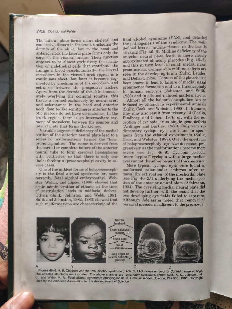
Figure 46-7 Shows:
- A: Schematic of normal human embryo - nasal placodes (na) positioned normally relative to neural plate
- B: Altered development in embryo from ethanol-treated mother - nasal placodes (na) positioned much more closely to the midline
- C to E: Scanning electron micrographs of mouse embryos showing:
- Nasal (na), lens (l), otic (o) placodes
- Heart (h), buccopharyngeal membrane (b)
- Eye field (e), notochord (n), prechordal mesoderm (pm), primitive streak (ps)
- The oral (buccal) cavity (b) is also shown
Nasal placodes are positioned much more closely to the midline in ethanol-treated embryos
🔷 OTOCEPHALY ("EAR HEAD") (Fig. 46-8)
Defect in Mesodermal Programming:
- A defect in programming of the mesoderm is apparently responsible for the otocephaly spectrum ("ear head")
- Figure 46-8A (milder form): Mandible loss/agnathia - derivatives of distal portions of first arch (mandible, etc.) are absent
- Figure 46-8B (more severe form): Little more than external ears apparent - hence "otocephaly" or "ear head"
Mechanism:
- Least severe cases (agnathia) associated with breakdown of mesodermal cores in first visceral arch in mouse mutant (Juriloff, Sulik, and Roderick, 1980)
- Neural crest cells appeared normal in this study
- Failure of vascularization from mesodermal core → complete arch breakdown
- Mesodermal core cells normally break down AFTER vascularization of the neural crest cell mesenchyme
- More severe cases: malformations similar to FAS through cyclopia (rudimentary eyes only)
- Most severe: typical "ear head" form (Fig. 46-8B) associated with more widespread mesodermal cell death including mesoderm underlying developing fore- and midbrains → fail to develop further
- Even more severe specimens: virtually no head at all (acephaly)
🔷 EMBRYO FOLDING (TUBULATION)
Neural Tube Formation:
- Folding movements → formation of TWO embryonic tubes:
- Neural tube
- Endodermally lined gastrointestinal tube
- Mechanism of neural tube closure: primarily involves coordinated contraction of a filamentous actin-myosin meshwork (terminal web) just beneath the luminal lining cell surfaces (Sadler and associates, 1982)
- Terminal webs are involved in many epithelial foldings, including morphogenetic movements of the olfactory placode
- Cell proliferation and matrix formation in the mesenchymal tissue underlying the neural plate and folds seem also to be involved
Neural Tube Defects (NTDs):
- Failure of complete neural tube closure → variety of NTDs including:
- Anencephaly (Fig. 46-9) - in head
- Spina bifida - cervical and trunk regions
- Severe closure defects in brain → eversion of brain with degeneration of all but brainstem portion
- "Anencephaly" = misnomer because brain is NOT entirely absent (brainstem remains)
- Anencephaly can also be experimentally caused by postclosure rupture of the dorsal brain
- Weakening with failure to rupture → meningomyeloceles (protrusion of brain into surrounding tissue)
- Meningomyeloceles = fluid-filled cavities with walls of both brain tissue and meninges
- Meningocele walls that contain ONLY meninges = outcome of closure of meninges only, with failure of complete closure or secondary opening of underlying brain
🔷 SUMMARY TABLE: KEY STRUCTURES AND THEIR ORIGINS
| Structure | Embryonic Origin |
|---|---|
| Facial bones and cartilages | Neural crest |
| Voluntary muscles of face | Myoblasts (from neural tube-adjacent mesoderm) |
| Blood vessels (endothelium) | Lateral plate mesoderm |
| Cranial sensory ganglia neurons | Ectodermal placodes + neural crest |
| Dermis of face | Neural crest + sclerotomes |
| Cranial base and vault | Sclerotomes (from occipital somites) |
| Peripheral nerve Schwann cells | Neural crest |
| Melanocytes | Neural crest |
🔷 IMPORTANT SYNDROMES - EMBRYOLOGICAL BASIS
| Syndrome | Embryological Mechanism |
|---|---|
| Treacher Collins syndrome | Abnormal ectodermal placode contribution to peripheral nervous system |
| Fetal Alcohol Syndrome (FAS) | Ethanol → cell death in medial anterior neural plate → midline deficiency → small midface, deficient philtrum |
| Holoprosencephaly | Deficiency of medial anterior neural plate → failure of cerebral hemisphere separation |
| Otocephaly (Ear Head) | Failure of vascularization from mesodermal core of first visceral arch |
| Cyclopia | Failure of prechordal plate development → two eye fields fail to separate |
| Cleft lip | Failure of contact/fusion between medial nasal, lateral nasal, and maxillary prominences |
| Cleft palate | Failure of palatal shelves (from maxillary prominences) to unite in midline |
| Neural tube defects | Failure of neural tube closure (anencephaly, spina bifida, meningomyeloceles) |
🔷 HIGH-YIELD EXAM BULLETS
- Day 32 = most major craniofacial malformations have already occurred
- Gastrulation = migration of epiblast cells through primitive streak to form mesoderm
- Induction = exclusively embryonic; inducing cells no longer required once signal sent; responding cell's ability = "competence"
- Neural crest = forms almost ALL skeletal and connective tissue of face and anterior neck (this is UNIQUE to the head - trunk crest cannot do this)
- Epithelium vs. mesenchyme = descriptive histological terms, NOT germ layer terms
- FAS mechanism = ethanol → midline neural plate cell death → closely approximated olfactory placodes → small medial nasal prominences → small midface, deficient philtrum
- Primary palate = formed by medial nasal + maxillary prominences
- Secondary palate = formed by palatal shelves from medial aspect of maxillary prominences
- Merging (visceral arch union) = mesenchyme becomes confluent (no epithelial breakdown) - different from fusion (palate/lip) which requires epithelial breakdown
- Notochord = organizer of the overlying neural plate; prechordal plate has similar organizing ability
- Neural tube closure mechanism = actin-myosin terminal web contraction
- "Anencephaly" = misnomer; brainstem IS present (it's mainly the cerebral hemisphere that degenerates)
- Otocephaly = mesodermal core failure in 1st visceral arch → agnathia (mild) → ear head (severe)
- Lateral plate mesoderm in head = forms ONLY visceral arch cores (endothelial cells for blood vessels) - NOT bone or connective tissue (unlike in trunk)
🔷 CHAPTER 46 TOPIC LIST (for further study)
- Vascular development
- Origins of voluntary muscles
- Development of peripheral nervous system
- Pituitary and pharyngeal glands
- Carotid and aortic bodies and sinuses
- Development of the eye and associated structures
- Growth centers in the face and visceral arches
- Development of the ear
- Development of skeletal tissues
- Reexamination of pathogenesis of craniofacial microsomia
The embryogenesis of cleft lip and palate is covered in detail in Chapter 48 of McCarthy.
can you make it the same way in an pdf without mentioning the pages from the book. keep the content same, only remove the book pages
mkdir -p /tmp/workspace/cleft-lip-notes && ls /tmp/workspace/
~/cleft-lip-notes/generate_notes_pdf.py
from reportlab.lib.pagesizes import A4
from reportlab.lib import colors
from reportlab.lib.styles import getSampleStyleSheet, ParagraphStyle
from reportlab.lib.units import cm, mm
from reportlab.platypus import (
SimpleDocTemplate, Paragraph, Spacer, Table, TableStyle,
HRFlowable, KeepTogether, ListFlowable, ListItem
)
from reportlab.lib.enums import TA_LEFT, TA_CENTER, TA_JUSTIFY
from reportlab.platypus import BaseDocTemplate, PageTemplate, Frame
from reportlab.lib.colors import HexColor
# ── Color palette ──────────────────────────────────────────────────────────
DARK_BLUE = HexColor("#1a3a5c")
MED_BLUE = HexColor("#2563a8")
LIGHT_BLUE = HexColor("#dbeafe")
ACCENT_TEAL = HexColor("#0d9488")
ACCENT_AMBER= HexColor("#d97706")
ACCENT_RED = HexColor("#dc2626")
LIGHT_GREY = HexColor("#f1f5f9")
BORDER_GREY = HexColor("#cbd5e1")
WHITE = colors.white
BLACK = colors.black
YELLOW_HL = HexColor("#fef9c3")
GREEN_HL = HexColor("#dcfce7")
PINK_HL = HexColor("#fce7f3")
# ── Document setup ─────────────────────────────────────────────────────────
OUTPUT = "/tmp/workspace/cleft-lip-notes/Embryology_Head_Neck_Notes.pdf"
doc = SimpleDocTemplate(
OUTPUT,
pagesize=A4,
rightMargin=2*cm, leftMargin=2*cm,
topMargin=2.5*cm, bottomMargin=2.2*cm,
title="Embryology of the Head and Neck – Study Notes",
author="Orris AI"
)
# ── Styles ─────────────────────────────────────────────────────────────────
styles = getSampleStyleSheet()
def style(name, **kw):
s = ParagraphStyle(name, **kw)
return s
# Cover / Chapter title
TITLE = style("TITLE",
fontSize=26, leading=32, textColor=WHITE, alignment=TA_CENTER,
fontName="Helvetica-Bold", spaceAfter=6)
SUBTITLE = style("SUBTITLE",
fontSize=13, leading=18, textColor=LIGHT_BLUE, alignment=TA_CENTER,
fontName="Helvetica", spaceAfter=4)
# Section heading (blue band)
SEC_HEAD = style("SEC_HEAD",
fontSize=13, leading=17, textColor=WHITE,
fontName="Helvetica-Bold", spaceAfter=0, spaceBefore=0,
leftIndent=8, rightIndent=8)
# Sub-heading
SUB_HEAD = style("SUB_HEAD",
fontSize=11, leading=14, textColor=DARK_BLUE,
fontName="Helvetica-Bold", spaceBefore=10, spaceAfter=4)
# Normal body
BODY = style("BODY",
fontSize=9.5, leading=14, textColor=HexColor("#1e293b"),
fontName="Helvetica", spaceBefore=2, spaceAfter=2,
alignment=TA_JUSTIFY)
# Bullet
BULLET = style("BULLET",
fontSize=9.5, leading=13, textColor=HexColor("#1e293b"),
fontName="Helvetica", leftIndent=16, spaceBefore=1, spaceAfter=1)
# Key point box
KEY = style("KEY",
fontSize=9.5, leading=13, textColor=HexColor("#1e293b"),
fontName="Helvetica-BoldOblique", leftIndent=8, rightIndent=8,
spaceBefore=2, spaceAfter=2)
# Small table cell
CELL = style("CELL",
fontSize=9, leading=12, textColor=HexColor("#1e293b"),
fontName="Helvetica", spaceBefore=1, spaceAfter=1)
CELL_B = style("CELL_B",
fontSize=9, leading=12, textColor=HexColor("#1e293b"),
fontName="Helvetica-Bold", spaceBefore=1, spaceAfter=1)
CELL_HD = style("CELL_HD",
fontSize=9.5, leading=12, textColor=WHITE,
fontName="Helvetica-Bold", alignment=TA_CENTER)
# ── Helper builders ────────────────────────────────────────────────────────
def section_heading(text):
"""Blue header band for a major section."""
tbl = Table([[Paragraph(f"◆ {text}", SEC_HEAD)]], colWidths=[17*cm])
tbl.setStyle(TableStyle([
("BACKGROUND", (0,0), (-1,-1), DARK_BLUE),
("ROUNDEDCORNERS", [4]),
("TOPPADDING", (0,0), (-1,-1), 6),
("BOTTOMPADDING",(0,0),(-1,-1), 6),
("LEFTPADDING", (0,0), (-1,-1), 10),
]))
return [Spacer(1, 10), tbl, Spacer(1, 6)]
def sub_heading(text):
return [Paragraph(text, SUB_HEAD)]
def body(text):
return [Paragraph(text, BODY)]
def bullet(items, color=ACCENT_TEAL):
elems = []
for item in items:
elems.append(Paragraph(f"• {item}", BULLET))
return elems
def key_box(text, bg=YELLOW_HL, border=ACCENT_AMBER):
tbl = Table([[Paragraph(f"⭐ KEY POINT: {text}", KEY)]], colWidths=[17*cm])
tbl.setStyle(TableStyle([
("BACKGROUND", (0,0), (-1,-1), bg),
("BOX", (0,0), (-1,-1), 1, border),
("TOPPADDING", (0,0), (-1,-1), 6),
("BOTTOMPADDING",(0,0),(-1,-1), 6),
("LEFTPADDING", (0,0), (-1,-1), 8),
]))
return [Spacer(1,4), tbl, Spacer(1,4)]
def exam_box(text, bg=GREEN_HL, border=ACCENT_TEAL):
tbl = Table([[Paragraph(f"📝 EXAM POINT: {text}", KEY)]], colWidths=[17*cm])
tbl.setStyle(TableStyle([
("BACKGROUND", (0,0), (-1,-1), bg),
("BOX", (0,0), (-1,-1), 1, border),
("TOPPADDING", (0,0), (-1,-1), 6),
("BOTTOMPADDING",(0,0),(-1,-1), 6),
("LEFTPADDING", (0,0), (-1,-1), 8),
]))
return [Spacer(1,4), tbl, Spacer(1,4)]
def simple_table(headers, rows, col_widths=None):
"""Build a styled data table."""
if col_widths is None:
n = len(headers)
col_widths = [17*cm/n]*n
data = [[Paragraph(h, CELL_HD) for h in headers]]
for row in rows:
data.append([Paragraph(str(c), CELL) for c in row])
tbl = Table(data, colWidths=col_widths)
style_cmds = [
("BACKGROUND", (0,0), (-1,0), MED_BLUE),
("GRID", (0,0), (-1,-1), 0.5, BORDER_GREY),
("ROWBACKGROUNDS",(0,1),(-1,-1), [WHITE, LIGHT_GREY]),
("TOPPADDING", (0,0), (-1,-1), 5),
("BOTTOMPADDING",(0,0), (-1,-1), 5),
("LEFTPADDING", (0,0), (-1,-1), 6),
("VALIGN", (0,0), (-1,-1), "TOP"),
]
tbl.setStyle(TableStyle(style_cmds))
return [Spacer(1,4), tbl, Spacer(1,6)]
def divider():
return [HRFlowable(width="100%", thickness=0.5, color=BORDER_GREY), Spacer(1,4)]
# ══════════════════════════════════════════════════════════════════════════
# BUILD STORY
# ══════════════════════════════════════════════════════════════════════════
story = []
# ── COVER BANNER ──────────────────────────────────────────────────────────
cover_tbl = Table(
[[Paragraph("EMBRYOLOGY OF THE HEAD AND NECK", TITLE)],
[Paragraph("Chapter 46 · Malcolm C. Johnston", SUBTITLE)],
[Paragraph("Comprehensive Study Notes", SUBTITLE)]],
colWidths=[17*cm]
)
cover_tbl.setStyle(TableStyle([
("BACKGROUND", (0,0), (-1,-1), DARK_BLUE),
("TOPPADDING", (0,0), (-1,-1), 18),
("BOTTOMPADDING",(0,0),(-1,-1), 18),
("LEFTPADDING", (0,0), (-1,-1), 12),
("RIGHTPADDING",(0,0), (-1,-1), 12),
("ROUNDEDCORNERS", [6]),
]))
story += [cover_tbl, Spacer(1, 16)]
# ── OVERVIEW ──────────────────────────────────────────────────────────────
story += section_heading("OVERVIEW & INTRODUCTION")
story += body(
"Understanding of normal and abnormal craniofacial development has increased dramatically. "
"The probable pathogenesis of some of the more common, surgically important malformations has been "
"clarified by detailed correlation of experimentally induced malformations with those caused in "
"humans by identical teratogens or genetic defects. Technical advances have provided information "
"about normal mechanisms even at the molecular level, making it possible to determine the primary "
"actions of abnormal genetic and environmental factors."
)
story += bullet([
"New developmental information necessitates reassessment of pathogenesis of some human malformations.",
"Chapter follows a step-by-step sequence of normal development with discussion of abnormal alterations at each stage.",
"This format permits understanding of how later development is changed following a primary alteration at an earlier stage.",
"Embryogenesis of cleft lip and palate is covered in detail in a dedicated chapter."
])
# ── SECTION 1 ─────────────────────────────────────────────────────────────
story += section_heading("NORMAL DEVELOPMENT: AN OVERVIEW")
story += sub_heading("Developmental Timeline (Figure 46-1)")
story += body(
"Surface changes observed in development of the human embryo are illustrated below. "
"Underlying developmental changes such as cell migrations and rates of cell proliferation have been "
"determined from studies on subhuman vertebrates; surface morphologic changes are similar "
"through the sensitive stages, permitting extrapolation with considerable confidence."
)
timeline_rows = [
["Day 19", "Neural plate, yolk sac, primitive node and streak, buccopharyngeal membrane, amnion"],
["Day 22", "Neural folds, mandibular arch (Arch I), somites, posterior neuropore, body stalk"],
["Day 25", "Anterior neuropore, optic vesicle, cardiac swelling, otocyst, visceral arches I–III"],
["Day 32", "Eye, medial nasal prominence, mandibular prominence, limb buds, visceral arches I–IV"],
["Day 44", "Eyelid, lateral nasal prominence, auricular hillocks, maxillary prominence, hand plate"],
["Day 50", "External auditory meatus visible"],
]
story += simple_table(["Day", "Key Structures Visible"], timeline_rows, [3*cm, 14*cm])
story += exam_box(
"Most major craniofacial malformations have already occurred by Day 32. "
"Up to Day 32, human surface features resemble other higher vertebrates — indicating similar developmental mechanisms."
)
# ── SECTION 2 ─────────────────────────────────────────────────────────────
story += section_heading("GASTRULATION & GERM LAYER FORMATION")
story += sub_heading("Sequence of Early Development (Figure 46-2)")
story += body(
"Fertilization is followed by a series of cell divisions and the formation of a fluid-filled cavity. "
"Only some of the cells (the inner cell mass) in the resulting vesicle go on to form the embryo; the "
"remaining cells serve support functions (e.g., formation of the placenta)."
)
story += bullet([
"<b>Fertilization (A) → cell divisions → blastocyst (D)</b>: fluid accumulation within the interior.",
"Only the <b>inner cell mass</b> (dark stipple) forms the embryo; remainder forms support tissues.",
"Inner cell mass separates into two cell layers: <b>Epiblast (ep)</b> = upper layer and <b>Hypoblast (hy)</b> = lower layer.",
"<b>Gastrulation (F):</b> Cells from the epiblast migrate through the midline <b>primitive streak (ps)</b> into the space between epiblast and hypoblast → forms the <b>mesoderm</b> (middle germ layer).",
"Unmigrated epiblast cells remaining on the surface → become the <b>ectoderm</b>.",
"The lower layer (hypoblast) forms the <b>endoderm</b> (at least in part from cells migrating from epiblast).",
"<b>Notochord (n)</b>: formed from cells deposited by the caudally regressing primitive streak.",
"At stage H: three mesodermal areas — <b>notochord (nc)</b>, <b>somites (s)</b>, <b>lateral plate (lp)</b> — are recognizable; neural crest cells (nc) begin migrations from neural folds ('crests').",
])
story += sub_heading("The Three Germ Layers")
germ_rows = [
["Ectoderm", "Remaining (unmigrated) epiblast", "Skin, nervous system, neural crest"],
["Mesoderm", "Migrated epiblast cells through primitive streak", "Muscle, bone, connective tissue"],
["Endoderm", "Hypoblast + some migrating epiblast cells", "Gut lining, pharynx, respiratory tract"],
]
story += simple_table(["Layer", "Origin", "Major Derivatives"], germ_rows, [3.5*cm, 7*cm, 6.5*cm])
# ── SECTION 3 ─────────────────────────────────────────────────────────────
story += section_heading("EPITHELIA vs. MESENCHYME (Figure 46-3)")
story += body(
"Cell migrations bring different cell populations together, permitting interactions between them. "
"An important conceptual distinction must be made between the <b>descriptive tissue terms</b> and the "
"<b>germ layer terms</b>."
)
story += bullet([
"<b>Epithelium</b> = compactly arranged embryonic tissue (e.g., neural plate, somites).",
"<b>Mesenchyme</b> = loosely organized embryonic tissue (migrating cells).",
"These are <b>DESCRIPTIVE (histological) terms</b> — NOT the same as germ layer origins.",
"All three germ layers (ectoderm, mesoderm, endoderm) can give rise to BOTH epithelia and mesenchyme.",
"Loosely arranged mesoderm is mesenchymal WHILE migrating from epiblast; later it organizes into compactly arranged epithelial structures (somites), which break down again into loosely migrating <b>myoblasts</b> (embryonic muscle cells).",
])
story += key_box(
"Figure 46-3 shows that germ layer origin and descriptive tissue type are independent — neither determines the other. "
"Do NOT confuse 'mesenchyme' with 'mesoderm'."
)
# ── SECTION 4 ─────────────────────────────────────────────────────────────
story += section_heading("PRIMARY EMBRYONIC INDUCTION")
story += bullet([
"<b>First key cell interaction = Primary Embryonic Induction:</b> Mesodermal cells induce overlying ectodermal cells to form the <b>neural plate</b>.",
"Induction is exclusively embryonic: once the message is transferred, the <b>inducing cell population is no longer required</b>.",
"Explanted induced neural plate can continue differentiation on its own.",
"The ability of cells to respond to inductive signals = <b>'competence'</b>.",
"Chemical substances are likely involved but underlying mechanisms remain poorly understood.",
])
story += sub_heading("Neural Tube Formation")
story += bullet([
"Induced neural plate thickens → rolls up → forms the <b>neural tube</b>.",
"Simultaneously: lateral body walls fold under → form gut and associated structures.",
"Forebrain overgrows the buccopharyngeal membrane and heart.",
"Neural crest cells migrate from the neural folds just before or at the time of neural fold contact.",
])
# ── SECTION 5 ─────────────────────────────────────────────────────────────
story += section_heading("NEURAL CREST — Most Critical Concept in Craniofacial Embryology")
story += body(
"Neural crest cells leave the neural folds, dissociate, and migrate as individuals forming a loosely "
"arranged mesenchyme. They often follow long and complicated migrations, interacting with local tissues "
"to form a spectacular array of derivatives."
)
story += sub_heading("Derivatives of Neural Crest Cells")
nc_rows = [
["Peripheral nervous system", "Ganglia, Schwann cells, sensory neurons"],
["Pigment cells", "Melanocytes of skin"],
["Skeletal & connective tissue", "Virtually ALL bones, cartilages, and connective tissue of the face and anterior neck; considerable portions of cranium"],
["Meningeal tissue", "Cranial meninges"],
["Endocrine", "Adrenal medulla, carotid body cells"],
]
story += simple_table(["System", "Derivatives"], nc_rows, [5.5*cm, 11.5*cm])
story += exam_box(
"In the head and neck, skeletal and connective tissues are formed by NEURAL CREST — not by mesoderm "
"(as occurs in the trunk). This is UNIQUE to the head and anterior neck. Trunk crest cells CANNOT form "
"such tissues in higher vertebrates. This peculiar role of cranial crest cells is KEY to understanding craniofacial malformations."
)
story += sub_heading("Cranial vs. Trunk Neural Crest")
story += bullet([
"<b>Cranial neural crest</b> = forms bone, cartilage, connective tissue of the face and anterior neck.",
"<b>Trunk neural crest</b> = forms peripheral ganglia, melanocytes — CANNOT form skeletal/connective tissues in higher vertebrates.",
])
story += sub_heading("After Crest Cell Migration")
story += bullet([
"Crest cells surround the mesodermal cores in visceral arches → form the mesenchyme of the rest of the face.",
"Initial mesodermal cores are involved in formation of <b>vascular elements</b> (endothelial buds invade and vascularize crest cell mesenchyme).",
"After vascularization, remaining mesodermal core cells degenerate and are replaced by <b>myoblasts</b> (from cells near the neural tube).",
"These myoblasts → form contractile cells of voluntary (skeletal) muscles of face and anterior neck.",
"Myoblasts sometimes undergo long secondary migrations to reach final destinations.",
])
story += sub_heading("Ectodermal Placodes")
story += body(
"Major contributions to the peripheral nervous system are made by ectodermal thickenings (placodes) "
"that are NOT part of the neural plate. This feature is critical for understanding at least one craniofacial malformation "
"(Treacher Collins syndrome)."
)
story += bullet([
"<b>Olfactory placodes</b> — some evidence derived from neural folds.",
"<b>Otic placodes</b> — develop in close relation to neural plate and tube.",
"<b>Ganglionic placodes</b> — form at some distance from neural plate/tube; contribute neurons to cranial sensory ganglia.",
])
# ── SECTION 6 ─────────────────────────────────────────────────────────────
story += section_heading("CLEFT LIP AND PALATE — Brief Overview")
story += sub_heading("Primary Palate Formation")
story += body(
"After crest cell migration, facial development enters a phase dominated by 'growth centers' in which "
"high rates of cell proliferation are maintained. Three of these growth centers give rise to the primary palate:"
)
story += bullet([
"Medial nasal prominence (×2)",
"Maxillary prominence",
"Upon contact, these form the initial separation of the oral and nasal cavities.",
"Morphogenetic movements of the medial and lateral nasal prominences also play a major role.",
"<b>Failure of contact and fusion between these prominences → cleft lip</b> (both common and rare forms).",
])
story += sub_heading("Secondary Palate Formation")
story += bullet([
"Palatal shelves form from the <b>medial (inner) aspect of the maxillary prominences</b>.",
"<b>Failure to unite in the midline → clefts of the hard and soft palate</b>.",
"Regulation of growth in these and other centers may involve <b>epithelial-mesenchymal interactions</b>, possibly involving a mesenchymal cell process meshwork.",
])
story += sub_heading("Merging vs. Fusion — Important Distinction")
story += bullet([
"<b>Merging</b> (visceral arch union) = underlying mesenchyme becomes confluent — NO epithelial breakdown required.",
"<b>Fusion</b> (palate and lip) = requires breakdown of contracting epithelia.",
"Distal portions of 1st and 2nd visceral arches unite in the midline through <b>merging</b>.",
])
story += key_box("Merging ≠ Fusion. Palate and lip formation requires FUSION (epithelial breakdown). Visceral arch union uses MERGING (mesenchymal confluence).")
# ── SECTION 7 ─────────────────────────────────────────────────────────────
story += section_heading("GASTRULATION & ORGANIZATIONAL PLANS OF ECTODERM AND MESODERM")
story += sub_heading("Mesoderm's 'Blueprint' Function")
story += bullet([
"Much of the 'blueprint' of the head is determined during or immediately after <b>gastrulation</b>.",
"Structural relationships of the upper face are dependent on the organizational plans of the mesoderm and anterior neural plate.",
"Mesoderm first induces overlying ectoderm to differentiate into neural tissues.",
"Induced neural plates have considerable ability to <b>'self-organize'</b> even with artificial inducers.",
"Specificity for derivatives (eyes, olfactory placodes) is in the <b>competent ectoderm</b>.",
"Quantity and positioning of derivatives is erratic without mesoderm (Holtfreter and Hamburger, 1955).",
"Prospective neural plate structures such as the eye constitute <b>'fields'</b>, which become progressively more committed to form a particular structure.",
])
story += sub_heading("Components of Mesoderm")
meso_rows = [
["Notochord", "Organizer of the overlying neural plate; initially attached to endoderm"],
["Prechordal plate", "Cranial extension of notochord; similar organizing abilities; sometimes called 'prechordal plate'; never forms actual notochord"],
["Paraxial mesoderm", "Forms somites → dermatomes, myotomes, sclerotomes"],
["Lateral plate", "Mostly angiogenic (blood and blood vessels) in the head; forms only the cores of visceral arches"],
]
story += simple_table(["Component", "Role"], meso_rows, [4.5*cm, 12.5*cm])
story += sub_heading("Exogastrulation Experiments (Holtfreter, 1934)")
story += bullet([
"High salt concentrations caused gastrulating mesodermal cells to balloon outward instead of entering the interior.",
"Mass of meso-/endodermal cells could form notochord.",
"<b>Incomplete (partial) gastrulation → cycloptic eyes</b> (Holtfreter and Hamburger, 1955).",
"Experiments by Adelmann (1937): removal/inhibition of prechordal plate → failure of thinning in midline neural plate and a relatively large cycloptic eye.",
"Removal of the midline portion of the anterior neural plate → two small eyes.",
])
# ── SECTION 8 ─────────────────────────────────────────────────────────────
story += section_heading("ORGANIZATIONAL PLAN — ECTODERM AND MESODERM (Figure 46-5)")
story += sub_heading("Post-Gastrulation Organization")
story += bullet([
"At the post-gastrulation stage, many ectodermal structures are already 'determined' although not yet morphologically recognizable.",
"Further pattern formation is recognizable at the post-tubulation stage.",
"The neural tube is segmented into <b>neuromeres</b> (numbered 1–9 in Figure 46-5C, with cranial nerves V, VII, IX, X labeled).",
"Ganglionic placode cells add neuroblasts to developing cranial sensory ganglia.",
"Contact occurs between visceral endoderm and ectoderm at locations of all ganglionic placodes EXCEPT the trigeminal.",
])
story += sub_heading("Somite Organization")
story += bullet([
"Segmentation in mesoderm → <b>well-defined somites (s)</b> in post-otic region + <b>poorly defined somitomeres (se)</b> in pre-otic region.",
"Somites are divided into: <b>dermatomes (d)</b>, <b>myotomes (m)</b>, <b>sclerotomes (s)</b>.",
"Somitomeres in the visceral arch region do <b>NOT</b> have dermatomes.",
"Lateral plate becomes segmented → forms cores in each visceral arch → surrounded by neural crest cells.",
])
story += sub_heading("Occipital Somite Derivatives")
somite_rows = [
["Dermatome", "Dermis of skin immediately overlying the somite"],
["Myotome", "Contractile cells of voluntary (skeletal) muscle"],
["Sclerotome", "Skeletal and connective tissues, including most of cranial base and vault"],
]
story += simple_table(["Somite Division", "Derivative"], somite_rows, [4.5*cm, 12.5*cm])
story += sub_heading("Lateral Plate Mesoderm — Head vs. Trunk Difference")
story += bullet([
"<b>In the trunk:</b> lateral plate forms skeletal/connective tissues and dermis of skin.",
"<b>In the head and anterior neck:</b> lateral plate forms ONLY the cores of the visceral arches — almost exclusively endothelial cells that contribute the linings of blood vessels.",
"Apart from dermis of skin overlying occipital somites, essentially all tissue in the head is formed by <b>neural crest and sclerotomes</b>.",
"Somite-like somitomeres anterior to the otic placode do <b>NOT</b> have dermatomes.",
])
story += exam_box(
"In trunk: lateral plate mesoderm forms bone/connective tissue/dermis. In head: lateral plate mesoderm forms ONLY endothelial cells (blood vessel linings). All other connective/skeletal tissue = neural crest."
)
story += sub_heading("Notochord and Prechordal Plate")
story += bullet([
"Notochord and prechordal plate are initially firmly attached to or inserted into endoderm.",
"Attachment is lost (at about 5 or 6 somites in amphibia) when paraxial mesoderm becomes confluent across the midline.",
"Organization of mesoderm is at least partly dependent on the neural plate and tube, and possibly the notochord.",
"Formation of cartilage in culture is dependent on neural tube AND notochord; formation of muscle is dependent on presence of neural tube alone.",
])
# ── SECTION 9 ─────────────────────────────────────────────────────────────
story += section_heading("HOLOPROSENCEPHALIES & MIDLINE DEFECTS (Figure 46-6, 46-7)")
story += sub_heading("Medial Anterior Neural Plate Deficiency")
story += body(
"Variable degrees of deficiency of the medial portion of the anterior neural plate lead to a series of "
"malformations termed the 'holoprosencephalies.' The name derives from partial or complete failure of the "
"anterior neural tube to form cerebral hemispheres with ventricles — so that there is only one (holo) "
"forebrain (prosencephaly) cavity in severe cases."
)
story += sub_heading("Fetal Alcohol Syndrome (FAS) — Figure 46-6")
story += body(
"One of the mildest forms of holoprosencephaly is fetal alcohol syndrome (fetal alcohol embryopathy). "
"Acute administration of ethanol at the time of gastrulation leads to midfacial defects. "
"Sulik, Johnston, and Webb (1981) showed that such malformations are characteristic of FAS."
)
story += bullet([
"<b>Mechanism:</b> Ethanol → cell death in medial anterior neural plate → midline deficiency → more closely approximated olfactory placodes → small medial nasal prominences.",
"<b>Contact of the placode</b> → failure of medial nasal prominence formation → arhinencephaly.",
"Comparable midline defects seen in the developing brain.",
])
fas_rows = [
["Narrow forehead", "Midline neural plate deficiency → reduced forebrain"],
["Short palpebral fissures", "Reduced eye field development"],
["Small nose", "Small medial nasal prominences (due to approximated olfactory placodes)"],
["Small midface", "Midline mesodermal/neural plate deficiency"],
["Long upper lip with deficient philtrum", "Failure of normal medial nasal prominence development"],
]
story += simple_table(["FAS Feature", "Embryological Mechanism"], fas_rows, [5.5*cm, 11.5*cm])
story += sub_heading("Holoprosencephaly Spectrum")
story += bullet([
"Almost all holoprosencephalies can be induced by ethanol in experimental animals.",
"In humans: may result from <b>trisomy 13</b> or single gene defects.",
"Eye size <b>decreases progressively</b> as malformations become more severe.",
"<b>Cyclopia perfecta</b> (large median eye) CANNOT be part of the spectrum — it cannot be induced by the same mechanism.",
"Typical cycloptic eyes found in malformed salamander embryos after removal of prechordal plate (Adelmann, 1934).",
"Ethanol administration → cell death in medial anterior neural plate → two eye fields fail to separate.",
])
story += key_box(
"Figure 46-7 shows that in ethanol-treated embryos, nasal placodes (na) are positioned much more closely "
"to the midline than in normal embryos — directly explaining the small midface and deficient philtrum seen in FAS."
)
# ── SECTION 10 ────────────────────────────────────────────────────────────
story += section_heading("OTOCEPHALY ('EAR HEAD') — Figure 46-8")
story += body(
"A defect in the programming of the mesoderm is apparently responsible for the otocephaly spectrum "
"(literally 'ear head'). This represents deficiency/failure of the first visceral arch mesodermal core."
)
story += sub_heading("Spectrum of Severity")
oto_rows = [
["Mild (Agnathia)", "Breakdown of mesodermal cores in first visceral arch → mandible and related structures absent; neural crest cells appear normal but arch breaks down due to failure of vascularization"],
["Moderate", "Malformations similar to FAS through cyclopia (rudimentary eyes only)"],
["Severe (Ear Head)", "Little more than external ears apparent — hence 'otocephaly'; associated with widespread mesodermal cell death including mesoderm underlying developing fore- and midbrains"],
["Most Severe (Acephaly)", "Virtually no head at all"],
]
story += simple_table(["Severity", "Features & Mechanism"], oto_rows, [4*cm, 13*cm])
story += bullet([
"Mesodermal core cells normally break down AFTER vascularization of the neural crest cell mesenchyme.",
"Failure of vascularization from the mesodermal core → complete arch breakdown.",
"Neural crest cells appeared normal in studies — the defect is in the <b>mesoderm</b>, not neural crest.",
])
# ── SECTION 11 ────────────────────────────────────────────────────────────
story += section_heading("EMBRYO FOLDING (TUBULATION)")
story += sub_heading("Two Embryonic Tubes Formed")
story += bullet([
"<b>Neural tube</b>",
"<b>Endodermally lined gastrointestinal tube</b>",
])
story += sub_heading("Mechanism of Neural Tube Closure")
story += bullet([
"Primarily involves <b>coordinated contraction of a filamentous actin-myosin meshwork (terminal web)</b> just beneath the luminal lining cell surfaces (Sadler and associates, 1982).",
"Terminal webs are involved in many epithelial foldings, including morphogenetic movements of the olfactory placode.",
"Cell proliferation and matrix formation in the mesenchymal tissue underlying the neural plate and folds also involved.",
])
story += sub_heading("Neural Tube Defects (NTDs)")
ntd_rows = [
["Anencephaly", "Head (anterior neural tube)", "Failure of complete neural tube closure — 'anencephaly' is a misnomer because brainstem IS present; severe closure defects → eversion of brain with degeneration of all but brainstem"],
["Spina bifida", "Cervical and trunk regions", "Incomplete closure of posterior neural tube"],
["Meningomyelocele", "Any level", "Protrusion of brain/spinal cord into surrounding tissue; fluid-filled cavities with walls of BOTH brain tissue and meninges"],
["Meningocele", "Any level", "Walls contain ONLY meninges — outcome of closure of meninges alone, with failure of complete closure or secondary opening of underlying brain/cord"],
]
story += simple_table(["NTD", "Location", "Mechanism"], ntd_rows, [4*cm, 4*cm, 9*cm])
story += key_box(
"'Anencephaly' is a MISNOMER — the brain is NOT entirely absent. Brainstem remains. It is the cerebral hemispheres that are absent/degenerated due to eversion following severe closure defect."
)
story += body(
"Anencephaly can also be caused experimentally by postclosure rupture of the dorsal brain. "
"Weakening with failure to rupture → meningomyeloceles. "
"Failure of neural tube closure associated with cell death can be induced in guinea pigs."
)
# ── SECTION 12 ────────────────────────────────────────────────────────────
story += section_heading("SUMMARY: KEY STRUCTURES AND THEIR EMBRYOLOGICAL ORIGINS")
origin_rows = [
["Facial bones and cartilages", "Neural crest"],
["Voluntary muscles of face and anterior neck", "Myoblasts (from mesoderm near neural tube) — long secondary migrations"],
["Blood vessel endothelium (face)", "Lateral plate mesoderm → endothelial cells"],
["Cranial sensory ganglia neurons", "Ectodermal placodes + neural crest"],
["Dermis of face", "Neural crest + sclerotomes"],
["Cranial base and vault", "Sclerotomes (from occipital somites)"],
["Peripheral nerve Schwann cells", "Neural crest"],
["Melanocytes (skin)", "Neural crest"],
["Adrenal medulla", "Neural crest"],
["Gut lining", "Endoderm"],
["Meninges (cranial)", "Neural crest"],
]
story += simple_table(["Structure", "Embryonic Origin"], origin_rows, [9*cm, 8*cm])
# ── SECTION 13 ────────────────────────────────────────────────────────────
story += section_heading("CRANIOFACIAL MALFORMATIONS — EMBRYOLOGICAL BASIS")
synd_rows = [
["Treacher Collins Syndrome", "Abnormal ectodermal placode contribution to peripheral nervous system"],
["Fetal Alcohol Syndrome (FAS)", "Ethanol → cell death in medial anterior neural plate → midline deficiency → small midface, deficient philtrum, narrow forehead"],
["Holoprosencephaly", "Deficiency of medial anterior neural plate → failure of cerebral hemisphere separation; spectrum from mild (FAS-like) to cyclopia"],
["Otocephaly (Ear Head)", "Failure of vascularization from mesodermal core of 1st visceral arch → arch breakdown; agnathia (mild) to acephaly (most severe)"],
["Cyclopia (typical)", "Failure of prechordal plate development → two eye fields fail to separate (NOT part of the holoprosencephaly spectrum)"],
["Cleft lip", "Failure of contact/fusion between medial nasal, lateral nasal, and maxillary prominences"],
["Cleft palate", "Failure of palatal shelves (from maxillary prominences) to elevate and unite in midline"],
["Neural tube defects", "Failure of neural tube closure: anencephaly (head), spina bifida (trunk/cervical), meningomyelocele, meningocele"],
["Cyclopia in holoprosencephaly", "Medial neural plate cell death → closely approximated olfactory placodes → failure of eye field separation"],
]
story += simple_table(["Syndrome/Malformation", "Embryological Mechanism"], synd_rows, [6.5*cm, 10.5*cm])
# ── SECTION 14 ────────────────────────────────────────────────────────────
story += section_heading("HIGH-YIELD EXAM BULLETS")
exam_bullets = [
"<b>Day 32</b> = most major craniofacial malformations have already occurred.",
"<b>Gastrulation</b> = migration of epiblast cells through primitive streak to form mesoderm.",
"<b>Induction</b> = exclusively embryonic; inducing cells no longer required once signal is sent. Responding cell's ability = 'competence.'",
"<b>Neural crest</b> = forms VIRTUALLY ALL skeletal and connective tissue of face and anterior neck (unique to head — trunk crest CANNOT do this in higher vertebrates).",
"<b>Epithelium vs. mesenchyme</b> = descriptive histological terms, NOT germ layer terms. All three germ layers can form both.",
"<b>FAS mechanism</b> = ethanol → midline neural plate cell death → closely approximated olfactory placodes → small medial nasal prominences → small midface, deficient philtrum.",
"<b>Primary palate</b> = formed by medial nasal prominences + maxillary prominences.",
"<b>Secondary palate</b> = formed by palatal shelves from the medial (inner) aspect of maxillary prominences.",
"<b>Merging</b> (visceral arch union) = mesenchyme becomes confluent; NO epithelial breakdown. Different from <b>fusion</b> (palate/lip) which REQUIRES epithelial breakdown.",
"<b>Notochord</b> = organizer of overlying neural plate. Prechordal plate has similar organizing abilities but never forms actual notochord.",
"<b>Neural tube closure mechanism</b> = actin-myosin terminal web contraction.",
"<b>'Anencephaly'</b> = misnomer; brainstem IS present. Cerebral hemispheres absent due to eversion and degeneration.",
"<b>Otocephaly</b> = mesodermal core failure in 1st visceral arch → agnathia (mild) through acephaly (most severe). Neural crest is normal — defect is in mesoderm.",
"<b>Lateral plate mesoderm in head</b> = forms ONLY visceral arch cores (endothelial cells) — NOT bone or connective tissue (unlike in trunk).",
"<b>Treacher Collins</b> = linked to abnormal ectodermal placode contributions to peripheral nervous system.",
"<b>Cyclopia perfecta</b> (large median eye) CANNOT be part of the holoprosencephaly spectrum — mechanism is different.",
"<b>Trisomy 13</b> can cause holoprosencephaly in humans.",
"<b>Somites</b> = dermatome (skin dermis) + myotome (skeletal muscle) + sclerotome (bone/cartilage/connective tissue).",
"<b>Somitomeres</b> (preotic region) = do NOT have dermatomes; form visceral arch cores surrounded by neural crest cells.",
]
story += bullet(exam_bullets)
# ── SECTION 15 — Chapter Topics ────────────────────────────────────────────
story += section_heading("ADDITIONAL CHAPTER TOPICS (for further study)")
story += body("The full chapter also covers these topics:")
story += bullet([
"Vascular development",
"Origins of voluntary muscles",
"Development of peripheral nervous system",
"Pituitary and pharyngeal glands",
"Carotid and aortic bodies and sinuses",
"Development of the eye and associated structures",
"Growth centers in the face and visceral arches",
"Development of the ear",
"Development of skeletal tissues",
"Reexamination of pathogenesis of craniofacial microsomia and related malformations",
])
story += exam_box(
"The embryogenesis of cleft lip and palate is covered in full detail in the dedicated chapter on Cleft Lip and Palate. "
"Normal development of the primary and secondary palates, and cleft lip and palate formation, are covered there comprehensively."
)
# ── FOOTER NOTE ───────────────────────────────────────────────────────────
story += [Spacer(1, 16)]
footer_tbl = Table(
[[Paragraph("Study notes compiled from: Embryology of the Head and Neck · Chapter 46 · Malcolm C. Johnston · McCarthy's Plastic Surgery",
style("FOOT", fontSize=7.5, leading=10, textColor=HexColor("#64748b"), fontName="Helvetica-Oblique", alignment=TA_CENTER))]],
colWidths=[17*cm]
)
footer_tbl.setStyle(TableStyle([
("BACKGROUND", (0,0), (-1,-1), LIGHT_GREY),
("BOX", (0,0), (-1,-1), 0.5, BORDER_GREY),
("TOPPADDING", (0,0), (-1,-1), 6),
("BOTTOMPADDING",(0,0),(-1,-1), 6),
]))
story += [footer_tbl]
# ── BUILD PDF ──────────────────────────────────────────────────────────────
doc.build(story)
print(f"PDF created: {OUTPUT}")
cd /tmp/workspace/cleft-lip-notes && python generate_notes_pdf.py 2>&1
ls -lh /tmp/workspace/cleft-lip-notes/Embryology_Head_Neck_Notes.pdf
Embryology Head Neck Notes
PDF Document · PDF
- Cover banner with chapter title and author
- All 15 sections with color-coded formatting:
- Blue section headers
- Yellow ⭐ Key Point boxes
- Green 📝 Exam Point boxes
- Styled tables for timelines, derivatives, syndromes, and NTDs
- All the same content from the notes - no book page numbers mentioned anywhere
- A clean footer with just the chapter source reference


I'm planning to read cleft lip and palate from mccarthy. Kindly summaries and simplify this pages in an note form. Make sure you don't miss any vital points which are important to know and study. Explain me with relevant pictures given in the book. can you make it the same way in an pdf without mentioning the pages from the book. keep the content same, only remove the book pages.
~/cleft-lip-notes/generate_notes_pdf_v2.py
#!/usr/bin/env python3
"""
McCarthy Chapter 46 - Embryology of the Head and Neck
Comprehensive Study Notes PDF (Part 2: Neural Crest, Visceral Arches, Vascular Development)
"""
from reportlab.lib.pagesizes import A4
from reportlab.lib import colors
from reportlab.lib.styles import getSampleStyleSheet, ParagraphStyle
from reportlab.lib.units import cm
from reportlab.platypus import (SimpleDocTemplate, Paragraph, Spacer, Table,
TableStyle, PageBreak, HRFlowable, ListFlowable, ListItem)
from reportlab.lib.enums import TA_LEFT, TA_CENTER, TA_JUSTIFY
# ── Colour palette ──────────────────────────────────────────────────────────
DARK_BLUE = colors.HexColor('#1a3a5c')
MID_BLUE = colors.HexColor('#2e6da4')
LIGHT_BLUE = colors.HexColor('#d6e8fa')
TEAL = colors.HexColor('#006e6e')
LIGHT_TEAL = colors.HexColor('#d0f0ee')
ORANGE = colors.HexColor('#c85000')
LIGHT_ORANGE= colors.HexColor('#fdecd5')
GREEN = colors.HexColor('#1a6b1a')
LIGHT_GREEN = colors.HexColor('#d4edda')
PURPLE = colors.HexColor('#5b2d8e')
LIGHT_PURPLE= colors.HexColor('#ede4f5')
YELLOW_BG = colors.HexColor('#fff9d6')
YELLOW_BOR = colors.HexColor('#e6b800')
RED = colors.HexColor('#8b0000')
LIGHT_RED = colors.HexColor('#ffe4e4')
GREY_BG = colors.HexColor('#f4f6f8')
GREY_BORDER = colors.HexColor('#cccccc')
WHITE = colors.white
BLACK = colors.black
# ── Page setup ───────────────────────────────────────────────────────────────
OUTPUT = '/tmp/workspace/cleft-lip-notes/Embryology_Head_Neck_Notes_Part2.pdf'
PAGE_W, PAGE_H = A4
MARGINS = dict(leftMargin=2*cm, rightMargin=2*cm, topMargin=2*cm, bottomMargin=2*cm)
DOC = SimpleDocTemplate(OUTPUT, pagesize=A4, **MARGINS)
styles = getSampleStyleSheet()
def style(name, **kwargs):
s = ParagraphStyle(name, **kwargs)
return s
# ── Custom styles ─────────────────────────────────────────────────────────────
S = {
'cover_title': style('cover_title',
fontName='Helvetica-Bold', fontSize=20, textColor=WHITE,
alignment=TA_CENTER, leading=26, spaceAfter=6),
'cover_sub': style('cover_sub',
fontName='Helvetica', fontSize=13, textColor=LIGHT_BLUE,
alignment=TA_CENTER, leading=18, spaceAfter=4),
'cover_note': style('cover_note',
fontName='Helvetica-Oblique', fontSize=10, textColor=colors.HexColor('#aaccee'),
alignment=TA_CENTER, leading=14),
'h1': style('h1',
fontName='Helvetica-Bold', fontSize=14, textColor=WHITE,
backColor=DARK_BLUE, borderPadding=(6,8,6,8),
leading=18, spaceBefore=14, spaceAfter=6),
'h2': style('h2',
fontName='Helvetica-Bold', fontSize=12, textColor=DARK_BLUE,
leading=16, spaceBefore=10, spaceAfter=4),
'h3': style('h3',
fontName='Helvetica-BoldOblique', fontSize=10.5, textColor=TEAL,
leading=14, spaceBefore=6, spaceAfter=3),
'body': style('body',
fontName='Helvetica', fontSize=9.5, textColor=BLACK,
leading=14, spaceBefore=2, spaceAfter=2, alignment=TA_JUSTIFY),
'bullet': style('bullet',
fontName='Helvetica', fontSize=9.5, textColor=BLACK,
leading=13, spaceBefore=1, spaceAfter=1,
leftIndent=14, firstLineIndent=-10),
'bullet2': style('bullet2',
fontName='Helvetica', fontSize=9, textColor=colors.HexColor('#222222'),
leading=12, spaceBefore=1, spaceAfter=1,
leftIndent=28, firstLineIndent=-10),
'table_hdr': style('table_hdr',
fontName='Helvetica-Bold', fontSize=9, textColor=WHITE, alignment=TA_CENTER),
'table_cell': style('table_cell',
fontName='Helvetica', fontSize=8.5, textColor=BLACK, leading=12),
'table_cell_c': style('table_cell_c',
fontName='Helvetica', fontSize=8.5, textColor=BLACK, leading=12, alignment=TA_CENTER),
'key_title': style('key_title',
fontName='Helvetica-Bold', fontSize=9.5, textColor=DARK_BLUE),
'key_body': style('key_body',
fontName='Helvetica', fontSize=9, textColor=BLACK, leading=13),
'exam_title': style('exam_title',
fontName='Helvetica-Bold', fontSize=9.5, textColor=GREEN),
'exam_body': style('exam_body',
fontName='Helvetica', fontSize=9, textColor=BLACK, leading=13),
'warn_title': style('warn_title',
fontName='Helvetica-Bold', fontSize=9.5, textColor=ORANGE),
'warn_body': style('warn_body',
fontName='Helvetica', fontSize=9, textColor=BLACK, leading=13),
'fig_caption': style('fig_caption',
fontName='Helvetica-Oblique', fontSize=8.5, textColor=colors.HexColor('#444444'),
leading=12, alignment=TA_CENTER),
}
# ── Helper builders ──────────────────────────────────────────────────────────
def h1(text): return Paragraph(text, S['h1'])
def h2(text): return Paragraph(text, S['h2'])
def h3(text): return Paragraph(text, S['h3'])
def body(text): return Paragraph(text, S['body'])
def sp(n=4): return Spacer(1, n)
def hr(color=MID_BLUE, t=0.5): return HRFlowable(width='100%', thickness=t, color=color)
def bullet(text, level=1):
prefix = '• ' if level == 1 else ' \u2013 '
sname = 'bullet' if level == 1 else 'bullet2'
return Paragraph(prefix + text, S[sname])
def key_box(title, items):
rows = [Paragraph('<b>' + title + '</b>', S['key_title'])]
for it in items:
rows.append(Paragraph('• ' + it, S['key_body']))
inner_table = Table([[r] for r in rows], colWidths=['100%'])
inner_table.setStyle(TableStyle([
('BACKGROUND', (0,0), (-1,-1), YELLOW_BG),
('BOX', (0,0), (-1,-1), 1, YELLOW_BOR),
('LEFTPADDING', (0,0), (-1,-1), 8),
('RIGHTPADDING', (0,0), (-1,-1), 8),
('TOPPADDING', (0,0), (-1,-1), 3),
('BOTTOMPADDING', (0,0), (-1,-1), 3),
('ROWBACKGROUNDS', (0,0), (-1,-1), [YELLOW_BG]),
]))
return inner_table
def exam_box(title, items):
rows = [Paragraph('<b>' + title + '</b>', S['exam_title'])]
for it in items:
rows.append(Paragraph('• ' + it, S['exam_body']))
inner_table = Table([[r] for r in rows], colWidths=['100%'])
inner_table.setStyle(TableStyle([
('BACKGROUND', (0,0), (-1,-1), LIGHT_GREEN),
('BOX', (0,0), (-1,-1), 1, GREEN),
('LEFTPADDING', (0,0), (-1,-1), 8),
('RIGHTPADDING', (0,0), (-1,-1), 8),
('TOPPADDING', (0,0), (-1,-1), 3),
('BOTTOMPADDING', (0,0), (-1,-1), 3),
]))
return inner_table
def warn_box(title, items):
rows = [Paragraph('<b>' + title + '</b>', S['warn_title'])]
for it in items:
rows.append(Paragraph('• ' + it, S['warn_body']))
inner_table = Table([[r] for r in rows], colWidths=['100%'])
inner_table.setStyle(TableStyle([
('BACKGROUND', (0,0), (-1,-1), LIGHT_ORANGE),
('BOX', (0,0), (-1,-1), 1, ORANGE),
('LEFTPADDING', (0,0), (-1,-1), 8),
('RIGHTPADDING', (0,0), (-1,-1), 8),
('TOPPADDING', (0,0), (-1,-1), 3),
('BOTTOMPADDING', (0,0), (-1,-1), 3),
]))
return inner_table
def fig_box(caption_text):
"""A styled figure description box."""
cap = Paragraph('<i>' + caption_text + '</i>', S['fig_caption'])
t = Table([[cap]], colWidths=['100%'])
t.setStyle(TableStyle([
('BACKGROUND', (0,0), (-1,-1), GREY_BG),
('BOX', (0,0), (-1,-1), 0.5, GREY_BORDER),
('LEFTPADDING', (0,0), (-1,-1), 10),
('RIGHTPADDING', (0,0), (-1,-1), 10),
('TOPPADDING', (0,0), (-1,-1), 5),
('BOTTOMPADDING', (0,0), (-1,-1), 5),
]))
return t
def section_box(text, bg=LIGHT_BLUE, border=MID_BLUE):
p = Paragraph(text, S['body'])
t = Table([[p]], colWidths=['100%'])
t.setStyle(TableStyle([
('BACKGROUND', (0,0), (-1,-1), bg),
('BOX', (0,0), (-1,-1), 0.8, border),
('LEFTPADDING', (0,0), (-1,-1), 10),
('RIGHTPADDING', (0,0), (-1,-1), 10),
('TOPPADDING', (0,0), (-1,-1), 6),
('BOTTOMPADDING', (0,0), (-1,-1), 6),
]))
return t
def make_table(header, rows, col_widths, hdr_bg=DARK_BLUE):
data = [[Paragraph('<b>'+str(h)+'</b>', S['table_hdr']) for h in header]]
for row in rows:
data.append([Paragraph(str(c), S['table_cell']) for c in row])
t = Table(data, colWidths=col_widths, repeatRows=1)
t.setStyle(TableStyle([
('BACKGROUND', (0,0), (-1,0), hdr_bg),
('ROWBACKGROUNDS', (0,1), (-1,-1), [WHITE, GREY_BG]),
('BOX', (0,0), (-1,-1), 0.8, colors.HexColor('#888888')),
('INNERGRID', (0,0), (-1,-1), 0.4, GREY_BORDER),
('VALIGN', (0,0), (-1,-1), 'TOP'),
('LEFTPADDING', (0,0), (-1,-1), 6),
('RIGHTPADDING', (0,0), (-1,-1), 6),
('TOPPADDING', (0,0), (-1,-1), 4),
('BOTTOMPADDING', (0,0), (-1,-1), 4),
]))
return t
# ============================================================
# BUILD CONTENT
# ============================================================
story = []
W = PAGE_W - 4*cm # usable width
# ── COVER PAGE ───────────────────────────────────────────────────────────────
cover_data = [[
Paragraph('McCarthy\'s Plastic Surgery', S['cover_sub']),
Paragraph('Chapter 46', S['cover_title']),
Paragraph('Embryology of the Head and Neck', S['cover_title']),
Spacer(1, 10),
Paragraph('Study Notes — Part 2', S['cover_sub']),
Spacer(1, 6),
Paragraph('Neural Crest · Visceral Arches · Vascular Development · Teratology', S['cover_note']),
Spacer(1, 16),
Paragraph('Comprehensive revision notes with all figures explained', S['cover_note']),
]]
cover_table = Table(cover_data, colWidths=[W])
cover_table.setStyle(TableStyle([
('BACKGROUND', (0,0), (-1,-1), DARK_BLUE),
('BOX', (0,0), (-1,-1), 2, MID_BLUE),
('TOPPADDING', (0,0), (-1,-1), 22),
('BOTTOMPADDING', (0,0), (-1,-1), 22),
('LEFTPADDING', (0,0), (-1,-1), 20),
('RIGHTPADDING', (0,0), (-1,-1), 20),
('ALIGN', (0,0), (-1,-1), 'CENTER'),
('VALIGN', (0,0), (-1,-1), 'MIDDLE'),
]))
story.append(cover_table)
story.append(PageBreak())
# ── TABLE OF CONTENTS ────────────────────────────────────────────────────────
story.append(h1(' TABLE OF CONTENTS'))
story.append(sp(6))
toc_items = [
('1', 'Neural Tube Defects (NTDs) & Hyperthermia', ''),
('2', 'NEURAL CREST — Overview', ''),
('3', 'Neural Crest Migration Mechanism (Fig. 46-10)', ''),
('4', 'Head Neural Crest Derivatives (Fig. 46-11)', ''),
('5', 'Crest Origin in Head & Neck Tissues (Fig. 46-12)', ''),
('6', 'Accutane (13-cis-Retinoic Acid) & Teratogenicity', ''),
('7', 'Retinoic Acid Syndrome (RAS)', ''),
('8', 'Other Teratogens Affecting Neural Crest', ''),
('9', 'Development of the Visceral Arches & Tongue (Table 46-1)', ''),
('10', 'Visceral Arch Development — Figures 46-14', ''),
('11', 'Vascular Development (Fig. 46-15)', ''),
('12', 'High-Yield Exam Bullets', ''),
]
toc_data = [[Paragraph('<b>#</b>', S['table_hdr']),
Paragraph('<b>Section</b>', S['table_hdr'])]]
for num, title, _ in toc_items:
toc_data.append([
Paragraph(num, S['table_cell_c']),
Paragraph(title, S['table_cell']),
])
toc_t = Table(toc_data, colWidths=[1.2*cm, W-1.2*cm])
toc_t.setStyle(TableStyle([
('BACKGROUND', (0,0), (-1,0), DARK_BLUE),
('ROWBACKGROUNDS', (0,1), (-1,-1), [WHITE, LIGHT_BLUE]),
('BOX', (0,0), (-1,-1), 0.8, MID_BLUE),
('INNERGRID', (0,0), (-1,-1), 0.3, GREY_BORDER),
('VALIGN', (0,0), (-1,-1), 'MIDDLE'),
('TOPPADDING', (0,0), (-1,-1), 5),
('BOTTOMPADDING', (0,0), (-1,-1), 5),
('LEFTPADDING', (0,0), (-1,-1), 6),
]))
story.append(toc_t)
story.append(PageBreak())
# ══════════════════════════════════════════════════════════════════════════════
# SECTION 1 — NTDs & Hyperthermia
# ══════════════════════════════════════════════════════════════════════════════
story.append(h1(' 1 · NEURAL TUBE DEFECTS (NTDs) & HYPERTHERMIA'))
story.append(sp(6))
story.append(h2('Hyperthermia and NTDs'))
story.append(body(
'Modest elevations of body temperature — only a few degrees centigrade — are sufficient to '
'interfere with neural tube closure. These temperature elevations often accompany infections '
'caused by viruses such as influenza. When such infections occur around <b>day 26</b> '
'(the time of cranial neural tube closure), they are particularly concerning.'
))
story.append(sp(4))
story.append(bullet('Hyperthermia principally causes <b>mental retardation</b> through brain effects'))
story.append(bullet('It also affects <b>neural crest and placodal cells</b> in humans'))
story.append(sp(6))
story.append(h2('Vitamin Supplementation — Evidence for NTD Prevention'))
story.append(section_box(
'The evidence that vitamin supplementation dramatically reduces the incidence of NTDs is '
'now convincing. Large doses of vitamins are employed, but even regular ("one-a-day") '
'vitamins appear helpful. <b>Folate supplementation</b> alone provides at least some '
'protection — folate is involved in nucleotide synthesis, and the demands of the embryo '
'and fetus are so great that maternal plasma levels fall during the latter part of pregnancy.',
bg=LIGHT_GREEN, border=GREEN
))
story.append(sp(4))
story.append(body('<b>Key mechanism:</b> Folate promotes cell proliferation aspects of neural tube closure.'))
story.append(sp(4))
story.append(exam_box('📝 Exam Points', [
'Vitamin supplementation (especially folate) reduces NTD incidence',
'Folate may also reduce incidence of cleft lip and palate',
'Day 26 = cranial neural tube closure (key vulnerable window)',
'Hyperthermia → affects neural crest + placodal cells, not just brain',
]))
story.append(sp(8))
story.append(h2('Figure 46-9: Anencephaly'))
story.append(fig_box(
'Figure 46-9 — Anencephaly. Two clinical photographs (frontal and lateral views) of an infant '
'specimen showing: complete absence of the cranial vault (no calvarium); bulging, abnormally '
'small eyes (microphthalmos); broad flat nose; gaping mouth. Despite the name implying "no-brain," '
'most subjects have intact, functional brain stems — only the cerebral and cerebellar hemispheres '
'are absent. The facial characteristics are typical of fairly severe holoprosencephaly. '
'(Lemire et al., Teratology, 23:297, 1981)'
))
story.append(sp(4))
story.append(key_box('⭐ Key Point', [
'Anencephaly = absent cerebral + cerebellar hemispheres, but brain stem intact',
'Facial features mimic severe holoprosencephaly (microphthalmos pattern)',
'Anencephaly is a severe neural tube defect — failure of cranial neuropore closure',
]))
story.append(sp(4))
story.append(PageBreak())
# ══════════════════════════════════════════════════════════════════════════════
# SECTION 2 — NEURAL CREST OVERVIEW
# ══════════════════════════════════════════════════════════════════════════════
story.append(h1(' 2 · NEURAL CREST — OVERVIEW'))
story.append(sp(6))
story.append(section_box(
'The neural crest plays a <b>dominant role</b> in embryonic development of the face and '
'anterior neck. Enormous advances have been made in understanding both normal and abnormal '
'neural crest development, particularly the cranial neural crest.',
bg=LIGHT_BLUE, border=MID_BLUE
))
story.append(sp(6))
story.append(h2('Origin of Cranial Neural Crest'))
story.append(body(
'Cranial neural crest arises from virtually all but the most anterior margins (folds) of the '
'portion of the neural plate that will give rise to the brain. In preparation for migration, '
'the crest cells separate from the neural plate and from each other.'
))
story.append(sp(6))
story.append(h2('Mechanism of Separation for Migration'))
story.append(bullet('<b>Loss of cell adhesion molecules</b> — cells detach from the neural plate'))
story.append(bullet('<b>Surface receptors for fibronectin</b> develop — used for make-and-break attachments'))
story.append(bullet('<b>Fibronectin</b> — a glycoprotein forming an open meshwork along which crest cells pull themselves'))
story.append(bullet('<b>Hyaluronic acid</b> — a highly hydrated polysaccharide filling the fibronectin meshwork, providing a "watery gel" for cell movement'))
story.append(sp(6))
story.append(key_box('⭐ Key Point — Fibronectin & Hyaluronic Acid', [
'Fibronectin = glycoprotein scaffold; crest cells grip and pull along it',
'Hyaluronic acid = hydrated gel filling the extracellular space for movement',
'After migration, hyaluronidase production allows cells to condense at target',
'Production of hyaluronidase → cells condense and interact with inducing tissues',
]))
story.append(sp(8))
story.append(h2('Arrival at Destination'))
story.append(body(
'When crest cells reach their final destination, production of <b>hyaluronidase</b> allows them '
'to condense and come into closer contact with inducing tissues and each other. This close contact '
'is essential — for example, crest cell contact with visceral arch endoderm permits the endoderm '
'to induce the formation of cartilages such as <b>Meckel\'s cartilage</b>.'
))
story.append(sp(4))
story.append(body(
'The type of cell ultimately formed (e.g., chondrocyte) is largely determined by interactions '
'between crest cells and other cell populations. Later development is largely dependent on the '
'initial <b>anteroposterior (cephalocaudal) positioning</b> of the crest cells in the neural folds.'
))
story.append(sp(4))
story.append(PageBreak())
# ══════════════════════════════════════════════════════════════════════════════
# SECTION 3 — NEURAL CREST MIGRATION MECHANISM (FIG. 46-10)
# ══════════════════════════════════════════════════════════════════════════════
story.append(h1(' 3 · NEURAL CREST MIGRATION MECHANISM'))
story.append(sp(6))
story.append(fig_box(
'Figure 46-10 — Mechanism of crest cell migration (still poorly understood). '
'Diagram shows a migrating cell "gliding" through the extracellular matrix. '
'KEY FEATURES: (1) An actin-myosin complex drives forward movement of the cytoplasm '
'relative to the cell surface membrane (arrows show direction). '
'(2) Cells make and break attachments to fibronectin. '
'(3) Membrane components from the trailing edge are internalized, transported to '
'the leading edge, and reincorporated — a "membrane recycling" mechanism. '
'(Johnston & Sulik, in Shields et al.: Craniofacial Dysmorphology, 1983)'
))
story.append(sp(6))
story.append(h2('Key Features of Crest Cell Movement'))
story.append(bullet('Cells adopt a <b>bipolar configuration</b> oriented in the direction of migration'))
story.append(bullet('<b>Actin-myosin meshwork</b> → moves cell membrane over the subsurface cytoplasm'))
story.append(bullet('<b>Fibronectin attachments</b> — make-and-break system for traction'))
story.append(bullet('Migration pathway: beneath the surface ectoderm, surrounding mesodermal cores of visceral arches'))
story.append(bullet('Crest constitutes all mesenchyme between surface ectoderm, underlying brain, and eye in the face'))
story.append(sp(6))
story.append(h2('Role of Retinoic Acid Binding Protein in Migration'))
story.append(body(
'The presence of <b>retinoic acid-binding protein</b> in migrating crest cells presumably has '
'something to do with regulating their movements. Crest cells appear to lose this binding protein '
'as soon as they have reached their destinations — suggesting the protein is specifically involved '
'in the migratory phase.'
))
story.append(sp(4))
story.append(body(
'Excess retinoids (such as vitamin A and 13-cis-retinoic acid) interfere with the movements of '
'many embryonic cells — the basis for the teratogenic effects of Accutane (discussed in Section 6).'
))
story.append(sp(4))
story.append(h2('Sensory Ganglia Formation'))
story.append(body(
'Cords of crest cells maintain contact with the neural tube to form the initial primordia of '
'each of the <b>cranial sensory ganglia</b> (the eighth nerve ganglion is a special case with '
'a different mechanism).'
))
story.append(sp(4))
story.append(PageBreak())
# ══════════════════════════════════════════════════════════════════════════════
# SECTION 4 — HEAD NEURAL CREST DERIVATIVES (FIG. 46-11)
# ══════════════════════════════════════════════════════════════════════════════
story.append(h1(' 4 · HEAD NEURAL CREST DERIVATIVES (Fig. 46-11)'))
story.append(sp(6))
story.append(section_box(
'Figure 46-11 is one of the most important diagrams in craniofacial embryology. It shows the '
'<b>full range of derivatives</b> of head (cranial) neural crest cells, expanded through cell-marking '
'techniques. Arrows indicate known or presumed inductive interactions. '
'(Johnston & Sulik, in Serafin & Georgiade: Pediatric Plastic Surgery, 1984)',
bg=LIGHT_PURPLE, border=PURPLE
))
story.append(sp(6))
story.append(h2('A. Neural Derivatives'))
neural_rows = [
('Nerve fibers', 'Sheath cells (Schwann cells)'),
('Perikarya (cell bodies)', 'Neuronal satellite cells'),
('Unknown mechanism (?)', 'Sensory neurons'),
('Endoderm interaction', 'Parasympathetic neurons'),
]
story.append(make_table(
['Inductive Source / Cell Type', 'Derivative'],
neural_rows, [W*0.5, W*0.5], hdr_bg=TEAL
))
story.append(sp(6))
story.append(h2('B. Endocrine Derivatives'))
endo_rows = [
('Pharyngeal endoderm (fifth pouch)', 'Calcitonin (C) cells (parafollicular cells of thyroid)'),
('Oral ectoderm (Rathke\'s pouch)', 'ACTH and MSH cells (anterior pituitary? — uncertain)'),
('Aortic arch vessels', 'Carotid body (chemoreceptors) + Carotid sinus (baroreceptors)'),
]
story.append(make_table(
['Inductive Source', 'Endocrine Derivative'],
endo_rows, [W*0.45, W*0.55], hdr_bg=PURPLE
))
story.append(sp(6))
story.append(h2('C. Skeletal and Connective Tissue Derivatives'))
skel_rows = [
('Pharyngeal endoderm, eye cup', 'CARTILAGE (Meckel\'s, etc.)'),
('Oral epithelium ("enamel organ")', 'Dental papilla → Odontoblasts (dentin)'),
('Various epithelia', 'BONE (facial skeleton)'),
('Epidermis, corneal epithelium', 'Dense connective tissue'),
('Brain', 'Meninges'),
('Corneal epithelium', 'Corneal "endothelium" (actually mesenchymal)'),
('Vascular endothelia', 'Vascular pericytes'),
('Myoblasts', 'Muscle satellite cells'),
('Glands, iris, blood vessels', 'Smooth muscle'),
('Unknown mechanism (?)', 'Cono-truncus (Septal mesenchyme) — heart septa'),
]
story.append(make_table(
['Inductive Source / Interaction', 'Skeletal / Connective Tissue Derivative'],
skel_rows, [W*0.45, W*0.55], hdr_bg=DARK_BLUE
))
story.append(sp(6))
story.append(h2('D. Pigment Derivatives'))
story.append(body(
'A permissive environment allows neural crest cells to become <b>pigment cells (melanocytes)</b>. '
'These migrate extensively through the dermis and epidermis.'
))
story.append(sp(6))
story.append(key_box('⭐ Key Points — Neural Crest Derivatives', [
'Neural, endocrine, skeletal/connective, AND pigment — all from one source!',
'Calcitonin (C) cells of thyroid = neural crest (fifth pharyngeal pouch endoderm)',
'Carotid body (chemoreceptors) and carotid sinus (baroreceptors) = neural crest',
'Dental odontoblasts = neural crest (via enamel organ induction)',
'Meninges overlying the face and anterior cranium = neural crest',
'Cardiac septa (conotruncus) = neural crest — explains conotruncal defects in NCC disorders',
'Corneal stroma and "endothelium" = neural crest',
]))
story.append(sp(4))
story.append(PageBreak())
# ══════════════════════════════════════════════════════════════════════════════
# SECTION 5 — CREST ORIGIN IN HEAD & NECK (FIG. 46-12)
# ══════════════════════════════════════════════════════════════════════════════
story.append(h1(' 5 · CREST ORIGIN IN HEAD & NECK TISSUES (Fig. 46-12)'))
story.append(sp(6))
story.append(section_box(
'Figure 46-12 uses <b>stippled (dotted) regions</b> to show tissues of neural crest origin '
'in three views. This is one of the most high-yield diagrams for understanding the extent '
'of neural crest contribution to the face, skull, and cranial base.',
bg=LIGHT_BLUE, border=MID_BLUE
))
story.append(sp(6))
story.append(h2('Diagram A — Connective Tissues (Lateral head view)'))
story.append(bullet('Stippled region covers the <b>entire face</b>: forehead, nose, lips, chin, jawline, anterior neck'))
story.append(bullet('Includes: dermis, subcutaneous tissues, voluntary muscles, glands, eyes, meninges, blood vessels'))
story.append(bullet('NON-crest: posterior scalp, back of head, back of neck (mesodermal)'))
story.append(sp(6))
story.append(h2('Diagram B — Skeletal Tissues (Lateral skull view)'))
story.append(bullet('Stippled region covers the <b>entire facial skeleton</b>: maxilla, mandible, nasal bones, zygomatic bones'))
story.append(bullet('Also includes anterior cranium (frontal bone)'))
story.append(bullet('NON-crest: posterior skull (occipital bone) and cervical vertebrae (mesodermal origin)'))
story.append(bullet('A horizontal cut line indicates the plane of section seen in Diagram C'))
story.append(sp(6))
story.append(h2('Diagram C — Cranial Cavity Floor (Superior / top-down view)'))
story.append(bullet('Top of skull removed along the line shown in Diagram B'))
story.append(bullet('Stippling shows <b>anterior and middle cranial fossae</b> floor = neural crest origin'))
story.append(bullet('Posterior cranial fossa = mesodermal (not crest)'))
story.append(sp(6))
story.append(key_box('⭐ Key Rule', [
'Of all the skeletal tissues in the FACE — only the enamel of teeth is NOT of crest origin',
'Connective tissues of face (dermis, subcutaneous, muscle fascia, glands, eyes, meninges, blood vessels) are crest-derived',
'Distribution of crest-derived connective tissues roughly coincides with skeletal tissue distribution',
'Both distributions coincide with crest cell distribution at the end of migration',
]))
story.append(sp(4))
story.append(PageBreak())
# ══════════════════════════════════════════════════════════════════════════════
# SECTION 6 — ACCUTANE (13-cis-Retinoic Acid) TERATOGENICITY
# ══════════════════════════════════════════════════════════════════════════════
story.append(h1(' 6 · ACCUTANE (13-cis-RETINOIC ACID) & TERATOGENICITY'))
story.append(sp(6))
story.append(warn_box('⚠️ Clinical Warning', [
'Accutane (isotretinoin, 13-cis-RA) is a Category X drug — absolutely contraindicated in pregnancy',
'Introduced in 1982 for severe cystic acne; ~500–1000 pregnancies exposed in the US',
'About 1 in 4 pregnancies carried to term results in SEVERE craniofacial and cardiovascular malformations',
'Most severely affected children do NOT survive beyond the first year of life',
'Perhaps the most serious drug-induced malformation since the thalidomide disaster of the 1960s',
]))
story.append(sp(6))
story.append(h2('What is Accutane?'))
story.append(body(
'Accutane is a <b>retinoid</b> — a group of compounds including vitamin A (retinol) and related '
'compounds. It is the <b>13-cis isomer</b> of retinoic acid. It differs from all-trans-retinoic acid '
'(all-trans-RA), which plays a normal role in embryonic development, only in the orientation of '
'the carboxyl group on the side chain.'
))
story.append(sp(6))
story.append(h2('Why is 13-cis-RA So Teratogenic?'))
story.append(bullet('Its <b>4-oxo metabolite</b> accumulates to approximately <b>3–5 times</b> the level of the parent compound'))
story.append(bullet('The 4-oxo metabolite is equally teratogenic with the parent compound in whole embryo culture'))
story.append(bullet('The drug has almost no effect at the serum levels found in treated patients — but if 4-oxo metabolite is added, the combination is <b>severely teratogenic</b>'))
story.append(bullet('Combined levels of the two compounds in patients\' sera is still only half of the teratogenic threshold'))
story.append(sp(6))
story.append(h2('Mechanism of Action'))
story.append(body(
'Retinoids (particularly retinol and all-trans-RA) have long been known to cause craniofacial '
'malformations. The main mechanisms include:'
))
story.append(sp(4))
story.append(bullet('<b>Membrane destabilization:</b> Retinoids are normal constituents of cell membranes involved in sugar molecule transfer. Excessive amounts destabilize lysosomal membranes, releasing cytolytic enzymes → cell death'))
story.append(bullet('<b>Cell migration interference:</b> Retinoids directly interfere with neural crest cell migration'))
story.append(bullet('<b>Programmed cell death disruption:</b> Normal apoptosis of cell populations is accelerated or disrupted'))
story.append(bullet('<b>Surface blebbing:</b> Major feature of retinoid toxicity, especially at higher doses'))
story.append(sp(6))
story.append(h2('Critical Time Window'))
story.append(section_box(
'The most vulnerable period is <b>during and just before crest cell migration</b> — approximately '
'<b>day 22 in humans</b> (equivalent to 27-somite stage in mouse models). This is when crest cells '
'are preparing to or beginning migration into the facial and visceral arch regions.',
bg=LIGHT_RED, border=RED
))
story.append(sp(6))
story.append(h2('Most Affected Crest Population'))
story.append(body(
'The most affected crest cell population is that of the <b>hyoid arch crest</b>, which appears '
'to undergo <b>complete cell death</b> before ever leaving the neural folds. For some reason '
'there is a considerable amount of normal programmed cell death in this portion of the hindbrain '
'neural plate — making these cells more vulnerable to 13-cis-RA.'
))
story.append(sp(4))
story.append(body(
'As a result of hyoid crest elimination, the distal rudiment mesenchyme migrates from the first '
'arch at a point where the two mesenchyme populations normally meet. This <b>prevents somitomeric '
'myoblasts</b> from entering the arch, aborting development of the <b>seventh nerve musculature</b>.'
))
story.append(sp(4))
story.append(PageBreak())
# ══════════════════════════════════════════════════════════════════════════════
# SECTION 7 — RETINOIC ACID SYNDROME (RAS)
# ══════════════════════════════════════════════════════════════════════════════
story.append(h1(' 7 · RETINOIC ACID SYNDROME (RAS)'))
story.append(sp(6))
story.append(h2('Clinical Features of RAS (Fig. 46-13)'))
story.append(section_box(
'Figure 46-13 shows craniofacial alterations from the retinoic acid syndrome. Upper panel: '
'Clinical photographs of a child with RAS showing severe facial and ear abnormalities. '
'Middle panel: Scanning electron micrographs of 8-somite control vs. Accutane-treated embryos — '
'note thinning of crest cell population and surface blebbing (arrowheads). '
'Lower panel: 27-somite mouse embryos comparing control and treated specimens showing '
'unilateral facial and visceral arch effects.',
bg=GREY_BG, border=GREY_BORDER
))
story.append(sp(6))
story.append(make_table(
['Feature', 'Detail'],
[
('Brain malformations', 'Largely limited to hindbrain — cerebellum and rhombic lip (entire rhombic lip is neural fold-derived)'),
('Mandibular abnormality', 'Moderate to severe underdevelopment (mandibular crest origin)'),
('External ear', 'Major abnormalities — pinna of ear and 2 of 3 middle ear ossicles are 2nd arch derived'),
('Middle ear ossicles', 'Stapes + incus + malleus affected; most are 2nd arch crest'),
('Thymus', 'Deficient or absent — connective tissue components largely from crest cells'),
('Cardiovascular', 'Outflow tract defects (conotruncal type) — neural crest origin'),
('Laterality', 'Alterations often UNILATERAL, more frequently on the RIGHT side'),
('Seventh nerve', 'Absence of facial muscle activity — but NOT true facial nerve paresis; hyoid crest elimination prevents muscle formation'),
],
[W*0.35, W*0.65], hdr_bg=RED
))
story.append(sp(6))
story.append(h2('Histopathology of RAS'))
story.append(bullet('<b>Neural crest cell thinning:</b> NC cell layer thinner; many cells show surface blebbing'))
story.append(bullet('<b>Cell death:</b> Programmed cell death of crest populations before they leave the neural plate'))
story.append(bullet('<b>Displacement/absence:</b> Crest cells are displaced or absent from normal positions'))
story.append(sp(6))
story.append(h2('Neural Crest Vulnerability to Retinoic Acid'))
story.append(body(
'Even before migration, crest cells take up large amounts of radiolabeled all-trans-RA and react '
'positively to an immunohistochemical stain for all-trans-RA binding protein — as do cells in the '
'neural plate at this stage. These characteristics are maintained during the entire migration of '
'cranial neural crest cells, then lost temporarily, only to reappear in selected populations '
'(such as those in the primary palate). This suggests all-trans-RA may be involved in establishing '
'<b>fields or pattern formations</b>, similar to its role in developing limb buds.'
))
story.append(sp(6))
story.append(exam_box('📝 Exam Points — RAS vs DiGeorge Syndrome', [
'RAS resembles DiGeorge syndrome (severe maternal alcoholism was documented in at least one DiGeorge case)',
'Both RAS and DiGeorge: ear defects + cardiovascular outflow tract defects + thymus deficiency',
'Maternal alcohol = rapidly lethal for migrating crest cells (membrane labilization mechanism)',
'Thalidomide malformations are virtually identical to those of 13-cis-RA at same developmental times',
'Maternal hyperthermia also produces similar malformations at same developmental ages',
]))
story.append(sp(4))
story.append(PageBreak())
# ══════════════════════════════════════════════════════════════════════════════
# SECTION 8 — OTHER TERATOGENS
# ══════════════════════════════════════════════════════════════════════════════
story.append(h1(' 8 · OTHER TERATOGENS AFFECTING NEURAL CREST'))
story.append(sp(6))
story.append(make_table(
['Teratogen', 'Mechanism', 'Effects'],
[
('Thalidomide', 'Direct effects on crest cells; same developmental window as 13-cis-RA', 'Facial, brain, cardiovascular, limb defects virtually identical to 13-cis-RA'),
('Maternal ethanol (alcohol)', 'Rapidly lethal for migrating crest cells; membrane labilization', 'Ear + facial defects + cardiovascular outflow tract defects; similar to RAS and DiGeorge'),
('Vitamin A (retinol) and all-trans-RA', 'Long known to cause craniofacial malformations in animals', 'Similar pattern to RAS; dose-dependent'),
('Maternal hyperthermia', 'Affects neural crest + placodal cells; mechanism not fully documented experimentally', 'Similar malformations to 13-cis-RA at same developmental ages'),
],
[W*0.22, W*0.38, W*0.4], hdr_bg=ORANGE
))
story.append(sp(6))
story.append(h2('Syndromes with Neural Crest Abnormalities'))
story.append(body(
'A number of syndromes that consistently or frequently have combinations of facial, auditory, '
'and cardiovascular system defects are thought to share neural crest cell development abnormalities '
'as their common denominator:'
))
story.append(sp(4))
story.append(bullet('<b>Craniofacial microsomia</b> — asymmetric facial underdevelopment'))
story.append(bullet('<b>Velocardiofacial syndrome</b> (DiGeorge sequence) — cardiac + palate + facial features'))
story.append(bullet('<b>CHARGE association</b> — Coloboma, Heart, Atresia choanae, Retardation, Genital, Ear defects'))
story.append(sp(4))
story.append(PageBreak())
# ══════════════════════════════════════════════════════════════════════════════
# SECTION 9 — VISCERAL ARCHES & TONGUE (TABLE 46-1)
# ══════════════════════════════════════════════════════════════════════════════
story.append(h1(' 9 · DEVELOPMENT OF THE VISCERAL ARCHES & TONGUE'))
story.append(sp(6))
story.append(section_box(
'The development of the lower face and anterior neck is dominated by the <b>visceral arches</b>. '
'Each arch has its own complement of blood vessel, voluntary muscle, nerve, and skeletal elements. '
'The term "branchial" (often used) is technically incorrect — it correctly describes only arches '
'with gill branches (in fish and amphibians). The mandibular and hyoid arches never have gill branches.',
bg=LIGHT_TEAL, border=TEAL
))
story.append(sp(6))
story.append(h2('Composition of Visceral Arches'))
story.append(bullet('Initially contain only <b>angiogenic mesoderm</b>'))
story.append(bullet('Neural crest cells migrate in, surrounding the mesodermal core'))
story.append(bullet('Aortic arch vessel endothelium = from mesoderm'))
story.append(bullet('Vessel wall pericytes = from neural crest'))
story.append(bullet('Other mesodermal capillary endothelial buds grow in to vascularize the crest cell mesenchyme'))
story.append(sp(6))
story.append(h2('Table 46-1: Visceral Arch–Derived Craniofacial Structures'))
story.append(make_table(
['Visceral Arch', 'Cranial Nerve', 'Muscles', 'Skeletal Structures'],
[
('First\n(Mandibular)',
'V (Mandibular branch)',
'Mastication: Temporalis, Masseter, Medial pterygoid, Lateral pterygoid\nMylohyoid + Anterior belly of digastric\nTensor tympani\nTensor veli palatini (palatine curtain)',
'Meckel\'s cartilage\nMalleus and Incus\nMandible\nSphenomandibular ligament'),
('Second\n(Hyoid)',
'VII (Facial nerve)',
'Facial expression: Cheek, Occipitofrontal, Auricular, Orbicularis\nStapedius\nStylohyoid\nPosterior belly of digastric',
'Reichert\'s cartilage\nStapes\nStyloid process\nLesser cornu of hyoid\nUpper part of body of hyoid\nStylohyoid ligament'),
('Third',
'IX (Glossopharyngeal)',
'Stylopharyngeus\nUpper pharyngeal muscles',
'Greater cornu of hyoid\nLower part of body of hyoid bone'),
('Fourth, Fifth, Sixth',
'X (Superior laryngeal + Recurrent laryngeal branch)',
'Pharyngeal muscles\nLaryngeal muscles',
'Thyroid cartilage\nArytenoid\nCorniculate\nCuneiform cartilages'),
],
[W*0.12, W*0.16, W*0.4, W*0.32], hdr_bg=TEAL
))
story.append(sp(4))
story.append(body('Source: Johnston, M. C., and Sulik, K. K.: Embryology of the head and neck. In Serafin, D., and Georgiade, N. G. (Eds.): Pediatric Plastic Surgery. St. Louis, C. V. Mosby Company, 1984.'))
story.append(sp(6))
story.append(exam_box('📝 High-Yield Arch Summary', [
'1st arch (V) → Muscles of mastication + Malleus + Incus + Meckel\'s cartilage',
'2nd arch (VII) → Muscles of facial expression + Stapes + Styloid process',
'3rd arch (IX) → Stylopharyngeus + Hyoid (greater cornu)',
'4th–6th arch (X) → Pharyngeal + laryngeal muscles + Laryngeal cartilages',
'Hyoid bone = both 2nd and 3rd arch contributions',
'"Branchial" is technically wrong — only use for fish/amphibians with gill branches',
]))
story.append(sp(4))
story.append(PageBreak())
# ══════════════════════════════════════════════════════════════════════════════
# SECTION 10 — VISCERAL ARCH DEVELOPMENT — FIGURES 46-14
# ══════════════════════════════════════════════════════════════════════════════
story.append(h1(' 10 · VISCERAL ARCH DEVELOPMENT — FIGURE 46-14'))
story.append(sp(6))
story.append(fig_box(
'Figure 46-14 — Scanning electron micrographs of developing visceral arches and tongue in mouse embryos. '
'A and C: Day 30 human equivalent — First arch (I) separating from heart (h); second aortic arch vessel still '
'carrying blood. B and D: Day 35 equivalent — dorsal views of visceral arches; visceral pouches and pharyngeal '
'clefts visible. E–G: Day 40–50 equivalent — tongue development sequence. Abbreviations: '
'e = eye; oc = oral cavity; mp = maxillary prominence; md = mandibular arch; hy = hyoid arch; '
'bv = blood vessel; ti = tuberculum impar; fc = foramen cecum; nt = neural tube; he = hypobranchial eminence. '
'(Johnston & Sulik, in Serafin & Georgiade: Pediatric Plastic Surgery, 1984)'
))
story.append(sp(6))
story.append(h2('Arch Structural Changes'))
story.append(bullet('The initial mesoderm of arches becomes partially segmented by ingrowth of pharyngeal cleft ectoderm'))
story.append(bullet('Outgrowth of pharyngeal pouch endoderm also penetrates from inside'))
story.append(bullet('The two epithelia contact each other between proximal portions of adjacent arches'))
story.append(bullet('In fish and amphibians → this contact breaks down to form <b>gill slits</b>'))
story.append(bullet('In humans → contact normally does NOT break down (occasional abnormal breakdown = branchial sinus/fistula)'))
story.append(sp(6))
story.append(h2('Tongue Development (Fig. 46-14 E to G)'))
story.append(body(
'The first sign of tongue formation is the appearance of the <b>lingual swellings</b>, which result '
'from the ingress of <b>hypoglossal cord myoblasts</b> — these form the contractile components of '
'tongue muscle.'
))
story.append(sp(4))
story.append(make_table(
['Region of Tongue', 'Embryological Origin', 'Innervation'],
[
('Anterior 2/3\n(body)', '1st arch neural crest (connective tissue); 1st arch mesodermal core (vessel endothelia)', 'Sensory: Lingual nerve (V3, mandibular branch of 1st arch)'),
('Posterior 1/3\n(root)', '2nd and 3rd arch contributions for noncontractile elements', 'Sensory: Glossopharyngeal (IX, 3rd arch)'),
('Muscle (all regions)', 'Hypoglossal cord myoblasts from somites (NOT crest)', 'Motor: Hypoglossal nerve (CN XII)'),
('Foramen cecum', 'Junction between anterior 2/3 and posterior 1/3; also represents invaginating thyroid epithelium', 'N/A — landmark structure'),
('Tuberculum impar (ti)', 'Contributes to tongue anterior body', 'Part of 1st arch floor of pharynx'),
('Posterior one-third', 'Formed by contributions from 3rd and 4th arches', '3rd arch → IX; 4th arch → X'),
],
[W*0.22, W*0.42, W*0.36], hdr_bg=TEAL
))
story.append(sp(6))
story.append(key_box('⭐ Key Point — Tongue Innervation', [
'The "taste" innervation (anterior 2/3) = chorda tympani (VII) via lingual nerve — 1st arch origin',
'The "common sensation" (anterior 2/3) = lingual branch of V3 — 1st arch nerve',
'Posterior 1/3 taste AND common sensation = IX — 3rd arch',
'Hypoglossal (XII) = motor to ALL tongue muscles (NOT visceral arch derived)',
'Foramen cecum = site of thyroid gland origin (thyroglossal duct descent)',
]))
story.append(sp(4))
story.append(PageBreak())
# ══════════════════════════════════════════════════════════════════════════════
# SECTION 11 — VASCULAR DEVELOPMENT (FIG. 46-15)
# ══════════════════════════════════════════════════════════════════════════════
story.append(h1(' 11 · VASCULAR DEVELOPMENT (Fig. 46-15)'))
story.append(sp(6))
story.append(fig_box(
'Figure 46-15 — Development of the facial arteries. Three stages illustrated: '
'3½ weeks: Visceral arches carry blood from heart to dorsal aorta through aortic arch vessels (numbered I–IV). '
'6 weeks: 1st and 2nd arches lose heart connections; stapedial artery (from 2nd arch vessel) supplies '
'entire facial region. '
'9 weeks: External carotid has fused with stapedial artery and takes over the face blood supply; '
'only a small remnant of stapedial artery remains. '
'(Ross, R. G., and Johnston, M. C.: Cleft Lip and Palate. Williams & Wilkins, 1972)'
))
story.append(sp(6))
story.append(h2('Three Major Phases of Facial Vascular Development'))
story.append(make_table(
['Stage', 'Age', 'Key Vessels', 'What Happens'],
[
('Phase 1\n(Initial)',
'3½ weeks',
'Aortic arch vessels I–IV; Dorsal aorta',
'Visceral arches act as conduits only — carry blood from heart to rest of embryo. No specialized facial circulation yet.'),
('Phase 2\n(Stapedial)',
'6 weeks',
'Stapedial artery (from 2nd arch remnant); Internal carotid; External carotid beginning',
'1st and 2nd arches have lost heart connections. Proximal 2nd arch vessel → stem of stapedial artery. Stapedial artery TEMPORARILY supplies virtually ALL the facial region in the human embryo.'),
('Phase 3\n(Definitive)',
'9 weeks',
'External carotid + branches; Small stapedial remnant',
'Proximal portion of 3rd aortic arch vessel grows forward and upward, fusing with the stapedial artery. External carotid + its branches become the definitive vascular system for most of the face. Only a small proximal portion of stapedial artery persists as a local vessel.'),
],
[W*0.12, W*0.1, W*0.3, W*0.48], hdr_bg=MID_BLUE
))
story.append(sp(6))
story.append(h2('Clinical Significance of Vascular Development'))
story.append(bullet('The <b>fusion between external carotid and stapedial arteries</b> may constitute a developmental weak point'))
story.append(bullet('This weak point may predispose to <b>hemorrhages</b> responsible for at least some aspects of unilateral craniofacial microsomia'))
story.append(sp(4))
story.append(h2('Vascular Contribution — Neural Crest vs Mesoderm'))
story.append(bullet('<b>Endothelium of aortic arch vessels</b> = from mesoderm'))
story.append(bullet('<b>Pericytes of vessel walls</b> = from neural crest'))
story.append(bullet('<b>Smooth muscle of vessel walls</b> = from neural crest'))
story.append(bullet('<b>Mesodermal capillary endothelial buds</b> grow in and vascularize crest cell mesenchyme'))
story.append(sp(6))
story.append(key_box('⭐ Stapedial Artery Summary', [
'3½ weeks — arches = simple conduits (I, II, III, IV aortic arch vessels)',
'6 weeks — 2nd arch vessel remnant = stem of stapedial artery',
'6 weeks — stapedial artery temporarily supplies ALL facial region',
'9 weeks — 3rd arch vessel (proximal) grows up and fuses with stapedial artery',
'9 weeks — External carotid takes over; stapedial regresses to small remnant',
'Pathological persistence of stapedial artery is a known rare anomaly',
]))
story.append(sp(4))
story.append(PageBreak())
# ══════════════════════════════════════════════════════════════════════════════
# SECTION 12 — HIGH-YIELD EXAM BULLETS
# ══════════════════════════════════════════════════════════════════════════════
story.append(h1(' 12 · HIGH-YIELD EXAM BULLETS'))
story.append(sp(6))
exam_bullets = [
('Neural Crest Fundamentals',
['Cranial neural crest arises from neural plate folds that will give rise to the BRAIN',
'Migration depends on: (1) fibronectin scaffold, (2) hyaluronic acid gel, (3) actin-myosin motility',
'After migration: hyaluronidase produced → cells condense → inductive interactions begin',
'Retinoic acid binding protein is present during migration, lost on arrival at destination']),
('Cranial Neural Crest Derivatives',
['Neural: Schwann cells, satellite cells, sensory neurons, parasympathetic neurons',
'Endocrine: Calcitonin C cells (thyroid), carotid body (chemoreceptors), carotid sinus (baroreceptors)',
'Skeletal: All facial cartilage + bone (EXCEPT enamel of teeth)',
'Connective: Dermis, meninges, corneal stroma, smooth muscle of vessels',
'Cardiac: Conotruncal septum (interventricular septum derivatives)',
'Pigment: Melanocytes throughout dermis/epidermis']),
('Neural Tube Defects',
['Folate supplementation → reduces NTD incidence AND cleft lip/palate',
'Day 26 = cranial neural tube closure — most vulnerable time for hyperthermia',
'Anencephaly = absent cerebral + cerebellar hemispheres; brain stem intact',
'Anencephaly facial features mimic holoprosencephaly (microphthalmos)']),
('Accutane / Retinoids',
['13-cis-RA (Accutane) = Category X; most dangerous drug teratogen since thalidomide',
'4-oxo metabolite accumulates 3–5× parent compound and is equally teratogenic',
'Most vulnerable population: hyoid arch crest (complete cell death before migration)',
'Result: absent 7th nerve musculature (absence of facial muscle activity ≠ 7th nerve paresis)',
'RAS features: brain (hindbrain), external/middle ear, mandible, thymus, cardiac outflow tract',
'Thalidomide + maternal alcohol + hyperthermia → similar defects at same developmental time']),
('Visceral Arches',
['1st arch = V (mandibular) → mastication muscles + malleus + incus + Meckel\'s cartilage',
'2nd arch = VII (facial) → facial expression muscles + stapes + styloid process',
'3rd arch = IX → stylopharyngeus + hyoid body (greater cornu + lower body)',
'4th–6th arch = X → pharyngeal/laryngeal muscles + laryngeal cartilages',
'Term "branchial" is INCORRECT for human arches (no gill branches in mandibular/hyoid)',
'Neural crest surrounds mesodermal core; endothelium = mesoderm; pericytes = crest']),
('Tongue Development',
['First sign: lingual swellings = ingress of hypoglossal cord myoblasts (somitic)',
'Anterior 2/3 connective tissue = 1st arch neural crest (sensory = V3)',
'Posterior 1/3 = 3rd + 4th arch (sensory = IX and X)',
'ALL tongue muscle (contractile) = hypoglossal cord myoblasts (motor = XII)',
'Foramen cecum = thyroid gland origin site',
'Tuberculum impar + lateral lingual swellings form anterior tongue body']),
('Vascular Development',
['3½ weeks: arches = simple blood conduits',
'6 weeks: stapedial artery (from 2nd arch) temporarily supplies ALL facial region',
'9 weeks: external carotid fuses with stapedial → becomes definitive facial supply',
'Stapedial-external carotid fusion = developmental weak point → craniofacial microsomia risk',
'Vessel endothelium = mesoderm; pericytes + smooth muscle = neural crest']),
]
col_w = [W*0.28, W*0.72]
for title, items in exam_bullets:
header = [[
Paragraph('<b>' + title + '</b>', S['table_hdr']),
Paragraph('<b>Key Points</b>', S['table_hdr'])
]]
rows = []
for it in items:
rows.append(['', Paragraph('• ' + it, S['table_cell'])])
all_rows = header + rows
t = Table(all_rows, colWidths=col_w)
style_cmds = [
('BACKGROUND', (0,0), (-1,0), DARK_BLUE),
('BACKGROUND', (0,1), (0,-1), LIGHT_BLUE),
('ROWBACKGROUNDS', (1,1), (1,-1), [WHITE, GREY_BG]),
('BOX', (0,0), (-1,-1), 0.8, MID_BLUE),
('INNERGRID', (0,0), (-1,-1), 0.3, GREY_BORDER),
('VALIGN', (0,0), (-1,-1), 'TOP'),
('TOPPADDING', (0,0), (-1,-1), 4),
('BOTTOMPADDING', (0,0), (-1,-1), 4),
('LEFTPADDING', (0,0), (-1,-1), 6),
('SPAN', (0,0), (0,-1)), # merge first column
]
t.setStyle(TableStyle(style_cmds))
story.append(t)
story.append(sp(8))
story.append(PageBreak())
# ── FOOTER PAGE ─────────────────────────────────────────────────────────────
story.append(hr(DARK_BLUE, 1.5))
story.append(sp(10))
footer_text = (
'<b>Source:</b> McCarthy, J. G. (Ed.): Plastic Surgery, Vol. 4: Cleft Lip and Palate and '
'Craniofacial Anomalies. Chapter 46: Embryology of the Head and Neck. W. B. Saunders Company.<br/>'
'<br/>'
'These notes cover Neural Crest development, migration, derivatives, teratology (Accutane/RAS), '
'Visceral Arch development (Table 46-1), and Facial Vascular Development (Fig. 46-15).<br/>'
'<br/>'
'<i>For educational use only. All figures described are referenced to original sources in the textbook.</i>'
)
story.append(Paragraph(footer_text, S['body']))
# ── BUILD PDF ─────────────────────────────────────────────────────────────────
DOC.build(story)
print('PDF created:', OUTPUT)
cd /tmp/workspace/cleft-lip-notes && python generate_notes_pdf_v2.py 2>&1
ls -lh /tmp/workspace/cleft-lip-notes/Embryology_Head_Neck_Notes_Part2.pdf
Embryology Head Neck Notes Part2
PDF Document · PDF
What's Inside the Notes
1. Neural Tube Defects & Hyperthermia
- Day 26 = cranial neural tube closure (most vulnerable window)
- Hyperthermia → affects neural crest AND placodal cells
- Folate supplementation reduces both NTDs and cleft lip/palate
- Figure 46-9 (Anencephaly) explained: absent cerebral + cerebellar hemispheres, brain stem intact; facial features mimic holoprosencephaly
2-3. Neural Crest — Origin & Migration
- Cranial crest arises from neural plate folds destined to form the brain
- Migration requires: fibronectin scaffold + hyaluronic acid gel + actin-myosin motility
- Figure 46-10 explained: actin-myosin complex, make-and-break fibronectin attachments, membrane recycling at cell trailing edge
- Retinoic acid binding protein present during migration, lost on arrival
4. Head Neural Crest Derivatives (Figure 46-11)
- Neural → Schwann cells, satellite cells, sensory + parasympathetic neurons
- Endocrine → Calcitonin C cells, carotid body/sinus, ACTH/MSH cells (?)
- Skeletal/Connective → All facial bone + cartilage, meninges, corneal stroma, cardiac septa, odontoblasts, melanocytes
5. Crest Distribution in Head & Neck (Figure 46-12)
- Diagram A (connective): entire face + anterior neck = crest origin
- Diagram B (skeletal): entire facial skeleton + frontal bone = crest origin
- Diagram C (cranial base): anterior + middle cranial fossae = crest; posterior fossa = mesoderm
- Only enamel of teeth is NOT crest-derived in the face
6-7. Accutane (13-cis-Retinoic Acid) & Retinoic Acid Syndrome (RAS)
- Category X; ~500-1000 US pregnancies exposed; 1 in 4 = severe defects
- 4-oxo metabolite accumulates 3-5× parent compound → main culprit
- Hyoid arch crest most vulnerable → complete cell death → absent 7th nerve musculature
- Figure 46-13 explained: clinical RAS photos + SEM embryo comparison
- RAS features: hindbrain, ear (external + middle), mandible, thymus, cardiac outflow tract
8. Other Teratogens
- Thalidomide, maternal alcohol, retinol, hyperthermia → all same pattern at same developmental window
9. Visceral Arches & Table 46-1
- Complete arch-by-arch table: nerve, muscles, skeletal structures (1st through 6th arches)
- "Branchial" = technically incorrect for humans
10. Tongue Development (Figure 46-14)
- Lingual swellings = first sign = hypoglossal myoblasts
- Anterior 2/3: crest connective tissue (V3 sensory); posterior 1/3: 3rd/4th arch (IX, X sensory); ALL muscle = XII motor
- Foramen cecum = thyroid origin site
11. Vascular Development (Figure 46-15)
- 3½ weeks → arches = simple conduits
- 6 weeks → stapedial artery temporarily supplies ALL facial region
- 9 weeks → external carotid fuses with stapedial → definitive facial supply
- Stapedial-external carotid fusion point = weakness → craniofacial microsomia risk


I'm planning to read cleft lip and palate from mccarthy. Kindly summaries and simplify this pages in an note form. Make sure you don't miss any vital points which are important to know and study. Explain me with relevant pictures given in the book. can you make it the same way in an pdf without mentioning the pages from the book. keep the content same, only remove the book pages.
~/cleft-lip-notes/generate_notes_pdf_v3.py
from reportlab.lib.pagesizes import A4
from reportlab.lib.styles import getSampleStyleSheet, ParagraphStyle
from reportlab.lib.units import mm
from reportlab.lib import colors
from reportlab.platypus import (
SimpleDocTemplate, Paragraph, Spacer, Table, TableStyle,
HRFlowable, KeepTogether
)
from reportlab.lib.enums import TA_CENTER, TA_LEFT, TA_JUSTIFY
# ── colours ───────────────────────────────────────────────────────────────────
C_NAVY = colors.HexColor("#1a3a5c")
C_TEAL = colors.HexColor("#0d6e8a")
C_AMBER = colors.HexColor("#e67e22")
C_GREEN = colors.HexColor("#27ae60")
C_RED = colors.HexColor("#c0392b")
C_PURPLE = colors.HexColor("#6c3483")
C_GREY = colors.HexColor("#f0f4f8")
C_YELLOW = colors.HexColor("#fef9e7")
C_GBOX = colors.HexColor("#e8f8f0")
C_OBOX = colors.HexColor("#fef5e4")
C_PBOX = colors.HexColor("#f4ecf7")
C_BORD_Y = colors.HexColor("#f1c40f")
C_BORD_G = colors.HexColor("#2ecc71")
C_BORD_O = colors.HexColor("#f39c12")
C_BORD_P = colors.HexColor("#9b59b6")
W, H = A4
def make_doc(path):
doc = SimpleDocTemplate(
path,
pagesize=A4,
leftMargin=18*mm, rightMargin=18*mm,
topMargin=18*mm, bottomMargin=18*mm,
)
base = getSampleStyleSheet()
body_font = "Helvetica"
def S(name, **kw):
return ParagraphStyle(name, **kw)
s_title = S("T", fontName="Helvetica-Bold", fontSize=22,
textColor=colors.white, alignment=TA_CENTER,
spaceAfter=4, leading=28)
s_subtitle = S("ST", fontName="Helvetica", fontSize=12,
textColor=colors.HexColor("#d0e8f5"), alignment=TA_CENTER,
spaceAfter=2)
s_h1 = S("H1", fontName="Helvetica-Bold", fontSize=14,
textColor=colors.white, spaceBefore=10, spaceAfter=3,
leading=18, leftIndent=0)
s_h2 = S("H2", fontName="Helvetica-Bold", fontSize=12,
textColor=C_NAVY, spaceBefore=8, spaceAfter=3, leading=16)
s_h3 = S("H3", fontName="Helvetica-BoldOblique", fontSize=11,
textColor=C_TEAL, spaceBefore=6, spaceAfter=2, leading=14)
s_body = S("B", fontName=body_font, fontSize=9.5,
leading=14, spaceAfter=4, alignment=TA_JUSTIFY)
s_bullet = S("BL", fontName=body_font, fontSize=9.5,
leading=13, leftIndent=12, firstLineIndent=-8,
spaceAfter=2, bulletIndent=4)
s_sub = S("SB", fontName=body_font, fontSize=9,
leading=13, leftIndent=22, firstLineIndent=-8, spaceAfter=2)
s_box = S("BX", fontName=body_font, fontSize=9.5, leading=13,
leftIndent=4, spaceAfter=2)
s_box_h = S("BXH", fontName="Helvetica-Bold", fontSize=10,
textColor=C_NAVY, spaceAfter=3, leading=14)
s_note = S("NT", fontName="Helvetica-Oblique", fontSize=9,
textColor=C_PURPLE, leading=12, leftIndent=4)
s_fig = S("FG", fontName="Helvetica-Oblique", fontSize=9,
textColor=colors.HexColor("#555555"), leading=13,
leftIndent=4, spaceAfter=2, alignment=TA_JUSTIFY)
def hr():
return HRFlowable(width="100%", thickness=0.5,
color=colors.HexColor("#cccccc"), spaceAfter=4)
def section_header(text, color=C_NAVY):
tbl = Table([[Paragraph(text, s_h1)]], colWidths=[W - 36*mm])
tbl.setStyle(TableStyle([
("BACKGROUND", (0,0), (-1,-1), color),
("TOPPADDING", (0,0), (-1,-1), 6),
("BOTTOMPADDING", (0,0), (-1,-1), 6),
("LEFTPADDING", (0,0), (-1,-1), 10),
("ROUNDEDCORNERS", [4,4,4,4]),
]))
return tbl
def box(paras, bg=C_YELLOW, border=C_BORD_Y, label=None):
items = []
if label:
items.append(Paragraph(label, s_box_h))
items += paras
tbl = Table([[items]], colWidths=[W - 36*mm])
tbl.setStyle(TableStyle([
("BACKGROUND", (0,0), (-1,-1), bg),
("BOX", (0,0), (-1,-1), 1.5, border),
("TOPPADDING", (0,0), (-1,-1), 6),
("BOTTOMPADDING", (0,0), (-1,-1), 6),
("LEFTPADDING", (0,0), (-1,-1), 10),
("RIGHTPADDING", (0,0), (-1,-1), 8),
]))
return tbl
def make_table(headers, rows, col_widths=None):
data = [[Paragraph(str(h), S("TH", fontName="Helvetica-Bold",
fontSize=9, textColor=colors.white,
leading=12))
for h in headers]]
for row in rows:
data.append([Paragraph(str(c), S("TC", fontName="Helvetica",
fontSize=9, leading=12))
for c in row])
if not col_widths:
cw = (W - 36*mm) / len(headers)
col_widths = [cw] * len(headers)
t = Table(data, colWidths=col_widths)
t.setStyle(TableStyle([
("BACKGROUND", (0,0), (-1,0), C_NAVY),
("BACKGROUND", (0,1), (-1,-1), colors.white),
("ROWBACKGROUNDS",(0,1), (-1,-1), [colors.white, C_GREY]),
("BOX", (0,0), (-1,-1), 0.5, colors.grey),
("INNERGRID", (0,0), (-1,-1), 0.25, colors.lightgrey),
("TOPPADDING", (0,0), (-1,-1), 4),
("BOTTOMPADDING", (0,0), (-1,-1), 4),
("LEFTPADDING", (0,0), (-1,-1), 5),
("VALIGN", (0,0), (-1,-1), "TOP"),
]))
return t
def B(text):
return Paragraph(f"<bullet>•</bullet> {text}", s_bullet)
def SB(text):
return Paragraph(f"<bullet>–</bullet> {text}", s_sub)
def P(text):
return Paragraph(text, s_body)
def H2(text):
return Paragraph(text, s_h2)
def H3(text):
return Paragraph(text, s_h3)
def Note(text):
return Paragraph(f"<i>{text}</i>", s_note)
def Fig(text):
return Paragraph(f"<i>{text}</i>", s_fig)
story = []
# ═══════════════════════════════════════════════════════════════════
# COVER / TITLE
# ═══════════════════════════════════════════════════════════════════
cover = Table([[
Paragraph("CLEFT LIP & PALATE", s_title),
Paragraph("Embryology of the Head & Neck — Study Notes Part 3", s_subtitle),
Paragraph("McCarthy's Plastic Surgery | Chapter 46", s_subtitle),
Paragraph("Voluntary Muscles · Peripheral Nervous System · Growth Centers · Craniofacial Microsomia", s_subtitle),
]], colWidths=[W - 36*mm])
cover.setStyle(TableStyle([
("BACKGROUND", (0,0), (-1,-1), C_NAVY),
("TOPPADDING", (0,0), (-1,-1), 14),
("BOTTOMPADDING", (0,0), (-1,-1), 14),
("LEFTPADDING", (0,0), (-1,-1), 16),
]))
story += [cover, Spacer(1, 8*mm)]
# ═══════════════════════════════════════════════════════════════════
# SECTION 1 — CONOTRUNCUS & OUTFLOW TRACT DEFECTS
# ═══════════════════════════════════════════════════════════════════
story.append(section_header("1 · Conotruncus & Outflow Tract Defects", C_NAVY))
story.append(Spacer(1, 3))
story.append(H2("What is the Conotruncus?"))
story += [
B("The conotruncus is the <b>conical outflow segment</b> of the embryonic heart"),
B("Longitudinal septa form on <b>opposite sides</b> → grow toward each other → fuse"),
B("The conotruncal septum aligns with the developing <b>interventricular septum</b>"),
B("Conotruncus then splits → forms stems of: (1) <b>Arch of the Aorta</b>, (2) <b>Pulmonary Artery</b> <i>(9-week specimen)</i>"),
]
story.append(H2("Neural Crest & Conotruncal Septum"))
story += [
B("<b>Most mesenchyme in the conotruncal septum</b> originates from a <b>small group of neural crest cells</b> originally in hindbrain neural folds behind the otic placode"),
B("Extirpation of this neural fold segment in chick → typical <b>outflow tract defect array</b>"),
B("Mildest expressions = <b>interventricular septal defects (VSD)</b>"),
B("<b>Neural crest does NOT contribute to the interventricular septum</b> — VSD defects are secondary to adjacent conotruncal septum abnormalities"),
]
story.append(H2("Associated Craniofacial Syndromes"))
story += [
B("<b>RAS</b> (Retinoic Acid Syndrome), <b>DiGeorge syndrome</b>, <b>Velocardiofacial syndrome</b>"),
B("<b>Unilateral craniofacial microsomia</b>, related <b>first and second arch defects</b>"),
B("<b>CHARGE association</b>"),
B("Neural crest derivatives associated with outflow tract defects are <b>highly suspect</b> in these syndromes' pathogenesis"),
]
story.append(Spacer(1, 3*mm))
# ═══════════════════════════════════════════════════════════════════
# SECTION 2 — ORIGINS OF VOLUNTARY MUSCLES
# ═══════════════════════════════════════════════════════════════════
story.append(section_header("2 · Origins of Voluntary Muscles", C_TEAL))
story.append(Spacer(1, 3))
story.append(H2("Source of Skeletal Myoblasts"))
story += [
B("With the exception of <b>extrinsic ocular muscles</b>, all voluntary myoblasts arise from <b>myotomes of somites</b> (Fig. 46-16)"),
B("In the preotic region — from structures <b>homologous to myotomes of postotic somites</b> (dermatomes)"),
B("Migration tracked using <b>3H-thymidine, Z-disc protein, desmin antibodies</b> in chick/mouse embryos"),
]
story.append(H2("Figure 46-16 — Visceral Arches & Voluntary Muscle Development"))
story.append(box(
[
Fig("KEY DIAGRAM: Lateral view of embryo showing visceral arches, otocyst, and hypoglossal cord."),
Fig("The original mesoderm forming the cores of the visceral arches is <b>replaced by presumptive myoblasts</b> that migrate in (shown by arrowheads)."),
Fig("Myoblasts migrate from proximal mesoderm = <b>myotomes of postotic somites</b>, or homologous preotic structures."),
Fig(""),
Fig("<b>First 3 postotic somites</b> → myotomes → <b>Hypoglossal cord</b> → supplies myoblasts for:"),
Fig(" • Tongue muscles"),
Fig(" • Other CN XII-supplied muscles"),
Fig(""),
Fig("<b>Extrinsic ocular muscle myoblasts</b> derived from <b>poorly defined myotomes</b> supplied by CN III, IV, and VI:"),
Fig(" a. Superior oblique b. Superior rectus c. Superior rectus d. Superior rectus"),
Fig(" e. Superior rectus f. Medial rectus g. Inferior oblique"),
],
bg=C_GREY, border=C_TEAL,
label="Fig 46-16: Development of Voluntary Muscles (Visceral Arches)"
))
story.append(Spacer(1, 2*mm))
story.append(H2("Myoblast Migration — Two Phases"))
story.append(make_table(
["Phase", "Description", "Mechanism", "Example"],
[
["Primary Migration",
"Move in tightly packed array, especially in hypoglossal cord",
"Mechanism unknown",
"Hypoglossal cord to tongue"],
["Secondary Migration",
"Single cells/small groups migrate to definitive muscle locations, 'bringing' nerve branches",
"Very long distances, poorly documented",
"Hyoid arch → occipitalis (scalp); 1st arch → temporalis"],
],
col_widths=[32*mm, 60*mm, 50*mm, 45*mm]
))
story.append(Spacer(1, 3))
story.append(H2("Visceral Arch Myoblast Behavior"))
story += [
B("Visceral arch myoblasts move into the <b>core of visceral arches</b>, replacing the angiogenic mesoderm"),
B("Last angiogenic cells undergo <b>massive degeneration</b> (Johnston & Listgarten, 1972)"),
B("After midline merging of mandibular and hyoid arches → <b>myoblast cores meet in the midline</b>"),
B("Cords of cells forming extrinsic ocular muscles: migration paths can be <b>very long and tortuous</b>"),
B("6th nerve cord (lateral rectus) path may partly explain <b>high incidence of internal strabismus</b>"),
B("During primary migration, myoblast cords <b>'bring' their nerve supply</b> — fibers penetrate through visceral arch myoblast cores, reaching ventral midline"),
]
story.append(H2("Myotube Formation & Post-Fusion Growth"))
story += [
B("Myoblasts fuse → <b>myotubes</b> with typical actin-myosin arrays appearing almost immediately after primary migration"),
B("Some myoblasts remain as <b>single cells or small groups</b> → undergo secondary migration"),
B("Once fused: myotube nuclei <b>can no longer divide</b>"),
B("Further growth by:"),
SB("Addition of nuclei from <b>satellite cells</b>"),
SB("Addition at ends of myotubes (at least into early postnatal life)"),
SB("<b>Hypertrophy</b> (exercise) — no increased nuclei"),
B("<b>Muscle spindles</b> (proprioceptive) = modified muscle fibers → same origins as muscle"),
]
story.append(H2("Non-Myoblast Components of Muscle"))
story.append(make_table(
["Component", "Origin"],
[
["Endothelial cells", "Angiogenic mesoderm"],
["Fibroblasts & connective tissue", "Crest mesenchyme or mesodermal mesenchyme (location-dependent)"],
["Satellite cell origin", "Unknown — possibly crest cells (quail crest nuclei found in occasional myotubes)"],
],
col_widths=[70*mm, 117*mm]
))
story.append(Spacer(1, 3*mm))
story.append(H2("Abnormalities of Muscle Development"))
story += [
B("Can result from alterations at <b>almost any developmental stage</b>"),
B("<b>RAS (Retinoic Acid Syndrome)</b>: failures of hyoid arch & hyoid arch formation → prevent <b>primary migration of paraxial mesoderm myoblasts</b> → abort 2nd arch facial expression muscle development → <b>'facial paresis'</b>"),
B("<b>Extra hypoglossal myoblast divisions</b> prior to fusion → responsible for <b>excessively large tongue in human cleft palate</b> (macroglossia — see Chap. 48)"),
]
story.append(Spacer(1, 3*mm))
# ═══════════════════════════════════════════════════════════════════
# SECTION 3 — PERIPHERAL NERVOUS SYSTEM DEVELOPMENT
# ═══════════════════════════════════════════════════════════════════
story.append(section_header("3 · Development of the Peripheral Nervous System", C_PURPLE))
story.append(Spacer(1, 3))
story.append(H2("Unique Features of the Head PNS"))
story += [
B("Head PNS has unique features vs neck/trunk — especially sensory and autonomic components"),
B("Special sense organs (ocular, acoustic-vestibular, olfactory) are <b>not discussed here</b>"),
B("Much of the PNS in the neck develops <b>according to the trunk pattern</b>"),
]
story.append(H2("First Elements of the Head PNS — Cranial Sensory Ganglia"))
story += [
B("First PNS elements = <b>primordia of cranial (general) sensory ganglia</b>"),
B("Consist of <b>cords of neural crest cells</b> that remain attached to the neural tube"),
B("Ganglia of CN V, VII, IX, X have <b>general sensory functions</b>"),
B("In the neck/trunk: most proprioceptive neurons migrate from neural folds with crest cells"),
B("In the head: most prospective proprioceptive neurons <b>remain in the neural tube</b>"),
]
story.append(H2("Trigeminal (CN V) Proprioceptive Neurons — Special Case"))
story += [
B("Dominating trigeminal system proprioceptive neurons remain in the <b>mesencephalic nucleus</b>"),
B("The mesencephalic nucleus migrates to the ventral brain stem in mammals, but remains <b>dorsal in birds</b>"),
B("Whether proprioceptive fibers from mesencephalic nucleus travel with <b>motor division of CN V</b> or first traverse the ganglion = unclear"),
B("<b>No muscle spindles</b> in muscles of facial expression"),
B("Some evidence that <b>12th nerve proprioceptive neuron cell bodies are in the brain stem</b>"),
]
story.append(H2("Figure 46-17 — Neural Crest & Ganglionic Placode Migration"))
story.append(box(
[
Fig("<b>6 panel diagram</b> showing migratory (a) and postmigratory (c, e) patterns of neural crest cells (stippled) and origins of cranial sensory ganglia."),
Fig(""),
Fig("<b>Legend symbols:</b>"),
Fig(" • Neural crest (stippled) • Mesoderm (white) • Endoderm (hatched)"),
Fig(" ○ Neurons of crest origin ● Neurons of placodal origin"),
Fig(" ⊛ Motor neuron ─── Motor fibers ........ Proprioceptive fibers -------- Cutaneous sensory fibers"),
Fig(""),
Fig("<b>Key finding shown:</b> Ganglionic primordia are initially formed by cords of neural crest cells remaining attached to the neural tube."),
Fig("Ectodermal 'thickenings' = <b>PLACODES</b> form adjacent to the distal ends of the ganglionic primordia:"),
Fig(" • <b>Two placodes</b> for the Trigeminal (V) ganglion"),
Fig(" • <b>One placode each</b> for Facial (VII), Glossopharyngeal (IX), and Vagal (X) ganglia"),
Fig("Placodes contribute <b>presumptive neuroblasts</b> that migrate into the previously purely crest primordia."),
],
bg=C_GREY, border=C_PURPLE,
label="Fig 46-17: Neural Crest Migration & Cranial Sensory Ganglia"
))
story.append(Spacer(1, 2*mm))
story.append(H2("Placodes — Key Facts"))
story += [
B("<b>Placodes</b> = thickenings of any surface or lining epithelium"),
B("Ganglionic placodes of higher vertebrates (except trigeminal) closely related to <b>contacting endodermal pouches</b> (extensions of pharyngeal endoderm)"),
B("Placodes appear to <b>induce or concentrate</b> prospective placodal neurons"),
B("<b>No pharyngeal endoderm contact</b> with surface ectoderm occurs for trigeminal placode induction"),
B("Trigeminal placodes are <b>thinner, spread over a greater area</b>"),
B("Trigeminal placode cells: migrate <b>individually or in small clusters</b> into the distal ophthalmic and maxillomandibular portions of the trigeminal ganglia"),
B("Other ganglia placodes: migrate as <b>solid cords</b>"),
B("Histologic appearance and early migrations are <b>similar in all vertebrates</b>, including mice and humans — except two trigeminal placodes are <b>confluent in mammals</b>"),
]
story.append(H2("Placodal vs. Crest Cells — Differentiation Timeline"))
story.append(make_table(
["Cell Type", "Differentiation at Migration?", "Timeline (Chick / Human)"],
[
["Placodal cells", "Partly differentiated into neurons — overt neuroblast characteristics", "Differentiate BEFORE migration begins"],
["Crest cells in ganglia", "Much less mature, do not begin differentiating into neurons", "~2 days later (chick) / ~8-10 days later (human)"],
],
col_widths=[45*mm, 80*mm, 62*mm]
))
story.append(Spacer(1, 3))
story += [
B("<b>All supporting cells</b> (Schwann's sheath cells for nerve fibers; satellite cells for neuronal bodies) = provided by the <b>neural crest</b>"),
]
story.append(Spacer(1, 3*mm))
story.append(H2("Placodal Neuron Functions — Taste & Sensory"))
story += [
B("<b>CN VII, IX, X</b> placodes are close to pharyngeal endoderm → likely <b>subserve taste functions</b>"),
B("<b>CN V neurons do NOT have taste functions</b> — but branches carry CN VII fibers"),
B("CN VII fibers: reach lingual nerve via <b>chorda tympani</b> → innervate taste buds in <b>anterior 2/3 of tongue</b>"),
B("<b>CN IX</b>: taste buds of <b>posterior 1/3 of tongue</b>"),
B("<b>CN X placodal neurons</b>: general sensory innervation of <b>most of the GI tract</b>"),
]
story.append(H2("Ganglionic Cell Survival & Nerve Growth Factor (NGF)"))
story += [
B("Sensory neurons depend on <b>retrograde transport of NGF</b> for survival"),
B("NGF is <b>synthesized by the target tissue</b>"),
B("Once target (NGF-producing) sites occupied: no new fibers can grow in → <b>excess neurons undergo cell death</b>"),
B("Estimated <b>~70% of neurons in spinal sensory ganglia undergo cell death</b>"),
B("Normal cell death in mouse trigeminal ganglion peaks on <b>day 10</b>, but has already begun by day 9"),
B("<b>Accutane (13-cis-RA)</b> given on day 9 greatly <b>accelerates cell death</b>"),
B("Dead cells in ganglion have same distribution as <b>marked ganglionic placodal cells</b>"),
B("Dead placodal cells accumulate in a mass under the placode — mass extends toward 1st arch; surrounding crest cells are seen <b>engulfing pyknotic debris (phagocytosis)</b>"),
]
story.append(Spacer(1, 3*mm))
# ═══════════════════════════════════════════════════════════════════
# SECTION 4 — TREACHER COLLINS SYNDROME (FIGURE 46-18)
# ═══════════════════════════════════════════════════════════════════
story.append(section_header("4 · Treacher Collins Syndrome & Placode Cell Death", C_RED))
story.append(Spacer(1, 3))
story.append(H2("How Placodal Cell Death Links to Treacher Collins"))
story += [
B("Distribution of dead placodal cells + secondary effects on neural crest cells can account for <b>almost all features</b> of Treacher Collins syndrome"),
B("Malformations of Treacher Collins include a <b>high incidence of wide posterior palatal clefts</b> (often soft palate only) — found in ~35% of patients"),
]
story.append(H2("Figure 46-18 — Treacher Collins Syndrome"))
story.append(box(
[
Fig("<b>Panels A, B</b>: Clinical photos of Treacher Collins malformation — bilateral abnormalities:"),
Fig(" • Abnormalities of external & middle ear"),
Fig(" • Mandibular underdevelopment"),
Fig(" • Deficiency or absence of zygomatic bone"),
Fig(" • ~1/3 of patients have cleft palate"),
Fig(" • Generalized underdevelopment in oropharyngeal region"),
Fig(""),
Fig("<b>Panels C, D</b>: Control mouse embryo SEM — showing section through corner of mouth through hindbrain, through the trigeminal placode and ganglion."),
Fig("<b>Panel E</b>: Murine Treacher Collins model (13-cis-RA treated) — scalloped-out depression lined with <b>trigeminal placode epithelium</b>, underlain with debris (arrowheads)."),
Fig("<b>Panel F</b>: Histologic section — similar to E but severely affected. Huge trigeminal ganglion (TG) with pyknotic cell debris throughout."),
Fig("<b>Panels G, H</b>: Higher magnification — presumed crest cells seen phagocytosing debris (from Ross & Johnston; Jarvis & Speight, 1987)."),
Fig(""),
Fig("<b>Mechanism:</b> Primary defect = excessive death of trigeminal placodal cells → secondary involvement of regional crest cell mesenchyme → Treacher Collins malformation pattern."),
],
bg=C_OBOX, border=C_RED,
label="Fig 46-18: Treacher Collins Syndrome — Clinical Features & Mechanism"
))
story.append(Spacer(1, 2*mm))
story.append(H2("Autonomic Innervation of the Head"))
story += [
B("Differentiation of neural crest cells for autonomic innervation requires <b>interaction with somatic mesoderm</b>"),
B("<b>Trunk crest cells</b> enter the anterior halves of somites"),
B("<b>Occipital somites do NOT</b> perform this function"),
B("First ganglion in the sympathetic chain = <b>superior cervical ganglion</b>"),
B("Superior cervical is intimately related to <b>common carotid artery</b>"),
B("Postganglionic fibers from superior cervical travel <b>throughout the head following the vascular tree</b>"),
B("Innervate: blood vessels, salivary & lacrimal glands, ciliary & iris muscles of the eye"),
]
story.append(H2("Parasympathetic Neurons of the Head"))
story += [
B("<b>Almost all parasympathetic neurons arise from cranial neural crest</b>"),
B("Exception: small number from most caudal trunk neural crest → innervate lowest GI tract"),
B("In the cranial region, prospective parasympathetic neurons reach the face and anterior neck by <b>migrating with crest cells</b> under the surface ectoderm"),
B("They interact with pharyngeal endoderm and other epithelia, taking up final positions <b>close to or within the organs they innervate</b>"),
B("Post-ganglionic neuronal fibers (otic, submaxillary, sublingual ganglia) usually have <b>only a short distance to travel</b>"),
B("Exception: <b>pterygopalatine and ciliary ganglia</b> — not particularly close to organs they innervate"),
B("<b>Greatest migrators</b>: vagal prospective parasympathetic neuroblasts — migrate from 4th visceral arch almost to the <b>caudal end of GI tract</b>"),
B("Specialized sensory receptors (e.g., taste buds) develop from <b>local tissues (epithelia)</b> — maintained by continued innervation"),
]
story.append(Spacer(1, 3*mm))
# ═══════════════════════════════════════════════════════════════════
# SECTION 5 — EYE & NEURAL CREST / PARAXIAL MESODERM CONTRIBUTIONS
# ═══════════════════════════════════════════════════════════════════
story.append(section_header("5 · Pituitary & Eye Development", C_GREEN))
story.append(Spacer(1, 3))
story.append(H2("Figure 46-19 — Pituitary Development (Rathke's Pouch)"))
story.append(box(
[
Fig("<b>Pituitary gland</b> develops as follows:"),
Fig(" • <b>Pars distalis (adenohypophysis)</b> develops from <b>Rathke's pouch</b> (upward evagination of oral ectoderm/roof of stomodeum)"),
Fig(" • <b>Pars nervosa (neurohypophysis)</b> develops as a downward evagination from the brain (infundibulum)"),
Fig(" • The two meet and fuse"),
Fig("The oral cavity, infundibulum, and notochord are all labeled in the diagram."),
],
bg=C_GBOX, border=C_GREEN,
label="Fig 46-19: Pituitary (Hypophysis) Development"
))
story.append(Spacer(1, 2*mm))
story.append(H2("Neural Crest Contributions to the Eye"))
story += [
B("Crest cells make <b>very extensive contributions</b> to the eye:"),
SB("Virtually ALL of the <b>sclera</b>"),
SB("ALL of the <b>corneal stroma and endothelium</b>"),
SB("<b>Ciliary muscles</b> that change lens shape"),
SB("Most other <b>periocular connective tissue</b>"),
B("Paraxial mesoderm forms:"),
SB("<b>Myoblasts of extrinsic ocular muscles</b>"),
SB("<b>Endothelia of blood vessels</b> (but NOT the corneal endothelium — better called 'mesothelium')"),
B("Pigmented cells derived from neural crest change iris color from blue to brown depending on density"),
B("Blood vessels crossing retinal surface enter the eye through the <b>choroid fissure</b>"),
]
story.append(H2("Microphthamia and Anophthalmia"))
story += [
B("Wide variety of earlier developmental alterations → <b>microphthalmia</b> (frequently with retinal colobomata) and <b>anophthalmia</b>"),
B("Most common = <b>underdevelopment of anterior neural plate</b> (including holoprosencephalies — Peters' anomaly)"),
B("Peters' anomaly results from <b>failure of the lens to separate completely from the corneal endothelium</b>"),
B("Ocular abnormalities sometimes associated with <b>unilateral craniofacial microsomia</b> and related syndromes"),
B("Include: <b>microphthalmos, anophthalmos</b> in severe cases, and <b>epibulbar dermoids</b> of the Goldenhar variant"),
]
story.append(Spacer(1, 3*mm))
# ═══════════════════════════════════════════════════════════════════
# SECTION 6 — GROWTH CENTERS IN THE FACE AND VISCERAL ARCHES
# ═══════════════════════════════════════════════════════════════════
story.append(section_header("6 · Growth Centers in the Face & Visceral Arches", C_AMBER))
story.append(Spacer(1, 3))
story.append(H2("Overview"))
story += [
B("At completion of neural crest migration → new phase begins: <b>regional growth centers predominate</b>"),
B("<b>Epithelial-mesenchymal interactions</b> are the primary regulators"),
B("Explants of facial prominence epithelia + mesenchyme show: continued mesenchyme proliferation depends on <b>presence of associated epithelium</b>"),
B("Analogy: facial outgrowth is similar to <b>limb bud development</b>"),
B("In limb bud: the <b>Apical Ectodermal Ridge (AER)</b> provides organizational capability — both epithelium + underlying mesenchyme are required"),
B("Serotonin stimulates cell proliferation in vitro — <b>serotonin neurotransmitter found in facial epithelium</b>; serotonin binding protein in underlying basement membrane"),
]
story.append(H2("Visceral Arch Growth"))
story += [
B("After crest cell migration + vascularization → remaining mesodermal core cells degenerate"),
B("Replaced by <b>paraxial mesoderm myoblasts</b>"),
B("At about the same time: <b>first and second aortic arch vessels shut down</b> → arches largely separate from heart"),
B("At arch tips: growth centers organized — similar to AER in limbs"),
B("Arches grow <b>ventrally over heart surface</b> → eventually contact each other in ventral midline → merge"),
B("Mesodermal cores also contact each other across the ventral midline"),
]
story.append(H2("Midline Clefts of the Mandible"))
story += [
B("Midline clefts of the mandible may/may not involve the lip and soft tissue of the chin"),
B("Most medial structures (e.g., incisor teeth) are often absent → suggests <b>final stages of mandibular arch formation were never completed</b>"),
]
story.append(H2("Maxillary Prominence (MxP) Growth"))
story += [
B("MxP grows similarly to mandibular arch but has <b>no mesodermal core</b>"),
B("First organized from mesenchyme and epithelia in the <b>proximal portion of the first arch and behind the eye</b>"),
B("MxP grows under eye and forward until it contacts the developing <b>MNP and LNP</b> (medial and lateral nasal prominences)"),
B("Most growth at the TIP — remains fused with underlying epithelium except when it 'jumps' a groove between itself and the LNP → leaves primordium of <b>lacrimal duct</b> underneath"),
B("MxP can develop as a free-ended structure (observed after crest extirpation or retinol administration in chick embryos)"),
]
story.append(Spacer(1, 3*mm))
# ═══════════════════════════════════════════════════════════════════
# SECTION 7 — MNP, LNP AND PRIMARY PALATE FORMATION
# ═══════════════════════════════════════════════════════════════════
story.append(section_header("7 · Nasal Prominences & Primary Palate Formation", C_TEAL))
story.append(Spacer(1, 3))
story.append(H2("MNP and LNP Formation"))
story += [
B("Prominences are organized around the <b>olfactory placode</b>"),
B("Radial symmetry of the placode is lost early as its lateral edge curls forward → initiating <b>LNP (Lateral Nasal Prominence) formation</b>"),
B("Rapid growth along the medial edge → distinct <b>MNP (Medial Nasal Prominence) formation</b>"),
B("Lower edge of MNP fuses with MxP at approximately <b>day 30</b> in the human embryo"),
B("LNP sweeps over MxP → contacts MNP at about <b>day 35</b> → completes formation of the <b>bilaminar epithelial seam</b>"),
B("Breakdown of the contacting epithelium → mesenchymes of the two sides become <b>confluent</b>"),
B("Behind the area of confluence: epithelium hollows out to form the <b>nasal passage</b> connecting nasal and oral cavities"),
B("All three prominences continue to grow forward → continued merging enlarges the area of mesenchymal confluence"),
B("<b>Primary palate formation completed by about day 37</b> in the human embryo"),
]
story.append(H2("EEC Syndrome — Important Cleft-Associated Syndrome"))
story.append(box(
[
B("<b>EEC = Ecto-Dermal Dysplasia-Ectrodactyly-Clefting Syndrome</b>"),
B("Combination of <b>cleft lip + absent digits (ectrodactyly)</b>"),
B("Indicates an <b>abnormal developmental step common to both primary palate and limb development</b>"),
B("Also: deficient sweat glands + abnormal tooth formation → <b>broad defect in epithelial-mesenchymal interactions</b>"),
],
bg=C_OBOX, border=C_AMBER,
label="EEC Syndrome (Bixler et al., 1972)"
))
story.append(Spacer(1, 2*mm))
story.append(H2("Palatal Shelf Development"))
story += [
B("Palatal shelves develop in a <b>limb bud-like fashion</b>"),
B("Medial edge of the shelf functions similar to the <b>AER</b>"),
B("There are syndromes where there appear to be <b>multiple failures of palatal shelf development</b>: mandibular clefts (with missing incisors), clefts of the palate, and ectrodactyly are combined in the same patients"),
]
story.append(H2("Auricular Hillocks & Ear Development"))
story += [
B("After merging of mandibular and hyoid prominences in the ventral midline → number of centers appear on their lateral surfaces"),
B("Most of which are <b>auricular hillocks</b> that form the external ear"),
B("Initially: part of mandibular arch between hillocks is rudimentary — but this region undergoes <b>incredible growth</b> to form the mandible and associated structures"),
B("Any deficiencies in growth → ears remaining relatively close to ventral midline"),
B("Such ears are often described as <b>'low set'</b> but usually in their <b>normal position relative to rest of the face and cranium</b>"),
]
story.append(H2("Secondary Growth Centers — Van der Woude Syndrome & Others"))
story += [
B("Two centers contributing to rapid growth of the arch are adjacent to the midline — termed <b>'md'</b> to distinguish them from the rest of the arch"),
B("Growth is predominantly outward from the arch surface → groove between these centers and the rest of the mandibular arch becomes quite deep before being smoothed out through merging"),
B("Failure of merging = <b>bilateral clefts</b> (whistling face syndrome)"),
B("Incomplete merging = <b>lip pits in Van der Woude's syndrome</b>"),
B("Variable combinations with clefts of lip or palate in Van der Woude's = more generalized problem of <b>fusion or merging</b>"),
B("Conditions with apparent <b>multiple merging problems</b> (not fusion): <b>oro-facio-digital (OFD) syndromes</b> — medial clefts of upper lip, syndactyly, etc."),
B("<b>Globular process of His</b> at end of MNP = another growth center, complicated by a morphogenetic movement"),
]
story.append(Spacer(1, 3*mm))
# ═══════════════════════════════════════════════════════════════════
# SECTION 8 — CRANIOFACIAL MICROSOMIA (CFM)
# ═══════════════════════════════════════════════════════════════════
story.append(section_header("8 · Craniofacial Microsomia (CFM) — Pathogenesis", C_RED))
story.append(Spacer(1, 3))
story.append(H2("Figure 46-27 — Unilateral CFM"))
story.append(box(
[
Fig("<b>Major features of unilateral craniofacial microsomia:</b>"),
Fig(" • Small, deformed pinna (external ear)"),
Fig(" • Generalized hypoplasia of adjacent soft tissue and skeleton"),
Fig(" • <b>Temporomandibular joint</b> (reduced temporal bone)"),
Fig(" • <b>Middle ear ossicles</b> (abnormal ossicles)"),
Fig(" • <b>Muscles of mastication</b> (reduced)"),
Fig(" • <b>Parotid gland</b> (small or absent)"),
Fig(" • Reduction of malar bone + mandible"),
Fig("(Modified from Poswillo, 1973)"),
],
bg=C_GREY, border=C_RED,
label="Fig 46-27: Unilateral Craniofacial Microsomia Features"
))
story.append(Spacer(1, 2*mm))
story.append(H2("Clinical Features of CFM"))
story += [
B("<b>Minimal anomaly</b>: deficient dysplastic ear (ear may be normal while other features present)"),
B("Features usually present:"),
SB("Abnormalities of middle ear"),
SB("Underdevelopment of mandibular ramus and condyle"),
B("Usually <b>unilateral</b>; if bilateral it is usually <b>asymmetric</b>"),
B("Epibulbar dermoids may be present"),
]
story.append(H2("Goldenhar Syndrome — Variant of CFM"))
story += [
B("Goldenhar considered CFM with epibulbar dermoids-lipodermoids = a variant (constitute 75-85% of Goldenhar cases)"),
B("Most Goldenhar cases have associated <b>vertebral (usually cervical)</b> and occasional rib abnormalities"),
B("Gorlin, Pindborg, and Cohen (1976): named it <b>Oculo-Auriculo-Vertebral (OAV) syndrome</b>"),
B("When patients carefully examined: ~<b>50% have associated cardiovascular defects</b> — virtually all of the <b>conotruncal (outflow tract) type</b>"),
B("Incidence of cardiac defects is about the same in Goldenhar and other variants of CFM"),
]
story.append(H2("Associated Anomalies in CFM"))
story.append(make_table(
["Finding", "Frequency"],
[
["Associated cleft lip (with or without cleft palate)", "Variable"],
["Isolated cleft palate", "7-25% in all types of CFM"],
["Renal anomalies", "~5-6% (sometimes called Branchio-Oto-Renal syndrome)"],
["Limb anomalies (almost invariably minor)", "Up to 50% of cases"],
["Cardiovascular defects (conotruncal type)", "~50% when carefully examined"],
],
col_widths=[90*mm, 97*mm]
))
story.append(Spacer(1, 3*mm))
story.append(H2("Reexamination of CFM Pathogenesis"))
story += [
B("Original hypothesis (Poswillo, 1973): CFM resulted from <b>focal hemorrhage</b> — now considered <b>unlikely to be the primary defect</b>"),
B("Recent studies suggest: <b>postotic crest cells form much of the mesenchymal septum of the conotruncal portion of the heart</b>"),
B("Extirpation of these cells → wide range of cardiovascular defects — not limited to conotruncal derivatives"),
B("Include: <b>interventricular septal defects</b> (possibly secondary to abnormal flow resistance from great vessel narrowing)"),
]
story.append(H2("Role of Retinoic Acid in CFM Pathogenesis"))
story += [
B("Increasing awareness that <b>all-trans-RA plays major and multiple roles</b> in regulating normal development"),
B("Crest cells have <b>large amounts of RA binding protein</b> both before and during migration"),
B("<b>Accutane (13-cis-RA) kills many crest cells</b> before they migrate AND interferes with crest cells during migration"),
B("Resultant malformations have many <b>similarities to CFM</b>"),
B("When Accutane administered later: kills <b>ganglionic placodal cells</b> → malformations similar to <b>Treacher Collins syndrome</b>"),
]
story.append(H2("Thalidomide — Connection to CFM"))
story += [
B("Thalidomide produces a range of malformations <b>virtually identical</b> to those produced by RA"),
B("Especially useful because of extensive work done on resulting malformations in humans and experimental animals"),
B("Used as a drug for nausea in pregnancy in most countries — most exposures were <b>too late for critical developmental stages</b>"),
B("In Germany: used as non-prescription antidepressant — <b>early use resulted in 1000-3000 infants with ear malformations</b> similar to CFM and Treacher Collins syndrome"),
]
story.append(Spacer(1, 3*mm))
# ═══════════════════════════════════════════════════════════════════
# SECTION 9 — HIGH-YIELD EXAM BULLETS
# ═══════════════════════════════════════════════════════════════════
story.append(section_header("9 · High-Yield Exam Points", C_GREEN))
story.append(Spacer(1, 3))
story.append(H2("Muscle Development"))
story.append(box(
[
B("All voluntary myoblasts (except extrinsic ocular) arise from <b>myotomes of somites</b>"),
B("<b>First 3 postotic somites → hypoglossal cord → tongue muscles (CN XII)</b>"),
B("Extrinsic ocular muscles from poorly defined myotomes → supplied by CN III, IV, VI"),
B("Migration: <b>Primary</b> (tightly packed) → then <b>Secondary</b> (single cells, bring nerve)"),
B("Fused myotubes <b>cannot divide</b> → growth by satellite cell nuclei addition"),
B("RAS → facial paresis due to failed hyoid arch myoblast migration"),
B("Macroglossia in cleft palate → extra hypoglossal myoblast divisions"),
],
bg=C_GBOX, border=C_GREEN
))
story.append(Spacer(1, 2))
story.append(H2("PNS Development"))
story.append(box(
[
B("<b>First PNS elements in head = cranial sensory ganglia primordia</b> (cords of crest cells attached to neural tube)"),
B("Placodes = ectodermal thickenings → contribute neuroblasts to cranial sensory ganglia"),
B("Trigeminal: 2 placodes (confluent in mammals); CN VII, IX, X: 1 placode each"),
B("Trigeminal proprioceptive neurons remain in <b>mesencephalic nucleus</b>"),
B("No muscle spindles in muscles of facial expression"),
B("~70% of spinal sensory ganglion neurons undergo <b>cell death</b> (NGF-dependent survival)"),
B("Accutane → accelerates trigeminal placodal cell death → <b>Treacher Collins-like</b> malformations"),
],
bg=C_GBOX, border=C_GREEN
))
story.append(Spacer(1, 2))
story.append(H2("Growth Centers & Primary Palate"))
story.append(box(
[
B("After crest migration → <b>epithelial-mesenchymal interactions</b> regulate growth"),
B("MNP + MxP fuse at <b>day 30</b>; LNP contacts MNP at <b>day 35</b>; Primary palate complete by <b>day 37</b>"),
B("<b>Lacrimal duct primordium</b> formed where MxP 'jumps' groove between itself and LNP"),
B("<b>EEC syndrome</b> = ectrodactyly + cleft lip = abnormal epithelial-mesenchymal interaction"),
B("<b>Van der Woude</b>: lip pits = incomplete merging of md growth centers"),
B("<b>OFD syndromes</b>: multiple merging (not fusion) problems"),
],
bg=C_GBOX, border=C_GREEN
))
story.append(Spacer(1, 2))
story.append(H2("CFM — Key Facts"))
story.append(box(
[
B("CFM = complex malformation; minimal anomaly = <b>deficient dysplastic ear</b>"),
B("<b>Unilateral</b> (if bilateral → usually asymmetric)"),
B("Goldenhar = CFM + epibulbar dermoids-lipodermoids = <b>OAV syndrome</b>"),
B("~50% CFM patients have <b>conotruncal cardiovascular defects</b>"),
B("Cleft palate in 7-25% of CFM"),
B("<b>Accutane</b> → kills crest cells → CFM-like malformations"),
B("<b>Accutane given later</b> → kills placodal cells → Treacher Collins-like malformations"),
B("<b>Thalidomide</b> produces malformations virtually identical to RA (1000-3000 ear malformations in Germany)"),
B("Focal hemorrhage theory (Poswillo 1973) → now <b>unlikely to be primary defect</b>"),
],
bg=C_PBOX, border=C_PURPLE
))
story.append(Spacer(1, 3*mm))
# ═══════════════════════════════════════════════════════════════════
# SECTION 10 — QUICK REFERENCE SUMMARY TABLE
# ═══════════════════════════════════════════════════════════════════
story.append(section_header("10 · Quick Reference Summary", C_NAVY))
story.append(Spacer(1, 3))
story.append(make_table(
["Topic", "Key Point", "Clinical Relevance"],
[
["Conotruncus", "Outflow tract of embryonic heart; split by conotruncal septum (from neural crest behind otic placode) into aorta + pulmonary artery",
"Crest extirpation → VSD, RAS, DiGeorge, CHARGE, Velocardiofacial"],
["Voluntary Muscles", "All skeletal myoblasts from somite myotomes; first 3 postotic somites → hypoglossal cord → CN XII tongue muscles",
"RAS → facial paresis; macroglossia in cleft palate"],
["Extrinsic Ocular Muscles", "From poorly defined myotomes; supplied by CN III, IV, VI; CN VI cord path explains internal strabismus",
"Strabismus; ocular muscle anomalies in craniofacial syndromes"],
["NGF & Neuronal Death", "~70% of sensory neurons die; excess neurons with no NGF-producing target sites die; Accutane accelerates trigeminal placodal death",
"Treacher Collins: primary defect = excess placodal cell death"],
["Treacher Collins", "Bilateral zygomatic/ear/mandible deficiency; wide posterior clefts in 35%; mechanism = excessive trigeminal placodal cell death + secondary crest effects",
"Accutane given late = Treacher Collins-like; cleft palate in 35%"],
["CFM", "Unilateral; minimal = dysplastic ear; +50% conotruncal cardiac defects; Goldenhar = OAV syndrome",
"Accutane kills crest → CFM-like; Thalidomide = identical to RA"],
["Primary Palate", "MNP+MxP fuse day 30; bilaminar seam day 35; nasal passage formed; complete day 37",
"EEC syndrome; Van der Woude (lip pits); OFD syndromes"],
["Parasympathetics", "Cranial neural crest origin; post-ganglionic fibers travel short distances except pterygopalatine, ciliary; vagal neuroblasts travel furthest",
"Autonomic innervation of face, glands, eye"],
],
col_widths=[38*mm, 85*mm, 64*mm]
))
story.append(Spacer(1, 6*mm))
# footer note
story.append(box(
[Paragraph(
"Part 3 of 3 — Covers: Conotruncus, Voluntary Muscle Origins (Fig 46-16), "
"PNS Development (Fig 46-17), Treacher Collins Mechanism (Fig 46-18), "
"Pituitary Development (Fig 46-19), Eye Contributions, Growth Centers, "
"Nasal Prominences & Primary Palate, Craniofacial Microsomia (Fig 46-27), "
"CFM Pathogenesis & Thalidomide Connections.",
s_note
)],
bg=C_GREY, border=colors.grey
))
doc.build(story)
print(f"PDF written: {path}")
make_doc("/tmp/workspace/cleft-lip-notes/Embryology_Head_Neck_Notes_Part3.pdf")