
In detail about thyroid from anatomy till its assessment and managment
In detail information on thyroid for my PG VIVA 。
thyroid gland anatomy diagram
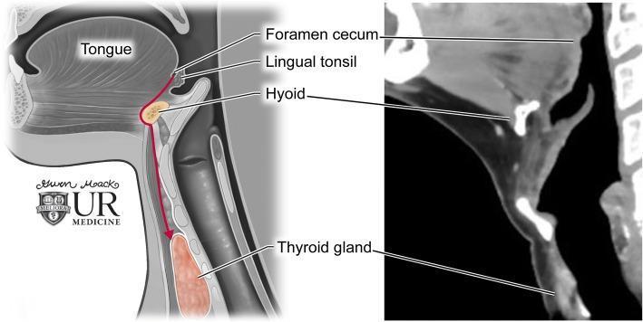
This dual-panel educational graphic illustrates thyroglossal duct anatomy and the embryological descent of the thyroid gland. On the left is a sagittal anatomical diagram showing the path of descent (indicated by a red arrow) from the foramen cecum at the base of the tongue, passing through the hyoid bone, to its final position at the thyroid gland anterior to the trachea. Key labeled structures include the tongue, lingual tonsil, foramen cecum, hyoid, and thyroid gland. The right panel displays a corresponding sagittal contrast-enhanced CT image of the neck. The CT scan highlights the radiologic appearance of these landmarks: the hyoid bone appears as a high-attenuation U-shaped structure, and the thyroid gland exhibits homogenous enhancement inferior to the thyroid cartilage. This comparison demonstrates the clinical relevance of embryological pathways in understanding the location of thyroglossal duct cysts, which can occur anywhere along this midline tract.
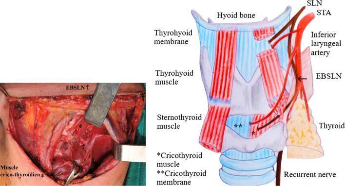
A composite medical visual consisting of a clinical intraoperative photograph (left) and a corresponding anatomical diagram (right) illustrating the anatomy of the neck and larynx. The content focuses on the anatomical variations of the External Branch of the Superior Laryngeal Nerve (EBSLN) in relation to the Superior Thyroid Artery (STA) and the thyroid gland, specifically representing Cernea Type IIb and Kierner Type III classifications. In the diagram, the EBSLN is shown descending alongside the STA and crossing it at a level below the upper pole of the thyroid gland, terminating in the cricothyroid muscle. Key labeled structures include the hyoid bone, thyrohyoid membrane/muscle, sternothyroid muscle, and cricothyroid membrane. The intraoperative photo displays the surgical field during thyroid surgery, with tissue retracted to expose the EBSLN in a clinical context. This material is designed for surgical education, highlighting the high-risk positioning of the nerve during the ligation of superior thyroid vessels.
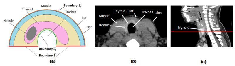
This composite educational graphic details the cross-sectional anatomy of the human neck for Finite Element (FE) thyroid simulation. (a) A 2D schematic diagram presents a simplified semicircular model of the neck. It labels the layers concentrically: skin (outermost), fat, and muscle. Inside the muscle layer, the thyroid gland is depicted in pink, containing an elliptical dark grey thyroid nodule. Boundaries are marked as Γ1 (skin surface), Γ2 (trachea boundary), and Γ3 (prescribed temperature base). (b) A corresponding axial CT scan image shows the actual anatomical cross-section, with labels identifying the skin, fat, muscle, thyroid gland, trachea (central air-filled void), and an internal nodule. The CT highlights varying tissue densities, from the dark fat layer to the intermediate density of muscle and thyroid tissue. (c) A sagittal CT view of the head and neck shows a red horizontal line indicating the anatomical plane of the axial slice in (b), positioned at the level of the thyroid gland. This visual serves as a reference for modeling heat transfer and infrared thermography in patients with thyroid nodules.
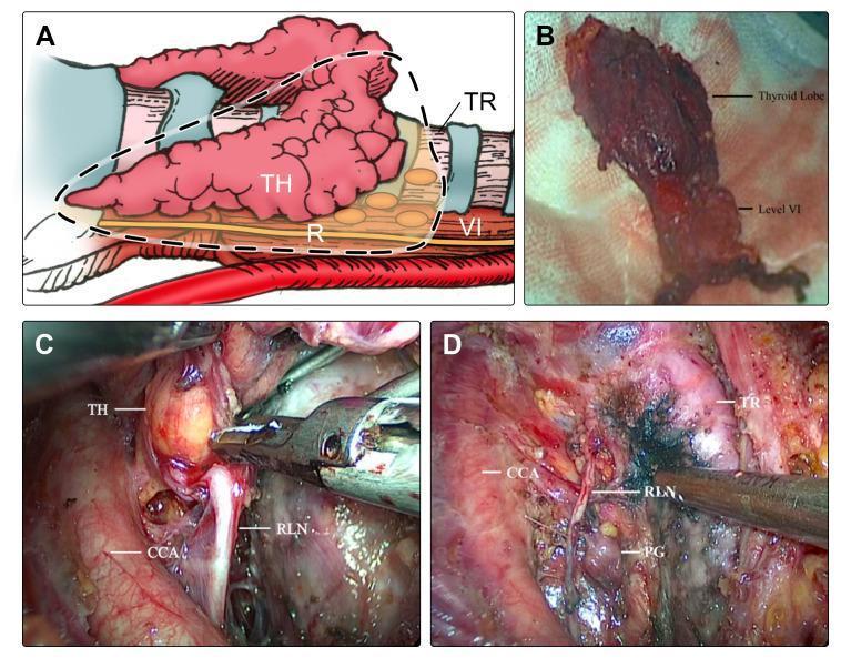
This composite figure illustrates a modified en-bloc resection technique for thyroid lobectomy and central neck dissection (CND). Panel A is an anatomical diagram showing the surgical area, including the thyroid (TH), trachea (TR), common carotid artery (CCA), and Level VI central compartment lymph nodes. Panel B displays a clinical photograph of a resected gross specimen, showing a thyroid lobe attached to a stalk of Level VI fibro-fatty tissue, demonstrating en-bloc removal. Panels C and D provide intraoperative endoscopic views during thyroidectomy. Panel C shows the surgical manipulation of the thyroid lobe (TH) in proximity to the recurrent laryngeal nerve (RLN) and the CCA. Panel D highlights the preservation of critical structures post-resection, including the RLN, parathyroid gland (PG), and trachea (TR) relative to the common carotid artery (CCA). The figure demonstrates the surgical anatomy and the objective of maintaining structural integrity of vital nerves and glands during oncologic resection of papillary thyroid carcinoma.
thyroid histology follicles colloid
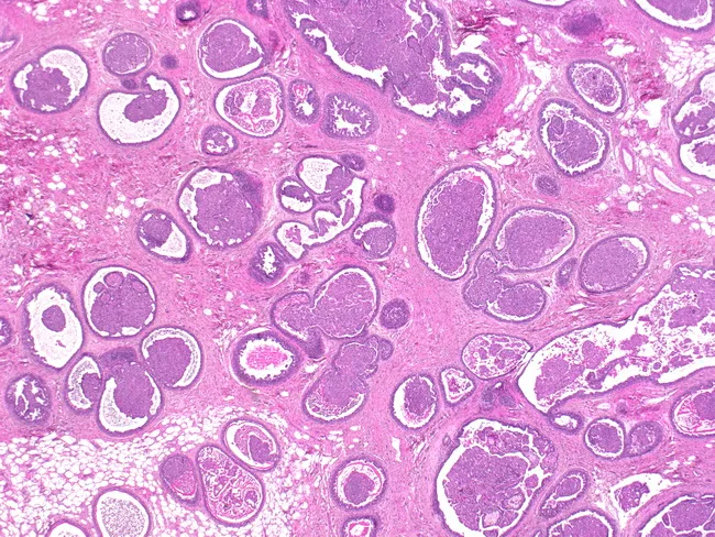
Imaging modality: Light microscopy of Hematoxylin and Eosin stained thyroid tissue. The section displays a diffuse, multilocular thyroid parenchyma with numerous round to oval follicles of varying diameter. Follicles are lined by a single layer of cuboidal to flattened epithelium, separating abundant eosinophilic colloid within expansive lumens. The colloid appears as homogenous pink material; some follicles are dilated with scant luminal epithelium, while others are more compact, creating a cobblestone-like appearance. The surrounding stroma is minimally fibrous, with occasional vascular spaces and sparse interfollicular connective tissue. There is no evident cellular atypia or overt inflammatory infiltrate in this field. Overall architecture shows a benign-appearing follicular pattern with preserved polarity and uniform follicle distribution, though follicle sizes vary, suggesting a multinodular or hyperplastic process. The image illustrates classical thyroid histology, showcasing normal thyroid parenchyma or a mild nodular change characterized by colloid-rich follicles. Diagnostic significance lies in identifying thyroid tissue with intact colloid-filled follicles, helping to differentiate benign thyroid tissue from neoplastic lesions such as follicular adenoma or follicular carcinoma when clinical data and adjunct studies are available. This image is applicable for educational demonstrations of thyroid histology, goiter pathology, and comparative endocrine pathology.
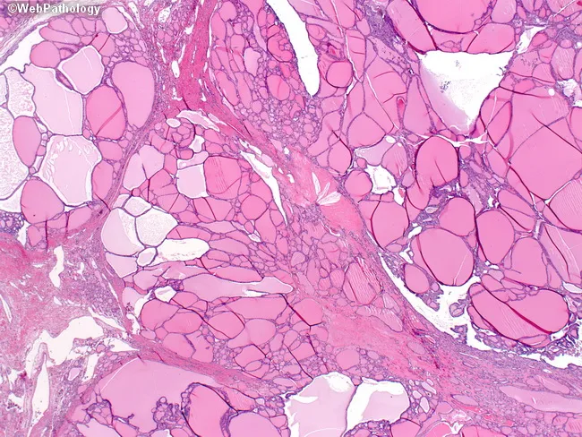
Imaging modality and technique: Bright-field light microscopy of a hematoxylin and eosin–stained thyroid tissue section. Specimen is a thyroid parenchymal biopsy/resection showing multinodular goiter with heterogenous follicle sizes. The micrograph reveals variably sized, dilated follicles interspersed with smaller hypercellular follicles; cells lining some follicles are flattened to cuboidal with scant colloid, while other follicles are large and colloid-rich, producing a biphasic appearance. The stroma is not heavily fibrotic; nodular architecture creates broad fibrous separations. Notable features include very large colloid-containing follicles, uneven distribution, and thickened septa in some areas. Overall thyroid architecture shows nodular hyperplasia without cytologic atypia; there is no invasion or malignant features evident. The image is representative of benign nodular goiter, reflecting long-standing iodine deficiency in some cases or chronic compensatory hyperplasia. Diagnostic significance: demonstrates classic histologic spectrum of multinodular goiter with variable follicle size and colloid content; important to distinguish from follicular neoplasms and inflammatory thyroid disease. Clinical correlation: correlates with clinically enlarging neck mass; usually euthyroid; histology informs management and follow-up. Feature set aligns with common teaching examples for medical education and pathology atlases, enabling search queries for terms such as multinodular goiter, colloid-rich follicles, flattened follicular epithelium, and nodular hyperplasia.
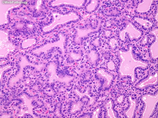
Imaging modality: light microscopy of hematoxylin and eosin–stained thyroid tissue sections. Primary subject: thyroid follicular epithelium with colloid-filled follicles. Anatomical context: thyroid gland parenchyma in the cervical region, showing follicles lined by tall columnar epithelium. Morphology: hypercellular epithelium with marked nuclear crowding creates papillary infoldings that protrude into the glandular lumens. Pale colloid is present within the follicles and margins appear scalloped. In architectural detail, papillary projections resemble papillary thyroid carcinoma but lack true fibrovascular cores, a key differentiator on histology. The follicular lumens vary in size, and colloid shows scalloping margins, consistent with benign activity rather than malignant invasion. Cytologic features include elongated nuclei with ovoid contours and scant cytoplasm; mitotic activity is not conspicuous. Immunophenotype is not assessed in this image; histology alone raises diagnostic considerations of benign hyperplastic changes including nodular hyperplasia or benign follicular adenoma with papillary-like infoldings. Diagnostic significance: recognition of papillary-like architecture without fibrovascular cores reduces the likelihood of classic papillary thyroid carcinoma, and highlights the need for correlating with nuclear features and ancillary studies. This image is relevant for surgical pathology reference, education, and differential diagnosis training in endocrine pathology. These considerations are essential for resident education, diagnostic conundrums, and interdisciplinary tumor boards.
thyroid cancer papillary follicular medullary pathology
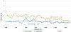
This line graph illustrates a joinpoint analysis of time trends for the age-standardized incidence of non-papillary thyroid cancer (including follicular, medullary, and anaplastic histological types) in Israel from 1980 to 2012. The data is stratified by gender within the Jewish population. The y-axis represents the rate per 100,000, while the x-axis denotes the calendar years. The visual demonstrates a statistically significant downward trend in the incidence for both Jewish women and Jewish men over the 32-year period. Jewish women (indicated by the upper green line) started with a higher incidence rate in 1980 compared to Jewish men (indicated by the lower blue line). Both groups show fluctuations year-over-year but exhibit a clear overall decline, with incidence rates narrowing and approaching lower, more similar values by 2012. This epidemiological data highlights shifting patterns in thyroid cancer pathology, contrasting the decreasing incidence of non-papillary types with the concurrent rise of papillary carcinoma observed in the same period.
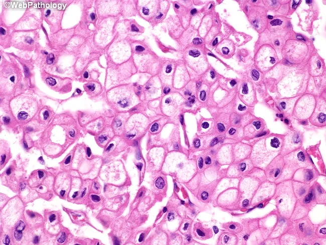
This is a histopathology slide of thyroid tissue prepared by hematoxylin and eosin staining, examined under brightfield light microscopy at high magnification. The specimen demonstrates epithelial neoplasm consistent with papillary thyroid carcinoma (PTC). The neoplastic cells are arranged in sheets and, where present, papillary fronds with fibrovascular cores are recognizable. The predominant cytoplasm is finely reticular and eosinophilic, with blander nuclear features. The nuclei show characteristic raisinoid appearance with nuclear grooves, giving an Orphan Annie eye look; the nuclear membranes are distinct and often thickened. Perinuclear halos, i.e., clear nuclear clearing around the chromatin, are evident. Some tumor cells exhibit voluminous cytoplasm, and in the plane of section, nuclei may be absent in those cells, a phenomenon of sectioning rather than true anuclear cells. The cytoplasmic clearing and grooves are diagnostic, aiding differentiation from benign nodules. The histology carries diagnostic significance, with PTC carrying generally favorable prognosis but potential for lymphatic metastasis; differential includes follicular carcinoma, medullary carcinoma, Hashimoto-related changes, and benign adenomas. Clinically, presence of these features correlates with occult metastasis risk and guides surgical planning, post-operative radioiodine therapy considerations, and molecular testing (e.g., BRAF V600E). This image is relevant for medical education, pathology teaching files, digital atlases, and research on thyroid cancer morphology.
thyroid ultrasound nodule assessment TIRADS
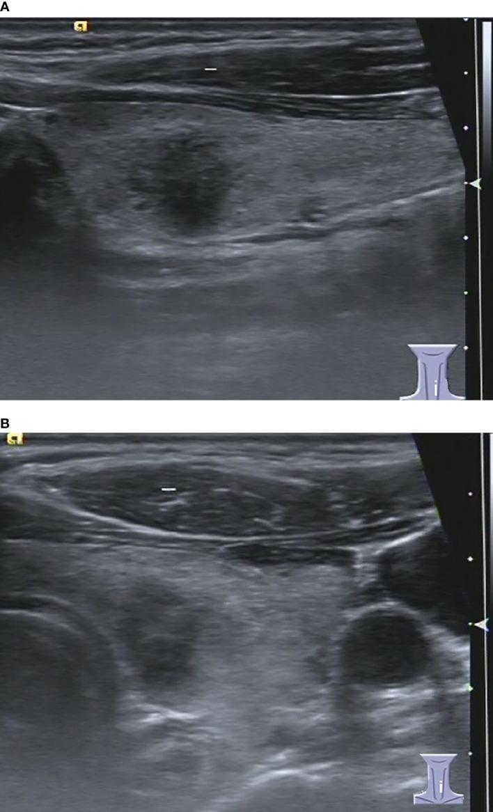
This diagnostic image displays two B-mode ultrasound scans (longitudinal view in A, axial view in B) of a human thyroid gland, demonstrating a focal nodule in the left lobe. The visual content showcases a solid, markedly hypoechoic nodule relative to the surrounding hyperechoic thyroid parenchyma. Key morphological features include an irregular shape and ill-defined margins. In the longitudinal scan (A), the nodule exhibits a 'taller-than-wide' orientation (anteroposterior diameter greater than the transverse diameter), which is a significant ultrasound finding in thyroid risk stratification. No posterior acoustic enhancement or shadowing is clearly evident. The surrounding thyroid tissue displays a heterogeneous echotexture, consistent with the patient's background of chronic lymphocytic thyroiditis. In the axial scan (B), the relationship of the thyroid lobe to adjacent cervical vascular structures is visible. This image serves as an educational example of TIRADS-based risk assessment (C-TIRADS 4C/K-TIRADS 5/ACR-TIRADS 5) for thyroid malignancies, despite the histopathological diagnosis of benign lymphocytic thyroiditis.
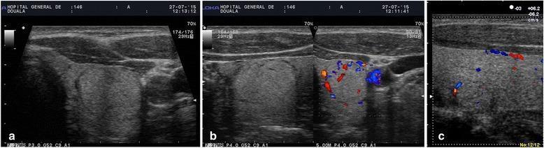
Diagnostic ultrasound imaging of the thyroid gland demonstrating characteristic TIRADS 3 thyroid incidentalomas. Panel (a) and (b) show a predominantly hyperechoic solid nodule located in the left thyroid lobe. The lesion features sharp, well-defined margins and is encircled by a peripheral hypoechoic halo. Color Doppler imaging in panel (b) reveals significant peripheral vascularization, appearing as a 'basket-weave' pattern of blood flow around the nodule's edge. Panel (c) illustrates a second nodule characterized as isoechogenic relative to the surrounding parenchyma, also exhibiting a peripheral hypoechoic halo and peripheral Doppler signals. These sonographic findings represent low-risk nodules (TIRADS 3) typically characterized by benign growth patterns, intended for medical education regarding thyroid cancer risk stratification and ultrasonographic morphological assessment.
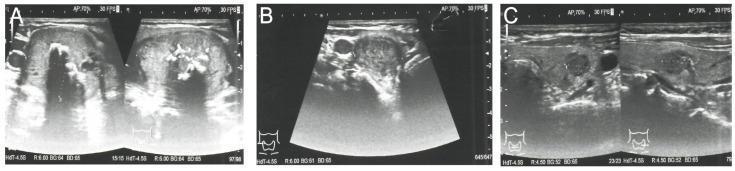
This set of grayscale ultrasound images (Figures A, B, and C) provides a diagnostic assessment of bilateral thyroid nodules in a patient with suspected medullary thyroid carcinoma (MTC). Image A displays the right thyroid lobe containing a large, dominant nodule (35 x 29 x 41 mm) characterized by heterogeneous echogenicity and internal macrocalcifications, which appear as bright echogenic foci with posterior acoustic shadowing. This lesion is classified as EU-TIRADS 4. Image B shows a second, smaller hypoechoic nodule (22 mm) in the right lobe, also classified as EU-TIRADS 4. Image C depicts the left thyroid lobe containing a 9 x 7 x 9 mm hypoechoic nodule with irregular margins, assigned a higher risk category of EU-TIRADS 5. These findings illustrate the sonographic presentation of multifocal thyroid lesions in the context of MEN2A syndrome. The variations in echogenicity, calcification patterns, and margin definition are key diagnostic features used for risk stratification and determining the necessity for fine-needle aspiration (FNA) biopsy.
Graves disease exophthalmos goiter hyperthyroidism clinical
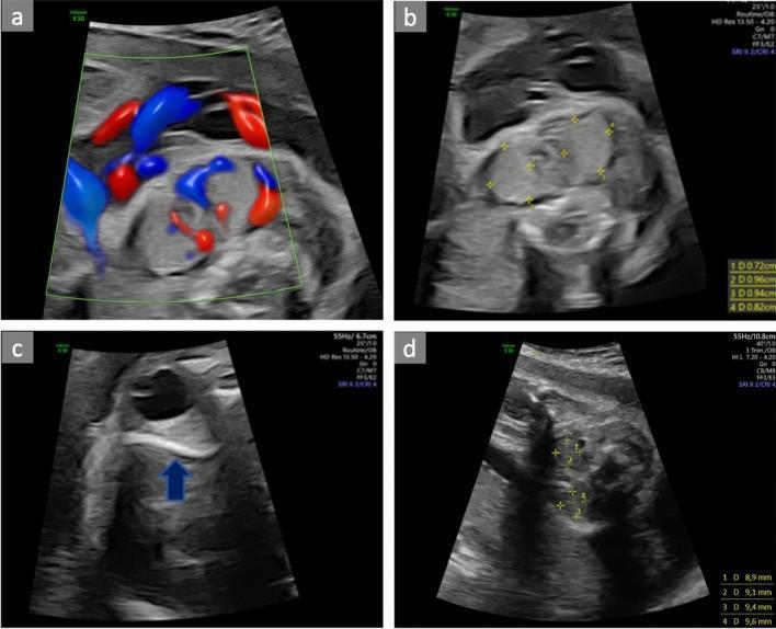
A multi-panel fetal ultrasound series demonstrating the diagnosis and monitoring of a fetal goiter associated with hyperthyroidism. Panel (a) shows a transverse view of the fetal neck utilizing color Doppler, revealing marked hypervascularity (the 'thyroid inferno' sign) within an enlarged thyroid gland. Panel (b) illustrates transverse biometry of the fetal thyroid, with calipers measuring an enlarged gland (dimensions approximately 0.72 cm to 0.96 cm). Panel (c) shows a fetal orbital view with a blue arrowhead pointing to retro-orbital tissue enlargement, indicative of fetal exophthalmos. Panel (d) displays a follow-up transverse ultrasound at 26 weeks, showing calipers measuring a reduced thyroid size (8.9 mm to 9.6 mm) following maternal propylthiouracil (PTU) treatment. The series illustrates the clinical management of fetal Graves' disease, highlighting key diagnostic markers: fetal goiter, increased vascularity, and associated signs like exophthalmos, as well as the utility of serial biometry for assessing therapeutic response in utero.
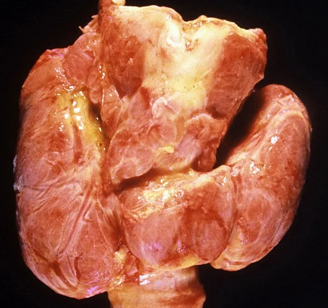
Imaging modality and technique: Gross autopsy photograph of the thyroid gland. The view is a frontal, macroscopic examination of the neck showing diffuse enlargement of both lobes with smooth surfaces and no discrete nodules. The tissue is pink to tan with subtle lobular architecture preserved. This gross appearance corresponds to diffuse hyperplasia and hypertrophy of thyroid follicles seen in Graves disease, producing a diffusely enlarged, symmetric goiter. There are no focal masses or inflammatory lesions evident in this specimen. Clinically, Graves disease is an autoimmune thyrotoxicosis characterized by endogenous hyperthyroidism, ophthalmopathy (exophthalmos), and dermopathy; the autopsy image reflects the thyroid component of that syndrome. Pathophysiology involves stimulation by thyroid-stimulating immunoglobulins that drive diffuse follicular hyperplasia and increased thyroid hormone synthesis. The gross findings support laboratory and clinical data such as suppressed TSH and elevated free T4/T3. Epidemiology notes peak incidence in females aged 20–40 years, with female-to-male ratio ≈10:1. This photograph is valuable for teaching gross pathology, correlating macroscopic thyroid changes with endocrinologic manifestations, and differentiating Graves disease from nodular goiters or thyroiditis in educational and research settings. It also highlights the importance of autopsy correlations in endocrine pathology. Educational utility.
thyroid FNA fine needle aspiration Bethesda cytology
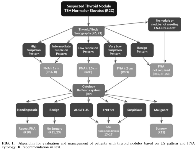
Summary : This figure presents an algorithmic flowchart for the evaluation and management of patients with suspected thyroid nodules, based on ultrasound (US) pattern and fine-needle aspiration (FNA) cytology, referencing the Bethesda system and specific recommendations.
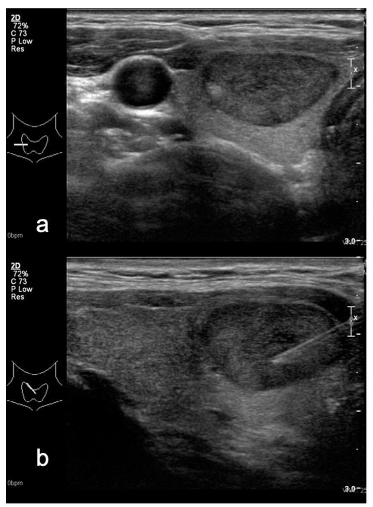
This diagnostic ultrasound image presents two views—transverse (a) and sagittal (b)—of a thyroid nodule, illustrating key features for TIRADS (Thyroid Imaging Reporting and Data System) risk stratification. In both planes, the nodule is characterized as mildly hypoechoic relative to the surrounding brighter (more echogenic) thyroid parenchyma. The nodule possesses a well-defined, ovoid-to-slightly-irregular shape without evidence of macrocalcifications. View (a) demonstrates the nodule's relationship to adjacent vascular structures, appearing as anechoic circular lumens. View (b) shows the nodule in its longitudinal axis, clearly depicting a biopsy needle positioned within the lesion, characteristic of a real-time ultrasound-guided fine needle aspiration (FNA) procedure. The image serves as an educational example of differentiating nodule echogenicity and performing diagnostic interventional procedures. Clinically, this nodule was classified as TIRADS 4A (Kwak system) and TIRADS 3 (M-TIRADS), with a subsequent Bethesda II cytology result indicating a benign process.
hashimoto thyroiditis lymphocytic infiltrate histology
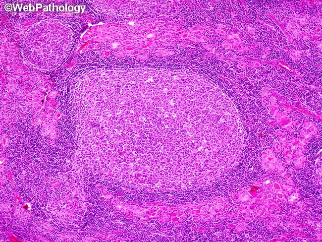
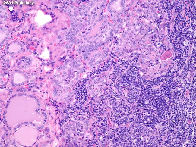
Imaging modality: Light microscopy of thyroid tissue; Hematoxylin and eosin stained; 400x magnification. The specimen shows thyroid parenchyma with dense mononuclear inflammatory infiltrate. There are well-developed germinal centers within lymphoid follicles indicating lymphoid folliculopathy; the normal thyroid follicles are small and atrophic and exhibit prominent Hurthle cell (oxyphilic) change with abundant granular eosinophilic cytoplasm. The interface between infiltrate and residual follicles is irregular; the architecture is disrupted with loss of colloid and follicular cell atrophy. The infiltrate is predominantly lymphocytic with plasma cells; occasionally macrophages; formation of organized lymphoid follicles suggests autoimmune thyroiditis. Clinically Hashimoto's disease is associated with anti-thyroid antibodies (anti-TPO, anti-thyroglobulin); this histology correlates with autoimmune hypothyroidism in adults, typically female, around age 60; pediatric cases occur. The diagnostic significance lies in identifying chronic lymphocytic thyroiditis and distinguishing from acute or granulomatous thyroiditis; Hashimoto's increases risk for thyroid lymphoma (MALT, diffuse large B-cell). Differential considerations include subacute thyroiditis, de Quervain's granulomatous thyroiditis, and radiation-induced changes. This image exemplifies classic Hashimoto histopathology: lymphoid follicle formation, Hurthle cell metaplasia, and thyroid parenchymal destruction. Potential clinical applications include educational reference for medical students and residents, differential diagnosis practice, and correlation with serologic autoimmune markers (anti-TPO, anti-Tg) and hypothyroid management.
This histopathology image shows thyroid parenchyma with dense lymphocytic infiltration in the interstitium and prominent germinal center formation, captured on a hematoxylin and eosin stained section. The infiltrate is predominantly CD3+ T cells, with a minority of CD20+ B cells and scattered mature plasma cells. Hyperplastic follicles and well-developed germinal centers are evident in the top-center region, accompanied by preserved thyroid parenchyma interspersed with lymphoid aggregates. The histology demonstrates features consistent with autoimmune thyroiditis, classically seen in Graves disease: chronic lymphocytic infiltration with follicular hyperplasia and organized lymphoid architecture. There may be mild disruption of thyroid follicles, with colloid depletion? In Graves, hyperplasia and lymphoid infiltration; However, the image emphasizes lymphoid follicle formation and interstitial T-cell predominance. Diagnostic significance: correlates with autoimmune thyroid disease activity, supports a diagnosis of Graves-related thyroiditis or Hashimoto-like lymphocytic thyroiditis; appropriate differential considerations include Hashimoto thyroiditis with germinal centers, follicular lymphoma should be considered in unusual clonal proliferation, and other inflammatory processes. This image is valuable for education on thyroid histology, autoimmune pathology, and the morphologic spectrum of lymphoid-rich thyroiditis. Potential clinical use cases include medical education, pathology exam review, and correlation with thyroid function tests and autoantibody profiles to evaluate disease activity and treatment response.
THYROID GLAND - Comprehensive PG VIVA Guide
1. EMBRYOLOGY
- Arises as a thickening of endodermal epithelium in the floor of the pharynx (foramen cecum - at junction of anterior 2/3 and posterior 1/3 of tongue)
- Descends caudally, anterior to the hyoid bone and laryngeal cartilages
- Reaches final position anterior to the 2nd-3rd tracheal rings by week 7
- The path of descent forms the thyroglossal duct, which normally obliterates
- Remnants persist as: thyroglossal duct cyst (most common midline neck cyst), lingual thyroid, or pyramidal lobe
- Derived from the 4th pharyngeal pouch (ultimobranchial bodies)
- Contributes parafollicular C-cells (calcitonin-secreting) which make up 1-2% of thyroid cells
Embryological pearl for VIVA: The foramen cecum marks the site of origin. Before operating on a midline neck mass, always confirm the presence of normal thyroid tissue elsewhere - a lingual thyroid may be the patient's only functioning thyroid tissue.
2. ANATOMY
Gross Anatomy
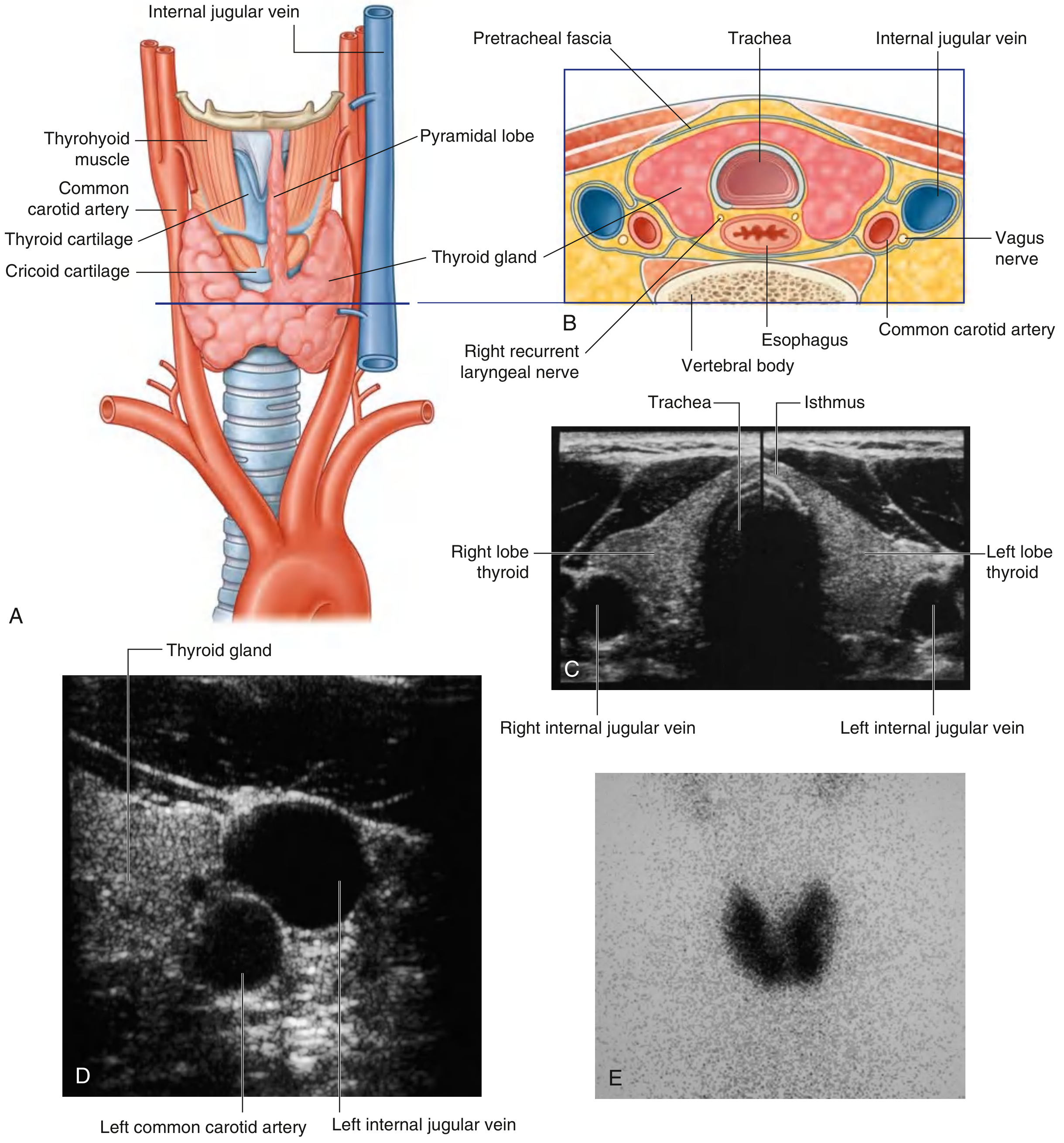
- Location: Anterior neck, posterior to strap muscles (sternohyoid, sternothyroid, omohyoid), within the pretracheal fascia (visceral compartment)
- Shape: H-shaped / butterfly-shaped; two lateral lobes connected by an isthmus
- Isthmus: Crosses anterior to 2nd-3rd tracheal rings
- Pyramidal lobe: Present in ~50% of people; ascends from the isthmus, usually to the left, toward the hyoid - remnant of thyroglossal duct
- Size (adult): Each lobe ~4-5 cm long, 2-3 cm wide; total weight ~25-30 g
- Color and consistency: Brown, firm; highly vascular gland
- Tubercle of Zuckerkandl: Posterior extension of the lateral thyroid lobe near the cricoid cartilage - an important surgical landmark for the RLN
Fascial Relations
- Enclosed within the pretracheal fascia (visceral layer of deep cervical fascia)
- Berry's ligament (posterior suspensory ligament): Condensation of pretracheal fascia attaching the posterior thyroid to the cricoid cartilage - the RLN passes in close proximity here
- Strap muscles lie anterior; trachea and esophagus lie posterior; carotid sheath lies lateral
Blood Supply
| Artery | Origin | Supplies |
|---|---|---|
| Superior thyroid artery | First branch of external carotid artery | Superior pole; divides into anterior and posterior glandular branches |
| Inferior thyroid artery | Thyrocervical trunk (from 1st part of subclavian) | Posterior and inferior thyroid; both parathyroid glands |
| Thyroidea ima artery | Aorta or innominate (1-4% of people) | Isthmus or inferior poles; midline course |
- Superior thyroid veins: Drain into internal jugular vein (with superior thyroid arteries)
- Middle thyroid veins: Drain into internal jugular vein (most variable; no accompanying artery)
- Inferior thyroid veins: Form a plexus, drain into brachiocephalic veins
Nerve Supply - Critical Surgical Anatomy
- Left RLN: Arises from vagus at the aortic arch, loops around the ligamentum arteriosum, ascends medially in the tracheoesophageal groove
- Right RLN: Arises from vagus at the right subclavian artery, passes posterior to it, more oblique course
- Non-recurrent RLN: Right side 0.5-1% (associated with aberrant right subclavian artery); left side - extremely rare (situs inversus + right aortic arch)
- Both RLNs cross the inferior thyroid artery - the nerve may pass anterior, posterior, or interdigitate with its branches
- Injury causes: ipsilateral vocal cord paralysis (hoarseness); bilateral injury = respiratory obstruction
- Travels with the superior thyroid artery and is at risk during ligation of the superior thyroid vessels
- Supplies the cricothyroid muscle (voice pitch modulation)
- Injury: loss of high-pitched voice (Cernea classification guides risk)
Lymphatic Drainage
- Level VI (central compartment): First echelon nodes for thyroid; bounded superiorly by hyoid, inferiorly by innominate artery; includes prelaryngeal, pretracheal, paratracheal nodes
- Levels II, III, IV (lateral neck/jugular chain): Second echelon
- Level VII (superior mediastinal): Third echelon; important for low thyroid cancers
3. HISTOLOGY
- Follicles: Spherical units lined by a single layer of cuboidal follicular cells (thyrocytes); lumen filled with colloid (thyroglobulin)
- Follicular cells: Cuboidal at rest, columnar when active; synthesize T3/T4
- Colloid: Gelatinous, homogeneous eosinophilic material; reservoir of stored thyroid hormone
- Parafollicular C-cells: Located between follicles; secrete calcitonin; increase in medullary thyroid carcinoma
- Stroma: Minimal connective tissue septae; abundant fenestrated capillaries
4. PHYSIOLOGY - THYROID HORMONE SYNTHESIS
Steps of Synthesis (Wolf-Chaikoff effect, Plummer-Vinson mnemonic)
- Iodide trapping: Active transport of I- into follicular cells by Na+/I- symporter (NIS) on basolateral membrane - stimulated by TSH
- Oxidation: I- → I (active iodine) by thyroid peroxidase (TPO) using H₂O₂
- Organification: Active iodine binds to tyrosine residues on thyroglobulin → Monoiodotyrosine (MIT) and Diiodotyrosine (DIT)
- Coupling:
- MIT + DIT = T3 (triiodothyronine)
- DIT + DIT = T4 (thyroxine/tetraiodothyronine) Catalyzed by TPO
- Storage: T3 and T4 stored in colloid as thyroglobulin
- Secretion: Colloid reabsorbed by endocytosis → lysosomes cleave T3/T4 from thyroglobulin → secreted into bloodstream
Transport in Blood
-
99% bound to Thyroxine-Binding Globulin (TBG), transthyretin, albumin
- Free T3 and Free T4 are biologically active forms
- T4 is a prohormone; peripherally converted to T3 by deiodinases (mainly type 1 and 2)
Hypothalamic-Pituitary-Thyroid Axis
- TRH (hypothalamus) → stimulates TSH release from anterior pituitary
- TSH (anterior pituitary) → stimulates all steps of thyroid hormone synthesis, gland growth
- Negative feedback: T3/T4 suppress both TRH and TSH
- TSH is the most sensitive test of thyroid function
Drugs Affecting Synthesis
| Drug | Mechanism |
|---|---|
| Propylthiouracil (PTU) | Inhibits TPO (organification + coupling); inhibits peripheral T4→T3 conversion |
| Methimazole/Carbimazole | Inhibits TPO (organification + coupling) |
| High-dose iodine (Wolff-Chaikoff) | Transiently inhibits organification |
| Lithium | Inhibits thyroglobulin proteolysis and release |
| Amiodarone | Iodine overload + direct thyrocyte toxicity; causes both hypo and hyperthyroidism |
5. THYROID FUNCTION TESTS (Assessment)
| Test | Normal Range | Significance |
|---|---|---|
| TSH | 0.5-4.5 mIU/L | Most sensitive; first-line screening test |
| Free T4 | 0.8-1.8 ng/dL | Confirms primary thyroid dysfunction |
| Free T3 | 2.3-4.1 pg/mL | More potent hormone; used in T3 toxicosis |
| Total T4/T3 | Affected by TBG changes | Less clinically useful |
| Anti-TPO antibody | Hashimoto's; also present in Graves' | |
| Anti-thyroglobulin Ab | Hashimoto's thyroiditis | |
| TSH receptor Ab (TRAb/TSI) | Graves' disease (diagnostic) | |
| Thyroglobulin (Tg) | Tumor marker post-total thyroidectomy for DTC | |
| Calcitonin | Medullary thyroid carcinoma (MTC) screening/marker |
- Low TSH + High T4/T3 = Hyperthyroidism (primary)
- High TSH + Low T4 = Hypothyroidism (primary)
- High TSH + Normal T4 = Subclinical hypothyroidism
- Low TSH + Normal T4 = Subclinical hyperthyroidism
- Low TSH + Low T4 = Central (secondary) hypothyroidism
6. THYROID IMAGING
Ultrasound (USG Neck)
- First-line imaging for all thyroid pathology
- Assesses: size, echogenicity, nodules, vascularity (Doppler), lymph nodes
- Features suggesting malignancy (ACR TI-RADS scoring system):
- Solid composition (vs. spongiform = benign)
- Very hypoechoic (more hypo than strap muscles)
- Taller-than-wide shape
- Irregular/lobulated margins
- Punctate echogenic foci (microcalcifications)
- Extrathyroidal extension (= definite malignancy)
- TR1 (benign) → TR2 (not suspicious) → TR3 (mildly suspicious) → TR4 (moderately suspicious) → TR5 (highly suspicious)
- FNA is recommended based on TR level and nodule size
Radionuclide Scintigraphy
- Technetium-99m (99mTc) pertechnetate: Trapped but not organified; quick and inexpensive
- Iodine-123 (¹²³I): Trapped and organified; physiological
- Iodine-131 (¹³¹I): Therapeutic doses; also used for Radioactive Iodine Uptake (RAIU)
- RAIU: Quantifies iodine uptake at 4 and 24 hours
- Increased RAIU = Graves' disease, toxic nodular goiter
- Decreased RAIU = thyroiditis (destructive), iodine excess, factitious hyperthyroidism
- "Hot" nodule (increased uptake) = almost always benign
- "Cold" nodule (decreased uptake) = 10-20% malignant risk → requires FNA
CT/MRI
- Not first-line; used for:
- Substernal (intrathoracic) goiter assessment (extent, tracheal compression)
- Locally advanced thyroid cancer (invasion assessment)
- If iodinated contrast used - wait 4-8 weeks before RAI treatment
- CT appearance: homogeneous, mildly hyperattenuating on non-contrast; bright enhancement with IV contrast
PET Scan (¹⁸F-FDG)
- Reserved for RAI-refractory differentiated thyroid cancer (DTC)
- Incidental FDG-avid thyroid nodule ("incidentaloma") has ~30-35% malignancy risk
7. FINE-NEEDLE ASPIRATION (FNA) - Bethesda System
- Most accurate, cost-effective first-line investigation for thyroid nodules
- Sensitivity >80%, specificity >90%
- Performed with 23-27 gauge needle; ultrasound-guidance improves accuracy
| Bethesda Category | Diagnosis | Malignancy Risk | Management |
|---|---|---|---|
| I | Non-diagnostic | 5-10% | Repeat FNA |
| II | Benign | <3% | Clinical/USG follow-up |
| III | AUS/FLUS (Atypia of undetermined significance) | 10-30% | Repeat FNA or molecular testing |
| IV | Follicular neoplasm / suspicious for FN | 25-40% | Lobectomy |
| V | Suspicious for malignancy | 50-75% | Total thyroidectomy or lobectomy |
| VI | Malignant | 97-99% | Total thyroidectomy |
8. THYROID DISORDERS AND MANAGEMENT
8A. HYPOTHYROIDISM
- Hashimoto's thyroiditis (most common worldwide; autoimmune; anti-TPO and anti-Tg Ab)
- Post-thyroidectomy / post-RAI (iatrogenic)
- Iodine deficiency (most common globally in developing world)
- Medications: lithium, amiodarone, interferon-alpha
- Central hypothyroidism (rare): pituitary/hypothalamic disease
- Fatigue, cold intolerance, weight gain, constipation
- Dry skin, hair loss, periorbital puffiness, macroglossia
- Bradycardia, diastolic hypertension
- Menorrhagia, infertility
- Delayed tendon reflexes (classic sign)
- Myxedema coma: hypothermia, respiratory failure, coma (life-threatening)
- Autoimmune destruction of thyroid follicular cells
- Histology: dense lymphocytic infiltration with germinal center formation, Hurthle cell (oxyphilic) metaplasia, atrophic follicles
- Increased risk of thyroid lymphoma (MALT type)
- Levothyroxine (L-T4) replacement; starting dose 1.6 mcg/kg/day
- Target TSH: 0.5-2.5 mIU/L (general population); 0.1-2.5 mIU/L (pregnant)
- Myxedema coma: IV T4 + T3, hydrocortisone, supportive ICU care
8B. HYPERTHYROIDISM
- Graves' disease (~70-80%)
- Toxic Multinodular Goiter (MNG) - Plummer's disease
- Toxic Adenoma (solitary autonomous nodule)
- Thyroiditis (subacute/De Quervain's, postpartum - destructive hyperthyroidism)
- Amiodarone-induced thyrotoxicosis (type 1 = iodine excess; type 2 = destructive)
- Rare: TSH-secreting pituitary adenoma, struma ovarii, factitious
- Palpitations, tachycardia/atrial fibrillation, heat intolerance, sweating
- Weight loss despite good appetite
- Tremor, hyperreflexia, anxiety, insomnia
- Oligomenorrhea, infertility
- Thyroid storm: hyperthermia (>40°C), tachycardia, altered consciousness, multiorgan failure (Burch-Wartofsky score used)
- Diffuse goiter (TRAb/TSI stimulate all follicular cells)
- Ophthalmopathy (exophthalmos/proptosis) - present in 25-50%
- Pretibial myxedema (dermopathy) - present in 5%
- (4th sign: Thyroid acropachy - clubbing-like periosteal reaction; rare)
| Option | Notes |
|---|---|
| Antithyroid drugs (ATD): Methimazole (preferred) or PTU | First-line; 30-40% remission rate after 12-18 months; PTU preferred in pregnancy first trimester and thyroid storm |
| Radioactive Iodine (RAI ¹³¹I) | Most commonly used in USA; destroys thyroid tissue; contraindicated in pregnancy, severe ophthalmopathy, large compressive goiter |
| Surgery (Total thyroidectomy) | Preferred when: severe ophthalmopathy, large goiter, suspicious nodule, pregnancy/breastfeeding, rapid control needed, failure/contraindication to other options; near 0% recurrence with total thyroidectomy |
8C. GOITER
- Diffuse enlargement without nodules; most commonly due to iodine deficiency
- Endemic goiter: iodine deficiency affecting >10% of population in a region
- Treatment: iodine supplementation; levothyroxine for suppression; surgery for compression
- Multiple nodules of varying size; most euthyroid
- Compression symptoms: dysphagia, stridor, SVC syndrome (substernal goiter)
- Pemberton's sign: facial congestion/arm elevation with arm raising (substernal goiter)
- CT/MRI for substernal extent; total thyroidectomy for compressive/retrosternal cases
- Clinical evaluation (history, physical exam - size, consistency, fixity, LN)
- TSH level (if low → scintigraphy; if normal/high → ultrasound)
- USG neck → TI-RADS scoring
- FNA for nodules meeting size/risk criteria → Bethesda classification
- Molecular testing if Bethesda III/IV
8D. THYROID CANCER
| Type | Origin | Frequency | Key Features |
|---|---|---|---|
| Papillary Thyroid Cancer (PTC) | Follicular cells | 70-80% | Most common; excellent prognosis; lymphatic spread; BRAF V600E mutation (60%) |
| Follicular Thyroid Cancer (FTC) | Follicular cells | 10-15% | Vascular/hematogenous spread; RAS mutation, PAX8-PPARγ; FNA cannot distinguish FTC from adenoma |
| Medullary Thyroid Cancer (MTC) | Parafollicular C-cells | 5-10% | Calcitonin elevated; associated with MEN 2A and 2B; RET proto-oncogene mutation |
| Anaplastic (Undifferentiated) | Follicular cells | <2% | Worst prognosis; rapidly fatal; not iodine-avid |
| Primary Thyroid Lymphoma | B lymphocytes | Rare | Background of Hashimoto's; MALT or DLBCL |
- Papillary structures with fibrovascular cores
- Orphan Annie eye nuclei (ground-glass/empty-looking nuclei with nuclear grooves)
- Nuclear pseudoinclusions
- Psammoma bodies (laminated calcifications) - pathognomonic
- Spreads via lymphatics (cervical LN metastases)
- Diagnosed only by capsular or vascular invasion (FNA cannot diagnose - shows follicular neoplasm)
- Spreads hematogenously → lungs, bone
- Minimally invasive FTC has excellent prognosis
- NIFTP (Non-Invasive Follicular Thyroid Neoplasm with Papillary-like nuclear features): essentially benign; lobectomy sufficient
- Calcitonin marker (serum calcitonin + CEA)
- 25% hereditary (MEN 2A: MTC + pheochromocytoma + parathyroid hyperplasia; MEN 2B: MTC + pheochromocytoma + mucosal neuromas + marfanoid habitus)
- RET mutation testing: screen all first-degree relatives
- Not iodine-avid; RAI not useful
- Amyloid deposits in stroma on histology
- Low risk: Intrathyroidal PTC <4 cm, no LN or distant mets
- Intermediate risk: Minor extrathyroidal extension, vascular invasion, >5 cervical LN mets
- High risk: Gross extrathyroidal extension, distant mets, >3 cm, incomplete resection
Surgical Management of Thyroid Cancer
| Scenario | Surgery |
|---|---|
| PTC/FTC ≥1 cm OR any high-risk features | Total thyroidectomy |
| Low-risk PTC 1-4 cm without ETE | Total thyroidectomy OR thyroid lobectomy (ATA 2015) |
| PTC <1 cm (microcarcinoma), low risk | Active surveillance OR lobectomy |
| Cervical LN metastases | Therapeutic central (Level VI) ± lateral neck dissection |
| Prophylactic central neck dissection | Controversial; considered for T3/T4 tumors |
-
Radioactive Iodine (RAI ¹³¹I):
- Ablates remnant thyroid tissue and residual/metastatic disease
- Required for high-risk patients; considered for intermediate risk; NOT for low-risk
- Require stimulation with TSH (stop levothyroxine → hypothyroid state OR rhTSH injection)
-
TSH Suppression with Levothyroxine:
- High-risk recurrence: TSH <0.1 mIU/L
- Intermediate risk: TSH 0.1-0.5 mIU/L
- Low risk (no evidence of disease): TSH 0.5-2.0 mIU/L (no suppression needed)
-
Surveillance:
- Serum thyroglobulin (Tg) every 6-12 months (tumor marker post-total thyroidectomy)
- Neck ultrasound at 6-12 months postoperatively, then per risk
- Anti-Tg antibodies: if elevated, Tg unreliable; use Ab titer as surrogate marker
- Whole-body RAI scan if Tg rising or structural recurrence suspected
-
Systemic Therapy (RAI-refractory disease):
- Multikinase inhibitors: Sorafenib (400mg BD) or Lenvatinib (24mg OD) - FDA approved
- BRAF inhibitors for BRAF V600E-mutated PTC
- RET-selective inhibitors: Selpercatinib, Pralsetinib for RET-altered MTC/DTC
- NTRK inhibitors: Larotrectinib, Entrectinib for NTRK fusion-positive tumors
9. THYROID SURGERY (Thyroidectomy)
Indications
- Thyroid cancer (or suspected malignancy on FNA)
- Compressive/obstructive goiter (dysphagia, dyspnea, Pemberton's sign)
- Graves' disease (selected cases)
- Toxic MNG / Toxic adenoma
- Cosmesis
Nomenclature
| Term | Definition |
|---|---|
| Hemithyroidectomy/Lobectomy | Removal of one lobe + isthmus |
| Near-total thyroidectomy | Leaves <1g of tissue (near Berry's ligament) |
| Total thyroidectomy | Complete removal of all thyroid tissue |
| Subtotal thyroidectomy | Leaves >1g of posterior thyroid tissue |
Complications
| Complication | Cause | Rate |
|---|---|---|
| Recurrent laryngeal nerve injury | Direct damage/stretch | Temporary: 2-4%; Permanent: 0-2% |
| Hypoparathyroidism | Inadvertent parathyroid removal or devascularization | Temporary: 10-30%; Permanent: 1-6% |
| Neck hematoma | Postoperative bleeding | 0.3-1%; can be life-threatening (airway) |
| Thyroid storm | Uncontrolled Graves' | Rare with proper preop preparation |
| Seroma | Fluid collection | Minor complication |
| Wound infection | Uncommon | <1% |
- Check PTH at 1-hour post-op; if low (<10 pg/mL) → high risk
- Symptoms: perioral numbness, Chvostek's sign (+ve face twitch), Trousseau's sign (+ve carpopedal spasm)
- Treat: calcium gluconate IV for acute; oral calcium + vitamin D for prophylaxis
Molecular Pathways in Thyroid Cancer (VIVA hotspot)
- MAPK pathway: BRAF V600E (most common, ~60% PTC), RAS mutations, RET/PTC rearrangements
- PI3K/AKT pathway: RAS mutations (follicular carcinoma predominantly), PTEN mutations
- RET point mutations: Hereditary MTC (codon-specific mutations predict aggressiveness)
- Cancer genome atlas (TCGA) 2014: >96% of PTCs can be molecularly characterized
10. QUICK VIVA REFERENCE POINTS
| Question | High-Yield Answer |
|---|---|
| Most sensitive test of thyroid function | TSH |
| Most common thyroid malignancy | Papillary thyroid carcinoma (70-80%) |
| Most common cause of hypothyroidism in developed countries | Hashimoto's thyroiditis |
| Most common cause globally | Iodine deficiency |
| Pathognomonic feature of PTC | Orphan Annie nuclei + psammoma bodies |
| Why can't FNA diagnose follicular carcinoma? | Cannot assess capsular/vascular invasion (cytological features are identical to adenoma) |
| Most common mutation in PTC | BRAF V600E (~60%) |
| Marker for MTC | Calcitonin (+ CEA) |
| Non-recurrent RLN - which side more common? | Right side (0.5-1%), associated with aberrant right subclavian artery |
| Surgery preferred in Graves' with severe eye disease | Total thyroidectomy (RAI worsens ophthalmopathy) |
| What is Berry's ligament? | Posterior suspensory ligament of thyroid to cricoid; RLN runs nearby |
| What is Tubercle of Zuckerkandl? | Posterior extension of thyroid lobe; surgical landmark for RLN |
| What is NIFTP? | Non-Invasive Follicular Thyroid Neoplasm with Papillary-like nuclear features; essentially benign; lobectomy sufficient |
| Bethesda IV risk and management | 25-40% malignancy risk; diagnostic lobectomy |
| Pemberton's sign | Facial flushing/SVC obstruction on arm raising - substernal goiter |
| Thyroid storm scoring | Burch-Wartofsky Point Scale |
| Best surgical option for <1 cm low-risk PTC | Active surveillance OR lobectomy (ATA 2015) |
Generate thyroid VIVA Q&A flash cards document
ls -la /tmp/workspace/ 2>/dev/null || mkdir -p /tmp/workspace && echo "workspace ready"
All the viva aspect only in very much detail