
Act as a World-Class Professor of General Medicine, Clinical Pharmacology, Pathology, and Medical Education. Teach me Malaria disease in a simple, exam-oriented, and clinically practical manner. For every disease, follow this format: 1. Definition - One-line exam definition - Simple explanation 2. Causes (Etiology) - Common causes - Important risk factors 3. Types / Classification - Brief explanation of each type 4. Pathophysiology - Step-by-step flowchart with arrows - Easy to understand 5. Clinical Features - Symptoms - Signs - Explain why important symptoms occur 6. Key Symptoms That Suggest the Diagnosis - Hallmark symptom - Classic clinical clues - Red flag findings
"malaria" AND "clinical features" AND "pathophysiology"
🦟 MALARIA - Complete Exam-Oriented Clinical Guide
1. DEFINITION
2. CAUSES (ETIOLOGY)
The Five Plasmodium Species
| Species | Type of Malaria | Severity |
|---|---|---|
| P. falciparum | Malignant tertian (falciparum) | Most dangerous - HIGH fatality |
| P. vivax | Benign tertian | Moderate - can relapse |
| P. ovale | Benign tertian | Mild - can relapse |
| P. malariae | Quartan malaria | Mild - chronic |
| P. knowlesi | Simian/zoonotic | Moderate-severe (misidentified as P. malariae) |
Exam tip: P. falciparum is responsible for almost all malaria deaths. P. knowlesi (Southeast Asia) looks like P. malariae on smear but behaves like falciparum.
Vector
- Only the female Anopheles mosquito transmits malaria
- Humans are the only natural reservoir
Other (Rare) Routes of Transmission
- Blood transfusion
- Needle sharing (IV drug users)
- Organ transplant
- Vertical (mother to fetus - congenital malaria)
Important Risk Factors
| Risk Factor | Reason |
|---|---|
| Travel to/residence in endemic zones (sub-Saharan Africa, Southeast Asia, South Asia, Central/South America) | Exposure to infected mosquitoes |
| No chemoprophylaxis taken | Loss of preventive protection |
| No bed nets or repellent use | Direct vector exposure |
| Children < 5 years | Immature immunity |
| Pregnant women | Immunosuppression + placental sequestration |
| Non-immune travelers | No prior partial immunity |
| Sickle cell trait (HbS heterozygous) | Protective against severe disease |
| Asplenia | Loss of red cell filtering - worse disease |
Exam pearl: Sickle cell trait, thalassemia, G6PD deficiency, and other red cell defects are most prevalent in malaria-endemic regions because heterozygous states confer protection from severe malaria - a classic example of balanced polymorphism.
3. TYPES / CLASSIFICATION
By Species (Most Clinically Important Classification)
- Fever every 48 hours (but often irregular in practice)
- Invades ALL ages of RBCs → high parasitemia
- Produces PfEMP1 → RBC sequestration in microvasculature
- Only species causing cerebral malaria, blackwater fever, multiorgan failure
- No hypnozoites → no true relapse (but recrudescence occurs)
- Drug resistance is a major problem
- Fever every 48 hours
- Only infect young/reticulocyte RBCs → lower parasitemia
- Contain hypnozoites (dormant liver stage) → TRUE RELAPSE after months/years
- Requires primaquine to eliminate liver stage
- P. ovale: found mainly in West Africa
- Fever every 72 hours ("quartan" = 4-day cycle)
- Infects old RBCs only
- Very chronic, low-grade
- Associated with nephrotic syndrome in children (immune complex deposition)
- No hypnozoites but can persist for decades
- 24-hour fever cycle (quotidian)
- Zoonotic from macaque monkeys in Southeast Asia (Borneo, Malaysia)
- Morphologically mimics P. malariae on blood smear
- Clinically can be severe and fatal
Fever Periodicity Summary
| Species | Fever Cycle | Type |
|---|---|---|
| P. falciparum | 48 hours | Tertian |
| P. vivax | 48 hours | Tertian |
| P. ovale | 48 hours | Tertian |
| P. malariae | 72 hours | Quartan |
| P. knowlesi | 24 hours | Quotidian |
4. PATHOPHYSIOLOGY
Life Cycle Diagram (from Robbins Pathology)
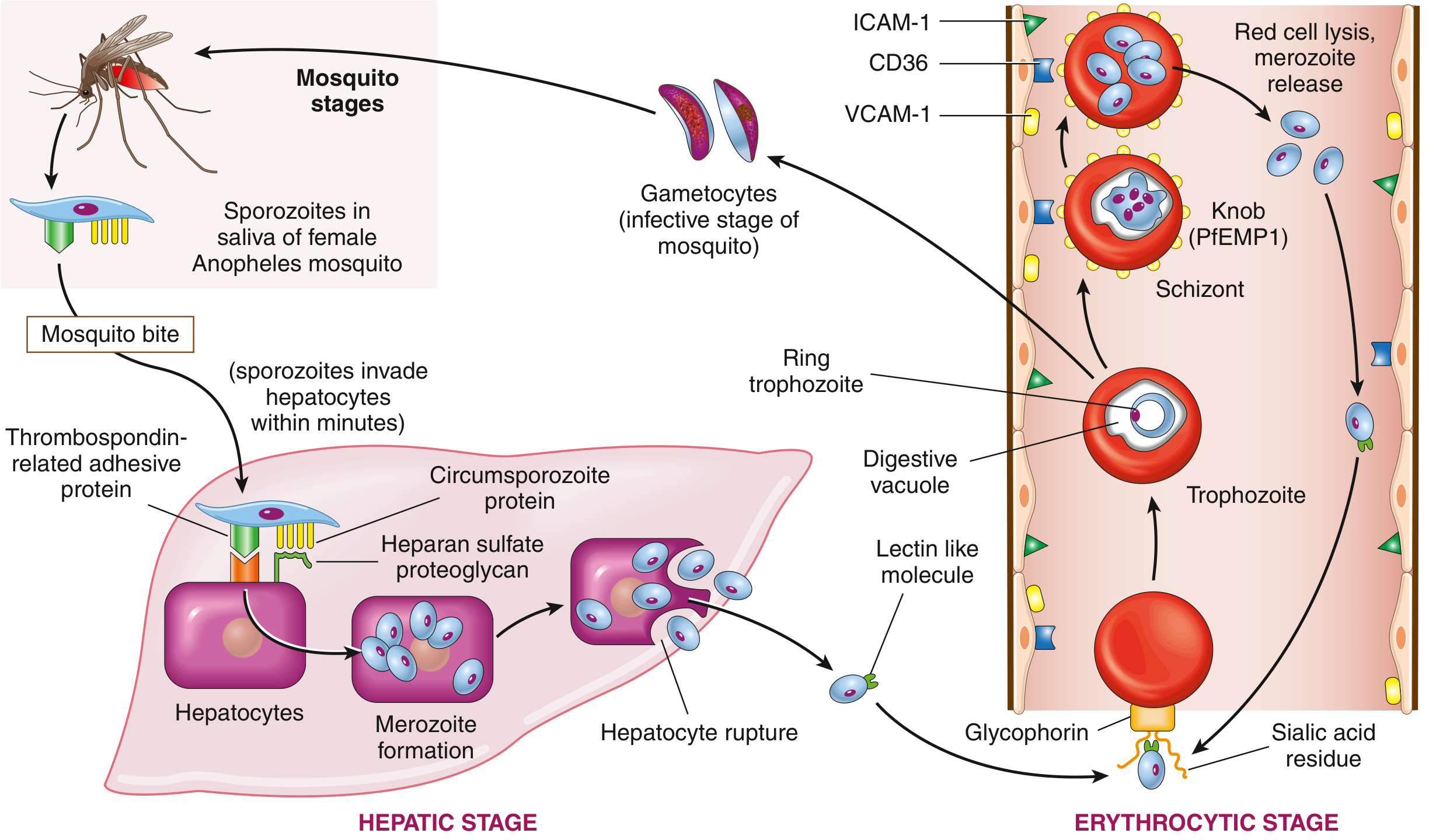
Step-by-Step Pathophysiology Flowchart
Female Anopheles mosquito bites human
↓
SPOROZOITES injected into bloodstream
↓
Sporozoites travel to LIVER within minutes
(bind via Thrombospondin-related adhesive protein +
Circumsporozoite protein → Heparan sulfate proteoglycan on hepatocytes)
↓
HEPATIC STAGE (clinically silent - 1-4 weeks incubation)
→ Sporozoites → differentiate into MEROZOITES inside hepatocytes
→ [P. vivax / P. ovale only: some form HYPNOZOITES = dormant stage → causes relapse later]
↓
Infected hepatocytes RUPTURE → release thousands of MEROZOITES into blood
↓
ERYTHROCYTIC STAGE begins (causes all clinical disease)
→ Merozoite surface lectin-like molecule binds GLYCOPHORIN (sialidated)
on RBC surface → merozoite invaginates into RBC inside a "digestive vacuole"
↓
Inside RBC: Merozoite → RING TROPHOZOITE → TROPHOZOITE → SCHIZONT
↓
SCHIZONT expresses PfEMP1 (P. falciparum erythrocyte membrane protein 1)
→ PfEMP1 forms KNOBS on RBC surface
→ Knobs bind endothelial adhesion molecules: ICAM-1, VCAM-1, CD36
→ Infected RBCs SEQUESTER in capillary beds (brain, lung, kidney, placenta)
→ This causes CEREBRAL MALARIA, ARDS, renal failure
↓
Schizonts → differentiate into MEROZOITES → RBC LYSES (every 48 or 72 hrs)
→ Merozoites released → infect new RBCs (cycle repeats)
→ RBC lysis triggers fever (TNF-α, IL-1, IL-6 release)
→ Hemozoin (brown malarial pigment = hemoglobin breakdown product) released
↓
Some trophozoites → GAMETOCYTES → ingested by next mosquito → sexual cycle restarts
Why Each Key Problem Occurs
| Manifestation | Mechanism |
|---|---|
| Cyclical fever | Synchronized RBC lysis releases merozoites + pyrogens (TNF-α, IL-1) |
| Hemolytic anemia | Destruction of parasitized RBCs + innocent bystander lysis |
| Splenomegaly | Hyperplasia of mononuclear phagocytes clearing parasitized RBCs; hemozoin deposition |
| Hepatomegaly | Same mechanism + hemozoin deposits in liver |
| Thrombocytopenia | Splenic sequestration + immune-mediated platelet destruction |
| Cerebral malaria | PfEMP1-mediated RBC sequestration in cerebral microvasculature → obstruction + inflammation |
| Blackwater fever | Massive intravascular hemolysis → hemoglobinemia + hemoglobinuria |
| Renal failure | Hemoglobinuria, hypovolemia, immune complex deposition, direct tubular toxicity |
| ARDS | Sequestration of RBCs in pulmonary microvasculature + inflammatory cytokines |
5. CLINICAL FEATURES
The Classic Malarial Paroxysm (Three Stages)
COLD STAGE (15-60 min)
→ Rigors (violent shaking chills), goose bumps, teeth chattering
→ Temperature RISING
↓
HOT STAGE (2-6 hours)
→ High fever (39-41°C / 102-106°F)
→ Flushing, headache, nausea, vomiting, body aches
→ No sweating yet
↓
SWEATING STAGE (2-4 hours)
→ Profuse sweating, temperature FALLS
→ Patient feels exhausted but better temporarily
→ Cycle repeats in 48 or 72 hours
Symptoms
| Symptom | Notes |
|---|---|
| Fever (cyclic paroxysms) | Hallmark - but early falciparum may be irregular/continuous |
| Chills and rigors | Especially at start of paroxysm |
| Headache | Common, can be severe |
| Myalgia and arthralgia | Generalized body aches |
| Nausea, vomiting, diarrhea | Can mimic gastroenteritis |
| Fatigue and malaise | Constant |
| Cough | Can occur; ARDS in severe cases |
Signs
| Sign | Notes |
|---|---|
| Fever | High, may be intermittent |
| Splenomegaly | Characteristic - "Tropical splenomegaly"; can be massive in chronic cases |
| Pallor | Due to hemolytic anemia |
| Jaundice | Hemolytic (unconjugated hyperbilirubinemia) |
| Hepatomegaly | Tender liver enlargement |
| Thrombocytopenia | Very common - platelets often <150,000 |
Signs of Severe/Complicated Malaria (P. falciparum)
| Severe Feature | Clinical Presentation | Mechanism |
|---|---|---|
| Cerebral malaria | Altered consciousness, seizures, coma | Microvessel sequestration in brain |
| Blackwater fever | Dark/black urine (hemoglobinuria) | Massive intravascular hemolysis |
| Severe anemia | Hb < 7 g/dL | RBC destruction |
| ARDS | Respiratory distress, bilateral lung opacities on CXR | Pulmonary sequestration + cytokines |
| Acute kidney injury | Oliguria/anuria, rising creatinine | Hemoglobinuria + sequestration |
| Hypoglycemia | Confusion, seizures | Parasite consumes glucose + quinine stimulates insulin |
| Circulatory collapse | Shock (algid malaria) | Dehydration + sepsis |
| Hyperparasitemia | >5% parasitized RBCs | Massive infection load |
| Abnormal bleeding | DIC | Coagulation cascade activation |
6. KEY SYMPTOMS THAT SUGGEST THE DIAGNOSIS
Hallmark Symptom
Cyclical paroxysms of chills → fever → profuse sweating in a patient from or traveling to an endemic area.
Classic Clinical Clues
| Clue | What It Suggests |
|---|---|
| Fever every 48 hours | P. falciparum, P. vivax, or P. ovale |
| Fever every 72 hours | P. malariae (quartan) |
| Travel history to Africa, South Asia, Southeast Asia, or Latin America | Endemic exposure |
| No malaria prophylaxis taken | Unprotected exposure |
| Fever + splenomegaly + anemia + thrombocytopenia | Classic triad - think malaria first |
| Dark cola-colored urine (hemoglobinuria) + jaundice + severe anemia | Blackwater fever (P. falciparum) |
| Fever returning months or years after travel | Relapse from hypnozoites (P. vivax or P. ovale) |
| Nephrotic syndrome in a child from endemic region | P. malariae nephropathy |
| Seizures + coma in febrile patient from Africa | Cerebral malaria (P. falciparum) until proven otherwise |
| Blood smear: "banana-shaped gametocytes" | P. falciparum - pathognomonic |
| Blood smear: "Schuffner's dots" in enlarged RBCs | P. vivax or P. ovale |
| Blood smear: "Maurer's clefts" + ring forms only | P. falciparum (late forms sequestered, not seen in blood) |
Red Flag Findings (Admit Immediately - Severe Malaria)
- 🚨 Altered consciousness / coma (cerebral malaria)
- 🚨 Respiratory distress (ARDS)
- 🚨 Severe anemia (Hb < 7 g/dL)
- 🚨 Hyperparasitemia (>5% parasitized RBCs)
- 🚨 Hypoglycemia (<2.2 mmol/L)
- 🚨 Blackwater fever (hemoglobinuria)
- 🚨 Circulatory collapse / shock
- 🚨 Acute kidney injury
- 🚨 Abnormal bleeding / DIC
- 🚨 Repeated convulsions
Clinical pearl: In falciparum malaria, peripheral smear may show ONLY ring forms and gametocytes - all other stages are sequestered in deep microvasculature and invisible on blood film. This is why very low parasitemia on smear does NOT rule out severe falciparum malaria.
Protective genetics pearl (from Murray & Nadel): Nearly all genetic RBC defects - sickle cell anemia, thalassemia, pyruvate kinase deficiency, G6PD deficiency - are most prevalent in malaria-endemic regions. Heterozygous sickle cell trait is protective against severe falciparum malaria. This is textbook balanced polymorphism.
Quick Exam Summary Table
| Feature | P. falciparum | P. vivax | P. malariae | P. knowlesi |
|---|---|---|---|---|
| Fever cycle | 48h (irregular early) | 48h | 72h | 24h |
| RBCs infected | All ages | Young | Old | All |
| Hypnozoites (relapse) | No | Yes | No | No |
| Severity | Severe/fatal | Moderate | Mild/chronic | Moderate-severe |
| Unique feature | Cerebral malaria, blackwater fever | Most common globally | Nephrotic syndrome | Zoonotic from monkeys |
| Blood smear hallmark | Banana gametocytes, Maurer's clefts, ring forms | Schuffner's dots, enlarged RBCs | Small RBCs, band trophozoites | Resembles P. malariae |
7. Laboratory Investigations of Malaria disease Create a table: Investigation | Expected Finding | Clinical Significance Include: - CBC - Biochemistry - Disease-specific tests - Imaging findings if relevant 8. Differential Diagnosis Create a comparison table: Disease | Similar Features | Differentiating Features 9. Short Clinical Case Example format: "A 55-year-old male presents with chest pain radiating to the left arm, sweating, and breathlessness. ECG shows ST elevation in leads II, III, and aVF. Troponin-I is elevated." Diagnosis: Reasoning: Provide only 1–2 short high-yield cases.
7. LABORATORY INVESTIGATIONS OF MALARIA
COMPLETE BLOOD COUNT (CBC)
| Investigation | Expected Finding | Clinical Significance |
|---|---|---|
| Hemoglobin (Hb) | Decreased - normochromic, normocytic anemia | Hemolytic destruction of parasitized RBCs + bystander lysis of uninfected RBCs + inhibited erythropoietin response |
| Total Leukocyte Count (TLC) | Normal or mildly decreased (leukopenia) | Atypical for most bacterial infections - helps point away from bacterial sepsis; leukocytosis suggests secondary bacterial infection |
| Platelets | Thrombocytopenia (often <150,000; may be severely low) | Splenic sequestration + immune-mediated destruction; thrombocytopenia is one of the most consistent CBC findings in malaria |
| MCV / RBC morphology | Normocytic; target cells may be seen | Reflects hemolytic process; RBC morphology on smear identifies species |
| Reticulocyte count | Low/inappropriately normal despite anemia | Erythropoietin suppression by parasite; blunted compensatory response |
| Peripheral blood smear (Giemsa/Wright stain) | Ring trophozoites, schizonts, gametocytes within RBCs | Gold standard for diagnosis - allows species identification and parasite density estimation |
Exam tip: Thrombocytopenia + normocytic anemia + fever in a traveler = malaria until proven otherwise.
BIOCHEMISTRY
| Investigation | Expected Finding | Clinical Significance |
|---|---|---|
| Serum bilirubin (Total + Indirect) | Elevated; predominantly unconjugated (indirect) | Hemolysis releases Hb → unconjugated bilirubin rises; jaundice is hemolytic in nature |
| LDH (Lactate Dehydrogenase) | Markedly elevated | Marker of hemolysis and cell destruction; also used in RDTs (plasmodial LDH) |
| Serum transaminases (AST/ALT) | Mildly to moderately elevated | Hepatocyte involvement from hemozoin deposition; rarely severe unless coinfection |
| Serum creatinine / BUN | Elevated in severe/falciparum malaria | Indicates acute kidney injury (AKI) from hemoglobinuria, hypovolemia, microvessel sequestration |
| Blood glucose | Hypoglycemia (severe falciparum malaria) | Parasite consumes glucose directly + quinine/quinidine stimulate excess insulin secretion; a medical emergency - may cause seizures and coma |
| Serum electrolytes | Hyponatremia common; electrolyte disturbances | From fluid shifts, vomiting, sweating, and inappropriate ADH secretion |
| Arterial blood gas / Lactate | Metabolic acidosis; elevated lactate | Tissue hypoperfusion from anemia and microvessel obstruction; high lactate = severe disease and poor prognosis |
| Coagulation profile (PT, aPTT, fibrinogen, D-dimer) | Deranged in severe disease | DIC in falciparum malaria - prolonged PT/aPTT, low fibrinogen, elevated D-dimer |
| ESR | Elevated | Non-specific marker of inflammation |
| Urinalysis | Hemoglobinuria (dark/black urine) in severe cases | Blackwater fever - massive intravascular hemolysis → free Hb in urine; predicts renal failure |
| Serum haptoglobin | Decreased | Consumed by free hemoglobin from hemolysis - confirms hemolytic process |
DISEASE-SPECIFIC TESTS
| Investigation | Expected Finding | Clinical Significance |
|---|---|---|
| Thick blood smear (Giemsa stain) | Parasitized RBCs; parasite count per WBC | Gold standard. One drop of blood, dried on slide. Most sensitive for detecting parasites even at low density. Standard in endemic areas. Done in triplicate over 12-24h if first smear negative |
| Thin blood smear (Giemsa/Wright stain) | Intraerythrocytic morphology visible | Species identification; RBC size changes and inclusions are seen. Preferred where malaria is uncommon and lab expertise is available |
| Rapid Diagnostic Test (RDT) | Detects parasite antigens: HRP-2 (falciparum-specific), pLDH and aldolase (all species) | Qualitative, results in 15-20 minutes. FDA-approved (BinaxNOW). Does NOT replace microscopy for species ID or density. HRP-2 false negatives exist in parts of South America and Horn of Africa where P. falciparum lacks the HRP-2 gene |
| PCR (Molecular assay) | Species-specific DNA amplification; detects even sub-microscopic parasitemia | Most sensitive and specific; distinguishes all 5 species including P. knowlesi. Not practical for routine use (time, cost, logistics). Used in reference labs and research |
| Serology (IgG/IgM antibodies) | Elevated malaria antibody titers | Slow to develop; persists for months-years. NOT useful for acute diagnosis. Used for epidemiological surveys and blood donor screening |
| Parasite density / Parasitemia % | >5% parasitized RBCs = severe malaria (WHO criterion) | Hyperparasitemia is a WHO severity criterion and indicates urgent parenteral treatment; in falciparum, late-stage parasites are sequestered and smear underestimates true burden |
IMAGING FINDINGS
| Investigation | Expected Finding | Clinical Significance |
|---|---|---|
| Chest X-ray (CXR) | Diffuse bilateral symmetric opacities (pulmonary edema pattern); lobar consolidation; pleural effusions in severe cases | Indicates malaria-associated ARDS (acute lung injury). Children and pregnant women most affected. Alveolar macrophages may contain hemozoin on BAL |
| Ultrasound abdomen | Splenomegaly (often massive); hepatomegaly; free fluid in severe disease | Confirms tropical splenomegaly; monitor for splenic rupture (most dangerous with P. vivax) - presents as sudden severe LUQ pain with hemodynamic collapse |
| CT Brain / MRI Brain | In cerebral malaria: petechial hemorrhages, diffuse cerebral edema, white matter changes | Indicates severe P. falciparum microvessel sequestration and cerebral ischemia |
Blood Smear Species Identification Quick Reference
| Feature | P. falciparum | P. vivax | P. ovale | P. malariae |
|---|---|---|---|---|
| RBC size | Normal/small | Enlarged | Enlarged, oval | Normal/small |
| Inclusions | Maurer's clefts | Schuffner's dots | Schuffner's dots | Ziemann's dots |
| Gametocyte shape | Banana/crescent (pathognomonic) | Round | Round | Round |
| Stages visible on smear | Ring forms only (schizonts sequestered) | All stages | All stages | All stages |
| Multiple rings/cell | Common (appliqué/accolé form) | Rare | Rare | Rare |
8. DIFFERENTIAL DIAGNOSIS
| Disease | Similar Features to Malaria | Differentiating Features |
|---|---|---|
| Typhoid fever (Salmonella typhi) | Prolonged fever, headache, hepatosplenomegaly, thrombocytopenia, travel history to endemic area | Fever is "step-ladder" pattern; relative bradycardia (Faget's sign); rose spots on trunk; positive Widal test / blood/stool culture; no cyclic paroxysms; no hemolytic anemia |
| Dengue fever | Fever, thrombocytopenia, headache, myalgia, travel to tropics | Classic "breakbone" pain (severe retro-orbital headache + severe myalgia); maculopapular rash; leukopenia more pronounced; positive NS1 antigen / dengue IgM; no splenomegaly in early disease; no blood smear parasites |
| Visceral Leishmaniasis (Kala-azar) | Prolonged fever, massive splenomegaly, anemia, weight loss, travel to endemic area | Very gradual onset over months; marked weight loss (cachexia); hyperpigmentation of skin; hypergammaglobulinemia; positive rK39 RDT / bone marrow smear for Leishman-Donovan bodies; no cyclic fever |
| Leptospirosis | Fever, chills, myalgia, jaundice, renal involvement, headache | Biphasic fever with "Weil's disease" (jaundice + AKI + hemorrhage); prominent calf muscle tenderness; conjunctival suffusion; uveitis; exposure to water/animals; positive MAT / IgM ELISA; no parasites on smear |
| Enteric (Typhoid) fever | Fever, splenomegaly, abdominal pain, headache | As above - see typhoid row |
| Bacterial Sepsis | High fever, chills, rigors, organ dysfunction | Leukocytosis (high TLC) rather than leukopenia; positive blood cultures; usually no splenomegaly or hemolytic anemia; no travel history; negative blood smear |
| Meningitis / Encephalitis | Fever, altered consciousness, seizures, headache (cerebral malaria mimics) | Meningism (neck stiffness, Kernig's/Brudzinski's signs); CSF analysis diagnostic; no parasitemia on blood smear; no hemolytic anemia; no travel history needed |
| Viral Hemorrhagic Fevers (Ebola, Dengue, Yellow Fever) | Fever, thrombocytopenia, hemorrhage, organ failure in travelers from endemic areas | All patients should have malaria smear done first (Rosen's); specific viral PCR/serology; different epidemiological exposure (Ebola = contact with infected body fluids) |
| Babesiosis | Fever, hemolytic anemia, thrombocytopenia, intraerythrocytic parasites on smear - the closest mimic | Exposure to tick bites in northeastern USA/New England (not mosquito); "Maltese cross" (tetrad) forms on smear (pathognomonic); no Schüffner's dots; positive Babesia PCR/serology; no travel to tropics required |
| Autoimmune Hemolytic Anemia (AIHA) | Hemolytic anemia, jaundice, splenomegaly, thrombocytopenia | Positive direct Coombs (DAT) test; no fever or travel history; no parasites on smear; may follow viral illness or drug exposure |
| Blackwater fever vs. Hemoglobinuria from other causes | Dark/black urine, hemolytic anemia, renal failure | Blackwater fever is specifically in the context of P. falciparum infection or quinine use; other causes include G6PD deficiency triggered by oxidant drugs, March hemoglobinuria, paroxysmal nocturnal hemoglobinuria (PNH) |
| Acute Schistosomiasis (Katayama fever) | Fever, eosinophilia, hepatosplenomegaly, travel to Africa/Asia | Marked eosinophilia (key differentiator); urticaria; exposure to fresh water; positive schistosoma serology; ova in urine/stool |
Key exam principle: In any febrile traveler returning from sub-Saharan Africa or South/Southeast Asia, malaria is the presumptive diagnosis until ruled out by at least three negative blood smears taken 12-24 hours apart. Do not wait for smear results before treating if clinical suspicion is high - P. falciparum kills within days.
9. SHORT CLINICAL CASES
Case 1 - Classic Uncomplicated Malaria
- Travel to high-endemic West Africa (Ghana) + no prophylaxis = classic exposure history
- 48-hour cyclic fever with rigors and sweating = the three-stage malarial paroxysm
- Splenomegaly from RBC destruction and mononuclear phagocyte hyperplasia
- Thrombocytopenia (62,000) + normocytic anemia = hallmark CBC findings
- Elevated indirect bilirubin = hemolytic anemia
- Leukopenia (not leukocytosis) differentiates from bacterial sepsis
- Next step: Thick and thin blood smear (Giemsa) immediately + RDT; IV artesunate or oral ACT (artemether-lumefantrine) without delay
Case 2 - Severe/Cerebral Malaria (Red Flag Case)
- Age < 5-6 years in sub-Saharan Africa = highest-risk group for severe falciparum malaria
- Seizures + coma + fever = cerebral malaria (PfEMP1 knob-mediated sequestration of RBCs in cerebral microvasculature causing ischemia + inflammation)
- Banana-shaped (crescent) gametocytes on smear = pathognomonic for P. falciparum
- Hyperparasitemia >5% = WHO severity criterion
- Severe anemia (Hb 5.1) = immediate transfusion territory
- Hypoglycemia (1.8 mmol/L) = parasite glucose consumption + quinine-stimulated insulin; explain the seizures and coma
- Next step: IV artesunate IMMEDIATELY (first-line for severe falciparum per WHO 2023 guidelines); IV dextrose for hypoglycemia; blood transfusion; airway protection; do NOT delay treatment for confirmatory tests
Exam memory hook: "The 3 H's of severe falciparum malaria" = Hypoglycemia + Hyperparasitemia + Hemolysis (anemia) + one bonus - Headache progressing to coma (cerebral malaria).
10. Treatment Of Malaria A. Non-Pharmacological Treatment B. Pharmacological Treatment Create a table: Drug | Drug Group | Mechanism of Action | Indication | Dose | Major Adverse Effects For every disease mention: - Drug of Choice (DOC) - First-line drugs - Alternative drugs - Emergency drugs (if applicable) 11. Clinical Pearls - Important bedside clues - High-yield exam facts - Viva points 12. Common Diagnostic Mistakes - Frequently missed findings - Common confusion with other diseases - How to avoid errors
10. TREATMENT OF MALARIA
A. NON-PHARMACOLOGICAL TREATMENT
| Intervention | Details | Rationale |
|---|---|---|
| Hospitalization | All suspected P. falciparum cases should be admitted - even if initially appear "uncomplicated." Severe malaria requires ICU-level care | Falciparum can deteriorate rapidly within hours of presentation; early organ failure must be detected |
| Strict bed rest | Enforce during febrile paroxysm | Reduces metabolic demand; prevents falls during rigor/seizure episodes |
| Oral hydration | Adequate fluid intake in uncomplicated cases | Counters dehydration from sweating and vomiting; prevents AKI |
| IV fluid management (severe cases) | Careful IV fluids - avoid fluid overload | Pulmonary edema risk is high in falciparum; overhydration can worsen ARDS. Fluid balance must be monitored strictly |
| Antipyretics | Paracetamol (acetaminophen) 500 mg every 6 hours | Controls fever and rigors; reduces discomfort. Avoid NSAIDs in thrombocytopenic patients |
| Blood glucose monitoring | Frequent glucose checks (especially in severe falciparum, pregnant women, patients on quinine) | Hypoglycemia is a life-threatening emergency; quinine stimulates insulin release; parasite consumes glucose |
| IV glucose (if hypoglycemic) | 50% dextrose IV bolus, then 10% dextrose maintenance | Immediately corrects hypoglycemia that may be driving coma/seizures |
| Blood transfusion | Indicated for Hb < 7 g/dL (severe anemia) or Hb < 10 g/dL with respiratory distress | Severe hemolytic anemia causes cardiovascular compromise; transfusion is life-saving, especially in children |
| Airway management | Intubation for cerebral malaria with GCS ≤ 8 | Protect airway; prevent aspiration; manage raised ICP |
| Seizure control | Benzodiazepines (diazepam/lorazepam) for active seizures | Cerebral malaria-associated seizures are common and must be treated aggressively |
| Mosquito net + insecticide | Permethrin-treated bed nets | Prevents reinfection and further transmission; core public health measure |
| Isolation from mosquitoes | Barrier nursing; avoid further mosquito bites while gametocytemic | Prevents transmission to other Anopheles mosquitoes during treatment |
| Nutritional support | High-calorie diet once tolerated | Malaria causes significant catabolism; nutritional support aids recovery |
| Monitor for complications | Urine output, creatinine, glucose, neuro status q4-6h | Early detection of AKI, ARDS, DIC, cerebral malaria progression |
Adjunctive treatments that are NOT recommended (per Harrison's): High-dose corticosteroids, heparin, dextran, urea, desferrioxamine, anti-TNF antibody - all have been studied and found ineffective or harmful in severe malaria. Do NOT use steroids in cerebral malaria.
B. PHARMACOLOGICAL TREATMENT
DRUG PRIORITY LEGEND
- 🥇 DOC / First-line
- 🥈 Alternative / Second-line
- 🚨 Emergency / Parenteral
- 🌿 Radical cure (anti-relapse)
PART 1: UNCOMPLICATED MALARIA
Chloroquine-Resistant / Unknown Resistance Areas (Most of the World)
| Drug | Drug Group | Mechanism of Action | Indication | Dose | Major Adverse Effects |
|---|---|---|---|---|---|
| 🥇 Artemether + Lumefantrine (Coartem) | Artemisinin combination therapy (ACT) | Artemether: endoperoxide bridge cleaved by heme iron → free radicals → oxidative stress kills parasite. Lumefantrine: inhibits β-hematin formation + fatty acid synthesis in parasite | DOC - Uncomplicated P. falciparum (chloroquine-resistant); also follow-on oral therapy after IV artesunate | Adults (>35 kg): 4 tablets (artemether 20 mg / lumefantrine 120 mg) twice daily × 3 days (6 doses total; first 2 doses 8h apart) | Nausea, vomiting, diarrhea; QTc prolongation (mild); headache; dizziness; hypersensitivity rash |
| 🥇 Artesunate + Amodiaquine | ACT | Artesunate: same as above. Amodiaquine: 4-aminoquinoline, blocks hemozoin formation | DOC alternative ACT - Uncomplicated P. falciparum | 2 adult tablets once daily × 3 days | Agranulocytosis with amodiaquine (rare but serious); nausea |
| 🥈 Atovaquone + Proguanil (Malarone) | Quinone + folate antagonist combination | Atovaquone: inhibits mitochondrial electron transport (cytochrome bc1 complex) → collapses mitochondrial membrane potential. Proguanil: inhibits dihydrofolate reductase (DHFR) → blocks folate synthesis | Uncomplicated P. falciparum (chloroquine-resistant); also chemoprophylaxis. Ideal for travelers | Adults: 4 adult tablets once daily × 3 days (with food or milk) | Nausea, vomiting, abdominal pain, headache; NOT for use in severe renal impairment |
| 🥈 Quinine + Doxycycline | Quinoline methanol + Tetracycline | Quinine: blocks hemozoin crystallization; intercalates DNA. Doxycycline: inhibits protein synthesis (30S ribosomal subunit) | Uncomplicated P. falciparum where ACT not available; chloroquine-resistant P. falciparum | Quinine sulfate 650 mg PO q8h × 3-7 days PLUS Doxycycline 100 mg PO twice daily × 7 days | Quinine: cinchonism (tinnitus, headache, nausea, visual disturbance, deafness); hypoglycemia; cardiac arrhythmia; hypotension. Doxycycline: photosensitivity; GI upset; esophageal ulceration; avoid in pregnancy and children <8 yrs |
| 🥈 Mefloquine | Quinoline methanol | Similar to quinine - accumulates in parasite food vacuole, disrupts hemozoin crystallization | Uncomplicated chloroquine-resistant P. falciparum; chemoprophylaxis | 750 mg PO loading dose, then 500 mg PO 6-12h later (total 1250 mg in 2 doses) | Severe neuropsychiatric effects - anxiety, vivid nightmares, psychosis, seizures, hallucinations; cardiac arrhythmia (avoid in epilepsy, psychiatric illness, cardiac conduction defects) |
Chloroquine-Sensitive Areas Only (Central America, Caribbean, parts of Middle East)
| Drug | Drug Group | Mechanism of Action | Indication | Dose | Major Adverse Effects |
|---|---|---|---|---|---|
| 🥇 Chloroquine phosphate | 4-Aminoquinoline | Concentrates in parasite food vacuole → inhibits heme polymerization → heme accumulates → toxic to parasite | DOC - Uncomplicated P. falciparum (sensitive strains only); ALL non-falciparum species (P. vivax, P. malariae, P. ovale, P. knowlesi) | 600 mg base PO, then 300 mg base at 6, 24, and 48h (total 1500 mg base over 3 days) | Pruritus (especially in Africans); nausea; headache; blurred vision; with prolonged use: retinopathy (irreversible), cardiomyopathy, myopathy. Avoid overdose - cardiac toxicity |
Radical Cure - Eliminating Hypnozoites (P. vivax and P. ovale ONLY)
| Drug | Drug Group | Mechanism of Action | Indication | Dose | Major Adverse Effects |
|---|---|---|---|---|---|
| 🌿 Primaquine phosphate | 8-Aminoquinoline | Interferes with mitochondrial electron transport in parasite's liver stage; active against hypnozoites AND gametocytes | Radical cure of P. vivax and P. ovale (eliminates dormant liver hypnozoites to prevent relapse); also gametocytocidal for P. falciparum (reduces transmission) | 15 mg base (30 mg salt) PO once daily × 14 days (with food to reduce GI side effects) | Hemolytic anemia in G6PD-deficient patients - MUST check G6PD status before use; methemoglobinemia; nausea; abdominal cramps. Contraindicated in pregnancy (G6PD status of fetus unknown) |
| 🌿 Tafenoquine | 8-Aminoquinoline | Same mechanism as primaquine; longer half-life | Radical cure of P. vivax - single dose alternative to 14-day primaquine | 300 mg PO single dose (in chloroquine-sensitive P. vivax: given with chloroquine) | Same hemolytic risk in G6PD deficiency - G6PD testing mandatory before use; psychiatric effects (anxiety, insomnia, suicidal ideation reported) |
PART 2: SEVERE / COMPLICATED MALARIA
Principle: IV artesunate is superior to quinine - reduces mortality by 35% in Asian adults and 22.5% in African children vs. quinine (Harrison's). It is now the WHO gold-standard for severe malaria.
| Drug | Drug Group | Mechanism of Action | Indication | Dose | Major Adverse Effects |
|---|---|---|---|---|---|
| 🚨🥇 Artesunate IV | Artemisinin derivative (water-soluble) | Endoperoxide bridge cleaved by intraparasitic heme iron → free radical oxidative damage → rapid parasite killing across all erythrocytic stages. Acts on ring, trophozoite, and schizont stages. Also reduces cytoadherence of infected RBCs | DRUG OF CHOICE - Severe/complicated falciparum malaria; patients unable to take oral medications | 2.4 mg/kg IV stat (children <20 kg: 3 mg/kg), then 2.4 mg/kg at 12h and 24h, then once daily until able to take oral treatment. Follow with full oral ACT course | Thrombocytopenia; hemolytic anemia (post-artesunate delayed hemolysis - can occur 2-3 weeks after treatment); elevated liver enzymes; hyperbilirubinemia; rare hypersensitivity |
| 🚨🥈 Artemether IM | Artemisinin derivative (oil-based) | Same mechanism as artesunate. Oil-based → erratic absorption; inferior to IV artesunate | Severe malaria when IV artesunate NOT available | 3.2 mg/kg IM stat, then 1.6 mg/kg IM once daily | Similar to artesunate; erratic absorption reduces reliability; injection site reactions |
| 🚨🥈 Quinine IV (now 3rd-line) | Quinoline methanol | Blocks hemozoin formation; also active against schizonts and gametocytes of non-falciparum species | Severe malaria when artesunate not available; ALSO used COMBINED with artesunate in confirmed or suspected artemisinin-resistant zones | Loading dose: 20 mg/kg salt IV infused over 4h; then 10 mg/kg over 2-8h every 8h until able to take oral | Cinchonism (tinnitus, deafness, visual disturbance, nausea); hypoglycemia (insulin release stimulation) - critical; cardiac arrhythmia, QTc prolongation; hypotension if infused too fast; avoid in prior quinine use in last 24h |
After parenteral treatment: Switch to full course of oral ACT (artemether-lumefantrine or atovaquone-proguanil) as soon as patient can swallow. Add primaquine/tafenoquine if P. vivax or P. ovale (after G6PD check).
PART 3: CHEMOPROPHYLAXIS FOR TRAVELERS
| Drug | Regimen | Notes |
|---|---|---|
| Atovaquone-proguanil (Malarone) | 1 adult tablet daily; start 1-2 days before, continue 7 days after travel | Best tolerated; excellent for short trips; expensive |
| Doxycycline | 100 mg daily; start 1-2 days before, continue 4 weeks after | Cheap; also protects against rickettsial diseases; photosensitivity common; not in pregnancy/children <8 yrs |
| Mefloquine | 250 mg weekly; start 2 weeks before, continue 4 weeks after | Neuropsychiatric side effects limit use; avoid in cardiac/psychiatric patients |
| Chloroquine | 300 mg base weekly; 1-2 weeks before, 4 weeks after | Only for chloroquine-sensitive areas (Central America, Caribbean) |
| Primaquine | 30 mg base daily; 1-2 days before, 7 days after | Also eliminates exoerythrocytic stages; G6PD check mandatory |
11. CLINICAL PEARLS
Bedside Clues
- Periodic spiking fever + chills + sweating + thrombocytopenia in any patient with tropical travel history = malaria until proven otherwise. Order the smear before any other test.
- In early P. falciparum malaria, fever is often continuous and irregular - the classic 48-hour cycle develops later. Don't be misled into ruling out malaria because the fever isn't cyclic yet.
- A patient returning from West Africa with "flu-like illness" has malaria until 3 negative smears prove otherwise. The most common missed diagnosis in returning travelers is malaria misdiagnosed as influenza or viral fever.
- Splenomegaly + thrombocytopenia is the classic pairing. If spleen is palpable and platelets are low in a febrile patient - think malaria first.
- Splenic rupture warning: Any patient with malaria who develops sudden, severe left upper quadrant pain with hemodynamic instability has a ruptured spleen (most common with P. vivax). Surgical emergency.
High-Yield Exam Facts
- P. falciparum is the ONLY species with banana/crescent-shaped gametocytes → pathognomonic on blood smear.
- P. vivax and P. ovale have Schüffner's dots in enlarged RBCs.
- P. malariae has band-form (ribbon-form) trophozoites and infects only old RBCs → lowest parasitemia.
- Blackwater fever = massive hemolysis + hemoglobinuria + AKI - exclusive to P. falciparum (or quinine-induced hemolysis in G6PD-deficient patients).
- Artesunate IV reduced mortality by 35% vs. quinine in Asian trials and 22.5% in African children - this is a major viva/exam statistic.
- Do NOT use steroids in cerebral malaria - randomized trials showed higher mortality with glucocorticoids. This is a classic "do NOT do" viva question.
- Primaquine is contraindicated in pregnancy - G6PD status of fetus is unknown and risk of fetal hemolysis is unacceptable. Use chloroquine suppression weekly throughout pregnancy for P. vivax instead.
- G6PD testing is mandatory before primaquine or tafenoquine - these 8-aminoquinolines cause fatal hemolysis in G6PD-deficient patients.
- Chloroquine resistance in P. falciparum is mediated by mutation in PfCRT (chloroquine resistance transporter gene). This is the molecular mechanism.
- P. knowlesi has a 24-hour (quotidian) fever cycle - the fastest of all species.
- Malaria exerts evolutionary pressure: Sickle cell trait (HbAS), thalassemia, G6PD deficiency all provide partial protection → textbook example of balanced polymorphism.
- WHO severe malaria criteria to memorize: Cerebral malaria, severe anemia (Hb<7), ARDS, hyperparasitemia (>5%), hypoglycemia, AKI, DIC, shock, repeated seizures, hemoglobinuria.
High-Yield Viva Points
- Q: Why does malaria cause thrombocytopenia? A: Splenic sequestration of platelets + immune-mediated destruction (antibody-coated platelets).
- Q: Why can P. falciparum cause cerebral malaria but P. vivax cannot? A: Only P. falciparum expresses PfEMP1 (knobs) that bind ICAM-1/VCAM-1/CD36 on brain endothelium → RBC sequestration in cerebral microvasculature.
- Q: Why does the fever occur cyclically? A: Synchronized RBC rupture releases merozoites and pyrogens (TNF-α, IL-1, IL-6) every 48 or 72 hours.
- Q: Why is repeat smear needed? A: Parasitemia fluctuates; in early infection synchrony is incomplete; in P. falciparum, mature stages are sequestered. A single negative smear does NOT rule out malaria. Repeat 3 times over 24-48h.
- Q: What is the difference between relapse and recrudescence? A: Relapse = reactivation of dormant hypnozoites in liver (P. vivax/ovale only). Recrudescence = re-emergence from subtherapeutic blood-stage parasitemia (any species, including P. falciparum). P. falciparum does NOT relapse but can recrudesce.
- Q: Why is P. falciparum peripheral smear "ring-form only"? A: Schizonts and trophozoites are sequestered deep in microvasculature via PfEMP1 - they are not circulating in peripheral blood and therefore not seen on smear. A low-parasitemia smear in falciparum does NOT mean mild disease.
12. COMMON DIAGNOSTIC MISTAKES
| Mistake | Why It Happens | How to Avoid It |
|---|---|---|
| "Only one negative smear rules out malaria" | Single smear misses low-density parasitemia, especially early in infection | Always repeat smear 3 times over 24-48h before excluding malaria. Treat if clinical suspicion is high regardless |
| Diagnosing "viral fever" or "influenza" in a returning traveler | Malaria symptoms (fever, myalgia, headache, GI upset) are non-specific and easily attributed to flu | Any febrile illness within 3 months of travel to endemic area = malaria until proven otherwise. Ask travel history on every febrile patient |
| Missing falciparum because parasitemia appears "low" | Mature P. falciparum stages are sequestered - peripheral blood smear underestimates true parasite burden | Low parasitemia on smear NEVER excludes severe falciparum. Clinical condition, not smear density alone, determines severity |
| Confusing malaria with typhoid fever | Both cause fever + splenomegaly + GI symptoms + thrombocytopenia | Typhoid has step-ladder fever, relative bradycardia, rose spots, no hemolytic anemia, no cyclic paroxysms. Send both blood culture AND blood smear |
| Confusing malaria with dengue fever | Both cause fever + thrombocytopenia + myalgia + travel history | Dengue: severe retro-orbital headache + maculopapular rash + leukopenia + no splenomegaly early. Send NS1 antigen and dengue IgM alongside malaria smear |
| Mistaking P. knowlesi for P. malariae on smear | Both have morphologically similar band/elongate trophozoites | P. knowlesi has 24-hour fever cycle (not 72h) and behaves clinically like severe falciparum. Southeast Asia exposure = send PCR to distinguish. Never dismiss a "P. malariae-looking" smear from Southeast Asia as benign |
| Using RDT alone without follow-up smear | RDT is quick but cannot estimate parasitemia or identify stage/species accurately | A positive RDT must always be followed by thick and thin blood smear for species ID and parasite density |
| Missing HRP-2-negative falciparum | P. falciparum strains in parts of South America and Horn of Africa have deleted HRP-2 gene → false negative HRP-2-based RDTs | If high clinical suspicion in a traveler from these regions with negative HRP-2 RDT → still do blood smear and PCR |
| Giving steroids for cerebral malaria | Intuitive to treat brain swelling with dexamethasone | Steroids are contraindicated - clinical trials showed increased mortality and complications. This is a life-threatening error |
| Skipping G6PD testing before primaquine | Urgency to prevent relapse | G6PD deficiency causes life-threatening hemolytic anemia with primaquine and tafenoquine. Always check G6PD status before prescribing |
| Treating P. falciparum with chloroquine in resistant areas | Chloroquine is cheap, familiar, and historically used | Chloroquine resistance is now near-universal for P. falciparum worldwide except a few areas (Central America, Caribbean). Always use ACT unless confirmed sensitive area |
| Failing to add primaquine after treating P. vivax | Treating only the blood stage seems sufficient as patient improves | Without primaquine, liver hypnozoites persist → relapse occurs weeks-months later. Always complete radical cure after confirming G6PD status |
| Diagnosing "gastroenteritis" when diarrhea and vomiting dominate | Malaria can present with prominent GI symptoms, especially in children | Include malaria in differential of any tropical febrile diarrheal illness, especially with splenomegaly or anemia |
| Assuming low/normal WBC count rules out serious infection | Clinicians expect high WBC in serious infection | Malaria typically causes leukopenia or normal WBC. Leukocytosis actually ARGUES AGAINST malaria and suggests bacterial superinfection |
Act as a World-Class Professor of General Medicine, Clinical Pharmacology, Pathology, and Medical Education. Teach me Typhoid disease in a simple, exam-oriented, and clinically practical manner. For every disease, follow this format: 1. Definition - One-line exam definition - Simple explanation 2. Causes (Etiology) - Common causes - Important risk factors 3. Types / Classification - Brief explanation of each type 4. Pathophysiology - Step-by-step flowchart with arrows - Easy to understand 5. Clinical Features - Symptoms - Signs - Explain why important symptoms occur 6. Key Symptoms That Suggest the Diagnosis - Hallmark symptom - Classic clinical clues - Red flag findings
🦠 TYPHOID FEVER (ENTERIC FEVER) - Complete Exam-Oriented Clinical Guide
1. DEFINITION
2. CAUSES (ETIOLOGY)
Causative Organisms
| Organism | Disease | Notes |
|---|---|---|
| Salmonella enterica serovar Typhi | Typhoid fever | Responsible for 80% of enteric fever; most severe |
| Salmonella enterica serovar Paratyphi A | Paratyphoid fever | Common in travelers; milder course |
| Salmonella enterica serovar Paratyphi B (Schotmuelleri) | Paratyphoid fever | Milder |
| Salmonella enterica serovar Paratyphi C (Hirschfeldii) | Paratyphoid fever | Rare |
Exam pearl: "Enteric fever" is the collective term for typhoid + paratyphoid. They are clinically indistinguishable. S. Typhi alone causes true typhoid fever. Vaccination protects only against S. Typhi - NOT Paratyphi, hence paratyphoid is more common in vaccinated travelers.
Transmission
- Primary route: Fecal-oral (contaminated food and water)
- Infective dose: 10⁵-10⁶ bacteria (intermediate - lower than most Salmonellae due to Vi capsule)
- Reservoir: Humans ONLY (no animal reservoir - strictly human disease)
- Chronic carrier: The most important source; bacteria persist in the gallbladder (especially with gallstones); 1-5% of patients become chronic carriers for >1 year
- "Typhoid Mary": Classic example of a healthy chronic carrier (Mary Mallon) who was a cook and infected >50 people
Important Risk Factors
| Risk Factor | Reason |
|---|---|
| Living in or traveling to endemic areas (South Asia - India, Pakistan; Southeast Asia; Africa; Latin America) | High environmental contamination |
| Poor sanitation and unsafe water supply | Main driver of endemic transmission |
| Eating street food or uncooked foods in endemic areas | Direct oral ingestion of contaminated food |
| Close contact with a known carrier | Person-to-person fecal-oral spread |
| No typhoid vaccination | Loss of partial protection |
| Children and adolescents in endemic areas | 90% of endemic cases; inadequate hygiene |
| Achlorhydria or antacid use | Reduced gastric acid barrier → lower infective dose needed |
| Immunocompromised state (HIV, sickle cell disease) | Increased susceptibility to intracellular pathogens |
| Returning immigrants visiting home countries | Often unvaccinated, travel to rural areas, lower precautions |
3. TYPES / CLASSIFICATION
By Causative Organism
- The "classic" and most severe form
- Responsible for 80% of enteric fever in endemic areas
- Full-blown syndrome with all classic features
- Risk of intestinal perforation and hemorrhage
- Clinically identical but generally milder and shorter course
- Lower mortality
- Increasingly common in travelers (not covered by most vaccines)
- Paratyphi A is the most common of the three
By Disease Course (Clinical Staging - Weeks)
| Stage | Timing | Key Features |
|---|---|---|
| Incubation period | Days 1-14 (mean 13 days; range 3-60 days) | Patient asymptomatic; bacteria multiplying silently in RES |
| Week 1 (Invasion/Bacteremic stage) | Days 1-7 | Step-ladder fever onset, headache, malaise, dry cough, constipation; blood culture most positive |
| Week 2 (Fastigium) | Days 8-14 | Sustained high fever (39-40°C); relative bradycardia; rose spots appear; hepatosplenomegaly; toxic appearance; diarrhea may develop |
| Week 3 (Complications) | Days 15-21 | Intestinal perforation and hemorrhage risk; severe toxemia; encephalopathy; DIC; death if untreated |
| Week 4 (Resolution or Relapse) | Days 22-28 | Gradual defervescence in survivors; relapse possible in 3-20% of patients |
By Carrier State
| Type | Definition | Clinical Note |
|---|---|---|
| Convalescent carrier | Stool positive for 3 weeks to 3 months after recovery | Common; usually self-limiting |
| Chronic carrier | Stool/urine positive for >1 year | 1-5% of cases; gallbladder is reservoir; food handlers are a public health hazard |
| Temporary carrier | Brief carriage after asymptomatic exposure | No clinical illness |
4. PATHOPHYSIOLOGY
Natural History Diagram (from Sherris & Ryan's Medical Microbiology)
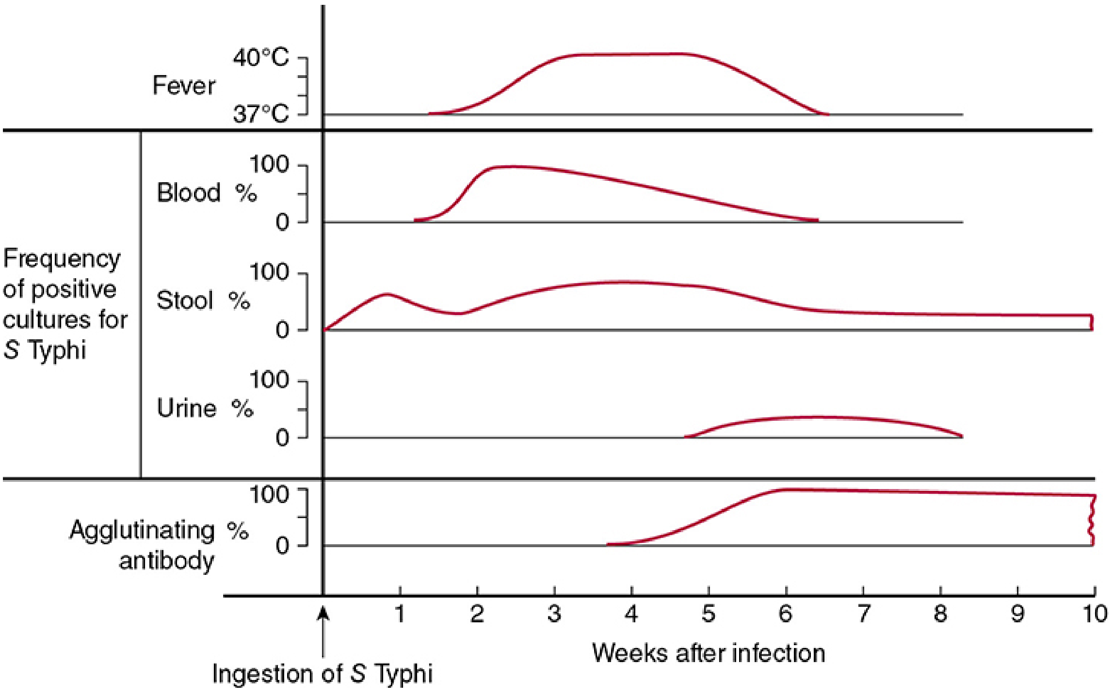
Step-by-Step Pathophysiology Flowchart
Ingestion of S. Typhi in contaminated food/water
↓
GASTRIC ACID BARRIER - Survived by:
→ pH homeostasis system + acid shock proteins expressed by S. Typhi
→ Vi capsular polysaccharide provides additional protection
↓
Reaches SMALL INTESTINE (terminal ileum - Peyer's patches)
↓
S. Typhi invades M CELLS (specialized epithelial cells overlying Peyer's patches)
↓
Engulfed by submucosal MACROPHAGES
→ Vi polysaccharide BLOCKS neutrophil phagocytosis (by interfering with complement deposition)
→ This FAVORS macrophage uptake over neutrophil killing
→ S. Typhi survives inside macrophage: INHIBITS oxidative burst (respiratory burst)
→ Continues to multiply inside membrane-bound vacuole within macrophages
↓
Bacteria (inside macrophages) travel via LYMPHATICS to:
→ Mesenteric lymph nodes → THORACIC DUCT → bloodstream →
→ LIVER, SPLEEN, BONE MARROW (all = reticuloendothelial system / RES)
↓
INCUBATION PERIOD (1-2 weeks): Silent multiplication in RES
→ No clinical symptoms yet
→ Mononuclear (not neutrophilic) inflammatory response in gut → minimal/no diarrhea early
→ Vi antigen downregulates innate Toll-like receptor responses in intestinal mucosa
↓
Burgeoning bacteria reach CRITICAL MASS in RES → flood into BLOODSTREAM
→ PRIMARY BACTEREMIA (Week 1)
→ LPS ENDOTOXIN enters blood → triggers fever (step-ladder pattern) and malaise
→ Fever rises SLOWLY over 72 hours → continuous high fever in Week 2
↓
Sustained bacteremia seeds other organs:
→ BILIARY TREE infected → bacteria re-enter intestine via bile → reinfects gut
→ PEYER'S PATCHES: hyperplasia → necrosis → ulceration (Week 3)
→ TERMINAL ILEUM / PROXIMAL COLON: ulcer perforates → intestinal perforation
or erosion of blood vessels → hemorrhage
→ Other sites: urinary tract, bone, joints, meninges, heart (myocarditis)
↓
ENDOTOXIN + CYTOKINE EFFECTS:
→ Continuous fever (TNF-α, IL-1, IL-6)
→ Relative bradycardia (direct myocardial depression / vagal stimulation)
→ Hepatic involvement → mild jaundice, elevated LFTs
→ Bone marrow depression → leukopenia, thrombocytopenia, anemia
→ Encephalopathy (severe cases)
↓
WITHOUT TREATMENT: Natural resolution in Week 4 (in 90% of survivors)
→ Immunity: both TH1 and TH2 mediated
→ 1-5% → CHRONIC CARRIER STATE (gallbladder reservoir)
Why the Gut Pathology Matters
Week 1: Peyer's patches HYPERPLASIA (lymphoid swelling) → no ulcer yet
↓
Week 2: Peyer's patches NECROSIS → central sloughing
↓
Week 3: ULCERATION of Peyer's patches in terminal ileum
→ Ulcers are TRANSVERSE (perpendicular to long axis of bowel)
→ Penetration = INTESTINAL PERFORATION (5% of untreated cases)
→ Erosion of vessels = INTESTINAL HEMORRHAGE (10% of untreated)
Exam pearl: Typhoid ulcers are TRANSVERSE (across the bowel wall, following Peyer's patch distribution). Compare with TB ulcers which are also transverse but have undermined edges; contrast with typhoid where edges are clean. Carcinoma ulcers are irregular; amoebic ulcers are flask-shaped.
5. CLINICAL FEATURES
The Week-by-Week Clinical Progression
WEEK 1 - "FLU-LIKE" (Bacteremic onset)
• Fever: begins low-grade, rises in STEP-LADDER pattern each day
• Frontal headache (prominent, often the first complaint)
• Myalgia and arthralgia
• Malaise, lethargy, anorexia
• DRY COUGH (in ~30% - misleads to respiratory diagnosis)
• Constipation (more common than diarrhea in early typhoid)
• Blood culture: MOST POSITIVE this week (>90% positive)
• Patient looks unwell but not critically ill yet
WEEK 2 - "CLASSIC TYPHOID" (Sustained fever)
• Fever: continuous, 39-40°C, peaking in the afternoon
• RELATIVE BRADYCARDIA - pulse slower than expected for the fever
• ROSE SPOTS - faint salmon-pink maculopapular lesions appear
• Hepatomegaly and SPLENOMEGALY develop
• Abdominal pain, distension, tenderness (lower quadrants)
• Diarrhea may develop (often "pea-soup" stools - mild, watery)
• "Typhoid tongue" - white coated with red tip and edges
• Toxemia increases; patient appears "toxic"
• Blood culture: still positive, begins declining
WEEK 3 - "DANGER ZONE" (Complications)
• Fever persists at plateau
• INTESTINAL COMPLICATIONS:
- Perforation: sudden severe abdominal pain, guarding, rigidity
- Hemorrhage: blood per rectum, sudden fall in temperature and BP
• Encephalopathy: confusion, delirium, stupor ("typhoid state" / "muttering delirium")
• DIC: petechiae, oozing
• Myocarditis: tachycardia, ECG changes
• Stool + urine culture: most positive now
WEEK 4 - RESOLUTION or RELAPSE
• Gradual defervescence (temperature falls by lysis, not crisis)
• Relapse in 3-20% (milder than original illness)
• Convalescence takes weeks to months
Symptoms
| Symptom | Timing | Notes |
|---|---|---|
| High fever (39-40°C) | Weeks 1-3 | Step-ladder rise in Week 1, sustained plateau in Week 2 |
| Frontal headache | Week 1 onwards | Often the most prominent early complaint |
| Anorexia | Throughout | Profound; significant weight loss |
| Malaise and myalgia | Week 1 onwards | Flu-like prodrome misleads clinicians |
| Constipation | Week 1 | More common than diarrhea early in adults |
| Dry cough | Week 1 (~30%) | Misleads to pneumonia or influenza diagnosis |
| Abdominal pain and distension | Week 2 onwards | Lower quadrant tenderness; may mimic appendicitis |
| Diarrhea | Week 2-3 | "Pea-soup" stools; late feature as disease progresses |
| Confusion/delirium | Week 3 | Indicates severe toxemia; "typhoid state" |
Signs
| Sign | Timing | Notes |
|---|---|---|
| Step-ladder fever | Week 1 | Temperature rises each day in increments |
| Relative bradycardia (Faget's sign) | Week 2 | Pulse slower than expected for given temperature; up to 50% of patients |
| Rose spots | End of Week 1 / Week 2 | 2-4 mm salmon-pink maculopapular lesions on abdomen and chest; blanch on pressure; last only 3-4 days; visible in ~30% of patients (missed in dark-skinned individuals) |
| Splenomegaly | Week 2 onwards | Soft, tender; present in ~50% of patients |
| Hepatomegaly | Week 2 onwards | Mild; tender; LFTs mildly elevated |
| Coated tongue | Week 2 | White coating with red tip and edges ("typhoid tongue") |
| Abdominal distension | Week 2-3 | From ileus and mesenteric lymphadenopathy |
| Dicrotic pulse | Occasionally | Double-beat pulse - associated with low peripheral vascular resistance from endotoxemia |
Why Important Signs Occur
| Sign | Mechanism |
|---|---|
| Step-ladder fever | Slow, progressive seeding of bloodstream by S. Typhi from RES - LPS endotoxin triggers cytokine cascade (TNF-α, IL-1) incrementally as bacterial load rises |
| Relative bradycardia | LPS endotoxin directly depresses SA node conduction + stimulates vagal tone + inhibits sympathetic response; fever raises temperature but pulse does not keep pace |
| Rose spots | Bacteria embolize to dermal capillaries during bacteremia → local inflammatory reaction → 2-4 mm erythematous lesions. S. Typhi can be cultured from biopsy of rose spots |
| Leukopenia | Bone marrow depression from intracellular S. Typhi infection + cytokine-mediated suppression of granulopoiesis |
| Splenomegaly | Hyperplasia of RES (macrophages) in spleen responding to intracellular bacteria + engorgement with activated lymphocytes and phagocytes |
| Constipation (early) | Inhibition of intestinal motility by endotoxin + vagal stimulation; Vi antigen downregulates mucosal inflammation preventing secretory diarrhea |
| Diarrhea (late) | Biliary re-seeding of gut in Week 2-3 → Peyer's patch ulceration → secretory and exudative diarrhea |
| Intestinal perforation | Progressive necrosis of Peyer's patches → transmural ulceration → perforation of terminal ileum (usually a single perforation in the antimesenteric border) |
6. KEY SYMPTOMS THAT SUGGEST THE DIAGNOSIS
Hallmark Symptom
Step-ladder fever for 1 week + relative bradycardia (pulse-temperature dissociation) in a patient with travel to or residence in a typhoid-endemic area (South Asia, Southeast Asia, Africa, Latin America).
Classic Clinical Clues
| Clue | What It Suggests |
|---|---|
| Fever rising 1°C per day over 5-7 days (step-ladder pattern) | Classic Week 1 typhoid pattern; not seen in most other infections |
| Pulse rate LOWER than expected for the degree of fever | Relative bradycardia / Faget's sign - hallmark of typhoid; also seen in brucellosis, leptospirosis, Q fever |
| Salmon-pink rose spots on abdomen/chest (2-4 mm, blanching, 3-10 in number) | Pathognomonic clinical appearance - appears end of Week 1/Week 2 |
| Constipation (not diarrhea) as the predominant GI symptom early | Counterintuitive - typhoid classically begins with constipation, not diarrhea |
| Dry cough in the first week before abdominal symptoms | Misleading respiratory prodrome - do not diagnose URTI without considering typhoid in travelers |
| Splenomegaly + tender hepatomegaly + fever | Enteric fever involvement of the reticuloendothelial system |
| Leukopenia (low white cell count) in a patient with high fever | Profound leukopenia contrasts with the leukocytosis expected in bacterial infections - a classic exam fact |
| Travel to India, Pakistan, Bangladesh, Nepal, Philippines, Mexico, Haiti | High-endemic zones; India has 18x higher risk than other endemic areas |
| Fever not responding to amoxicillin or common antibiotics | Classic history in case reports - S. Typhi is inherently resistant to amoxicillin (and MDR strains to chloramphenicol/cotrimoxazole) |
| "Pea-soup" diarrhea in Week 2-3 after initial constipation | Late-stage gallbladder re-seeding of gut |
| Sudden worsening with acute abdomen in Week 3 | Intestinal perforation - surgical emergency |
Red Flag Findings (Complicated/Severe Typhoid)
- 🚨 Intestinal perforation: Sudden severe abdominal pain, guarding, rigidity, paradoxical fall in fever (temperature drops when perforation occurs - a diagnostic trap)
- 🚨 Intestinal hemorrhage: Fresh blood per rectum, sudden hypotension, fall in temperature
- 🚨 Encephalopathy: Confusion, delirium, stupor, coma ("typhoid state")
- 🚨 Myocarditis: New arrhythmia, cardiac failure signs in typhoid patient
- 🚨 DIC: Bleeding from multiple sites, petechiae, ecchymosis
- 🚨 Prolonged fever >3 weeks without diagnosis in a returning traveler = must rule out typhoid with bone marrow culture
- 🚨 Typhoid in pregnancy: High risk of abortion, preterm labor, maternal death
Exam memory hook - "The TYPHOID mnemonic" for classic features:
- T - Temperature: step-ladder rise
- Y - (wh)Y is the pulse slow? Relative bradycardia
- P - Peyer's patches → ulceration → perforation in Week 3
- H - Hepatosplenomegaly
- O - Onset: constipation before diarrhea
- I - Invasion: RES (liver, spleen, bone marrow, lymph nodes)
- D - Diagnostic clue: Rose spots + leukopenia
7. Laboratory Investigations of typhoid disease Create a table: Investigation | Expected Finding | Clinical Significance Include: - CBC - Biochemistry - Disease-specific tests - Imaging findings if relevant 8. Differential Diagnosis Create a comparison table: Disease | Similar Features | Differentiating Features 9. Short Clinical Case Example format: "A 55-year-old male presents with chest pain radiating to the left arm, sweating, and breathlessness. ECG shows ST elevation in leads II, III, and aVF. Troponin-I is elevated." Diagnosis: Reasoning: Provide only 1–2 short high-yield cases.
7. LABORATORY INVESTIGATIONS OF TYPHOID FEVER
COMPLETE BLOOD COUNT (CBC)
| Investigation | Expected Finding | Clinical Significance |
|---|---|---|
| Total Leukocyte Count (TLC) | Leukopenia (WBC 2,000-4,000/µL) or normal WBC; leukocytosis is unusual | Bone marrow depression by intracellular S. Typhi + cytokine-mediated suppression. Leukopenia in a febrile patient is a strong pointer toward typhoid - most bacterial infections cause leukocytosis |
| Neutrophil count | Neutropenia (absolute); relative lymphocytosis | Same mechanism; neutropenia may be profound in severe disease |
| Hemoglobin / RBCs | Mild normochromic normocytic anemia; worsens with hemorrhage | Bone marrow depression + hemolysis; severe anemia if intestinal hemorrhage occurs |
| Platelets | Thrombocytopenia (mild-moderate); severe in complicated disease | Bone marrow suppression + increased consumption; DIC causes severe thrombocytopenia |
| ESR | Moderately elevated | Non-specific inflammatory marker |
Exam tip: The classic triad on CBC = Leukopenia + Anemia + Thrombocytopenia in a febrile patient returning from South Asia = typhoid fever until proven otherwise. Leukocytosis suggests intestinal perforation (secondary peritonitis) as a complication.
BIOCHEMISTRY
| Investigation | Expected Finding | Clinical Significance |
|---|---|---|
| Liver function tests (ALT, AST) | Mildly-moderately elevated (2-3× normal); rarely severe | S. Typhi invades hepatocytes and Kupffer cells; hemozoin-like pigment deposition. Hepatitis-like picture in ~50% of patients |
| Serum bilirubin | Mildly elevated (conjugated + unconjugated) | Hepatocellular involvement; hemolysis contributes to indirect bilirubin rise |
| Alkaline phosphatase | Mildly elevated | Non-specific hepatic involvement |
| Serum albumin | Low/borderline in prolonged illness | Negative acute-phase reactant; chronic catabolism and malnutrition from prolonged anorexia |
| CRP (C-reactive protein) | Elevated | Acute-phase response; useful to monitor therapeutic response |
| Serum sodium | Hyponatremia (in severe cases) | SIADH (inappropriate ADH from endotoxemia) + sweating + poor intake |
| Blood glucose | Hypoglycemia possible in severe/pediatric cases | Poor oral intake + hepatic dysfunction |
| Coagulation profile (PT, aPTT, D-dimer) | Deranged in complicated disease | DIC in severe/untreated typhoid; prolonged PT indicates liver dysfunction |
| Blood urea / Creatinine | Elevated in severe/complicated cases | Pre-renal AKI from dehydration; rarely intrinsic renal damage |
| Cardiac enzymes (Troponin, CK-MB) | Elevated if myocarditis develops | Myocarditis is a rare but serious complication; always ECG febrile patients with chest pain or arrhythmia |
DISEASE-SPECIFIC / MICROBIOLOGICAL TESTS
| Investigation | Expected Finding | Clinical Significance |
|---|---|---|
| Blood culture (2-3 sets from different sites) | Positive in 60-80% in Week 1-2; declines to ~40% if antibiotics already given | First-line diagnostic test. Most sensitive in Week 1 (bacteremic peak). Should be taken BEFORE starting antibiotics. Culture on bile-containing media (ox bile broth) increases yield. Minimum 10 mL blood per bottle |
| Bone marrow culture | Positive in 90% even in patients already on antibiotics | Gold standard for diagnosis. Remains positive despite prior antibiotic therapy (bacteria hidden inside macrophages in marrow). Reserved for culture-negative cases on antibiotics. Labor-intensive but invaluable |
| Stool culture | Positive in 30-50% in Week 2-3; may remain positive in carriers | Peaks during Weeks 2-3 as biliary re-seeding occurs. Less useful in Week 1. Essential for carrier detection. Negative stool does NOT rule out typhoid |
| Urine culture | Positive in ~25% in Week 3 | From urinary tract seeding during sustained bacteremia; useful in later stages |
| Duodenal string test (bile culture) | Positive in ~70% of cases | String capsule swallowed, retrieves duodenal/biliary contents for culture. Combined with blood culture gives 90% sensitivity. Not widely used but high-yield |
| Widal test (Serological agglutination test) | Rise in agglutinins against O antigen (somatic) ≥1:160 and H antigen (flagellar) ≥1:160; 4-fold rise in paired sera (acute + convalescent) is most reliable | Detects antibodies rising from Week 2. Limitations (critical exam fact): False positives with previous Salmonella infections, prior vaccination, cross-reactions with other Enterobacteriaceae; false negatives if sampled too early (Week 1) or if patient is immunocompromised. CDC does NOT recommend the Widal test for acute diagnosis - it should be interpreted with clinical context. A single titre is unreliable; paired titres 2 weeks apart are meaningful |
| Rapid diagnostic tests (RDTs) / TYPHIDOT / TUBEX TF | Detects IgM against S. Typhi O9 antigen (TUBEX) or 50 kDa outer membrane protein (Typhidot) | Useful in resource-limited endemic settings where culture is unavailable. Moderate sensitivity (~70-80%) and specificity. TUBEX TF has advantage of detecting IgM early (Week 1). Not recommended to replace culture |
| PCR (Molecular assay) | Detects S. Typhi-specific DNA | High sensitivity in early bacteremic phase; not yet validated against bone marrow culture; not practical for routine use due to cost and laboratory requirements |
| Vi antigen serology | Elevated anti-Vi antibody titers | Identifies chronic carriers (not acute disease diagnosis); used in outbreak investigation |
| Antimicrobial sensitivity testing (AST) | Tests for MDR strains: resistance to chloramphenicol, ampicillin, co-trimoxazole; fluoroquinolone resistance (nalidixic acid test as surrogate); XDR (extensively drug-resistant) strains | Mandatory on all isolates. MDR S. Typhi has been circulating since 1989 (plasmid-encoded resistance). XDR strains (also resistant to ceftriaxone + azithromycin) emerged in Pakistan 2016 onwards. Guides choice of antibiotic - culture without sensitivity testing is insufficient |
Culture Yield by Week and Site - Summary Table
| Culture Site | Week 1 | Week 2 | Week 3 | Week 4 | Notes |
|---|---|---|---|---|---|
| Blood | 80-90% | 60-70% | 40-50% | Declining | Best in Week 1; decreases with antibiotic use |
| Bone marrow | 90% | 90% | 90% | 90% | Gold standard; remains positive regardless of antibiotics |
| Stool | 10-30% | 40-60% | 50-75% | Declining | Rises as biliary re-seeding occurs |
| Urine | Rare | 10% | 25% | 15% | Late-phase finding |
| Bile/String test | High | High | High | - | ~70% across phases |
Exam pearl: Blood culture = best in Week 1; Stool culture = best in Week 3; Bone marrow culture = best at ANY week (even after antibiotics).
IMAGING FINDINGS
| Investigation | Expected Finding | Clinical Significance |
|---|---|---|
| Ultrasound abdomen | Hepatomegaly; splenomegaly; mesenteric lymphadenopathy; free fluid in complicated cases; gallbladder wall thickening in carriers | Non-invasive; confirms organomegaly; detects complications. Gallbladder stones + thickened wall = suspect chronic carrier state |
| Plain X-ray abdomen (erect) | Free air under the diaphragm (pneumoperitoneum) in intestinal perforation | Surgical emergency. Classic "air under diaphragm" = perforated viscus. Must be performed urgently if sudden worsening abdominal pain occurs in Week 3 |
| CT scan abdomen | Intestinal wall thickening (terminal ileum); perforation; peritoneal free air/fluid; hepatosplenomegaly; mesenteric lymph nodes enlarged | Superior to plain X-ray for detecting perforation and abscess formation; guides surgical planning |
| Chest X-ray | Usually normal; rarely pneumonia or pleural effusion in complicated cases | S. Typhi can cause pneumonia from hematogenous seeding; right lower lobe pneumonia reported |
| Echocardiogram | Wall motion abnormality; pericardial effusion in myocarditis/pericarditis | Indicated if arrhythmia or cardiac signs develop; typhoid myocarditis is rare but life-threatening |
| MRI/CT brain | Cerebral edema; encephalitis changes in typhoid encephalopathy | Indicated for altered sensorium; cerebral involvement from endotoxemia |
8. DIFFERENTIAL DIAGNOSIS
| Disease | Similar Features to Typhoid | Differentiating Features |
|---|---|---|
| Malaria | Fever, hepatosplenomegaly, anemia, thrombocytopenia, travel history to endemic area | Cyclic paroxysmal fever (48h or 72h) with rigors and sweating vs. typhoid's continuous step-ladder fever; no relative bradycardia; positive blood smear/RDT for Plasmodium; leukopenia less pronounced; no rose spots |
| Dengue fever | Fever, headache, myalgia, thrombocytopenia, leukopenia, travel history | Sudden onset high fever (breakbone fever); severe retro-orbital headache; maculopapular/petechial rash (not rose spots); positive NS1 antigen or dengue IgM/IgG; no splenomegaly early; no relative bradycardia; no step-ladder pattern; fever resolves faster (5-7 days) |
| Brucellosis | Undulant (relapsing) fever, malaise, hepatosplenomegaly, leukopenia, relative bradycardia | Exposure to animals (cattle, goats, sheep) or unpasteurized dairy; fever is UNDULANT (waves, not step-ladder); profuse sweating (especially nocturnal); positive Brucella serology (RBPT, SAT) or blood culture on specialized media (Castañeda); bone and joint involvement more prominent |
| Leptospirosis | Fever, headache, myalgia, jaundice, hepatosplenomegaly, renal failure | Exposure to fresh water or animal urine; biphasic fever (leptospiremic phase + immune phase/Weil's disease); conjunctival suffusion (red eyes); prominent calf muscle tenderness; uveitis; positive MAT or IgM ELISA; no rose spots; no leukopenia |
| Visceral Leishmaniasis (Kala-azar) | Prolonged fever, massive splenomegaly, anemia, weight loss, pancytopenia, endemic travel | Very gradual onset over months (weeks to months, not days); massive splenomegaly disproportionate to other findings; skin hyperpigmentation; positive rK39 RDT or bone marrow/spleen biopsy showing Leishman-Donovan bodies; high fever but no step-ladder pattern |
| Typhus (Rickettsia) | Fever, headache, myalgia, rash, thrombocytopenia | Exposure to lice (epidemic) or mites/ticks (scrub/endemic); maculopapular rash more generalized (not just abdomen) and appears in first few days; eschar at bite site in scrub typhus; positive Weil-Felix reaction or specific Rickettsia PCR/IFA; relative bradycardia also present |
| Miliary Tuberculosis | Prolonged fever, hepatosplenomegaly, pancytopenia, leukopenia, weight loss | Usually more insidious with prominent night sweats and weight loss; history of TB contact/HIV; "snowstorm" pattern on CXR (miliary nodules); raised ADA; positive Mantoux, IGRA, or sputum AFB/culture; no intestinal perforation risk; no rose spots |
| Infectious Mononucleosis (EBV) | Fever, pharyngitis, hepatosplenomegaly, lymphadenopathy, leukopenia/atypical lymphocytes | Mainly affects adolescents; prominent exudative tonsillitis + cervical lymphadenopathy; splenomegaly without hepatic predominance; positive Monospot test / EBV IgM VCA; atypical lymphocytes on smear; no rose spots; no relative bradycardia |
| Appendicitis | Abdominal pain (right iliac fossa), fever, leukocytosis | Typhoid can present with RIF pain (mimicking appendicitis) from mesenteric adenitis involving ileocaecal nodes; however appendicitis has leukocytosis (not leukopenia); Rovsing's sign, rebound tenderness; fever is lower-grade; no hepatosplenomegaly; ultrasound/CT distinguishes |
| Viral Hepatitis (A, E) | Fever, jaundice, hepatomegaly, elevated LFTs, anorexia, GI symptoms | Jaundice is more prominent and earlier; no splenomegaly; markedly elevated ALT (>10× normal); positive hepatitis A IgM or Hepatitis E IgM; no rose spots or relative bradycardia; no leukopenia |
| Influenza | Fever, headache, myalgia, dry cough, malaise | Typhoid Week 1 can look identical to flu; however influenza: rapid onset (hours); prominent respiratory symptoms; winter/outbreak clustering; positive rapid flu test or PCR; no hepatosplenomegaly; no step-ladder fever; no rose spots; resolves in 7-10 days |
Key rule: In any febrile patient returning from South Asia (especially India, Pakistan, Bangladesh, Nepal), typhoid must be the first differential considered. Blood cultures must be drawn before starting antibiotics - this single step is the most common preventable diagnostic error.
9. SHORT CLINICAL CASES
Case 1 - Classic Typhoid Fever
- Travel to Pakistan (highest-risk country for typhoid) + no vaccination mentioned
- Step-ladder fever rising incrementally over 5 days - the pattern most characteristic of typhoid
- Relative bradycardia (pulse 74 with fever 39.6°C - expected pulse would be ~110-120 bpm) is pathognomonic for typhoid/brucellosis/leptospirosis
- Constipation rather than diarrhea in Week 1 - classic feature (counterintuitive but examinable)
- Dry cough misleads toward respiratory diagnosis - but typhoid's early cough is from endotoxemia, not pulmonary infection
- Leukopenia + thrombocytopenia in a febrile patient = typhoid fever profile (bacterial infections cause leukocytosis)
- Next step: Blood cultures (3 sets) before any antibiotics; start empiric ceftriaxone or azithromycin while awaiting sensitivity
Case 2 - Complicated Typhoid (Intestinal Perforation)
- 3 weeks of untreated fever - classic time frame for intestinal perforation (Week 3 = danger zone)
- Paradoxical sudden fall in temperature at time of perforation - a diagnostic trap; the peritoneal cavity acts as a heat sink; simultaneously pulse shoots up (surgical peritonitis)
- Board-like rigidity + absent bowel sounds = peritonitis from perforation
- Pneumoperitoneum (free air under diaphragm on erect CXR/AXR) = confirms hollow viscus perforation
- Hemodynamic instability = septic shock from fecal peritonitis
- Perforation site: antimesenteric border of terminal ileum at a necrotic Peyer's patch (single perforation, transverse)
- Mortality of typhoid perforation: 10-32% even with surgery
- Next step: Emergency laparotomy (primary closure or resection + anastomosis depending on findings); IV ceftriaxone + metronidazole (cover gut flora); ICU monitoring; blood cultures before antibiotics if time permits
Exam trap: The sudden fall in temperature with worsening clinical condition in a Week 3 typhoid patient is NOT an improvement - it signals perforation. Never be falsely reassured by a falling temperature in this context.
10. Treatment Of Malaria A. Non-Pharmacological Treatment B. Pharmacological Treatment Create a table: Drug | Drug Group | Mechanism of Action | Indication | Dose | Major Adverse Effects For every disease mention: - Drug of Choice (DOC) - First-line drugs - Alternative drugs - Emergency drugs (if applicable) 11. Clinical Pearls - Important bedside clues - High-yield exam facts - Viva points 12. Common Diagnostic Mistakes - Frequently missed findings - Common confusion with other diseases - How to avoid errors
10. TREATMENT OF TYPHOID FEVER
A. NON-PHARMACOLOGICAL TREATMENT
| Intervention | Details | Rationale |
|---|---|---|
| Hospitalization | All confirmed or suspected typhoid cases should be admitted | Prevents complications; allows close monitoring for perforation/hemorrhage; prevents transmission |
| Isolation | Enteric precautions (contact precautions); isolated until 3 negative stool and urine cultures on 3 separate days post-recovery | S. Typhi is shed in stool and urine; prevents spread to contacts and healthcare workers |
| Strict bed rest | Enforce throughout febrile period | Reduces metabolic demand; prevents falls; limits risk of splenic trauma leading to rupture |
| Oral rehydration / IV fluids | Oral fluids for uncomplicated cases; IV fluids (normal saline or Ringer's lactate) for severe disease, vomiting, or altered consciousness | Counters dehydration from fever, sweating, vomiting, and diarrhea; maintains renal perfusion |
| Nutritional support | Soft, easily digestible, high-calorie diet (semi-solid/liquid); avoid high-fibre, spicy, or rough foods | Typhoid causes profound anorexia and catabolism; nutrition maintains gut mucosal integrity; rough food can aggravate Peyer's patch ulcers and risk triggering perforation |
| Antipyretics | Paracetamol 500-1000 mg every 4-6h as needed for fever | Controls fever and headache; avoid NSAIDs (risk of GI bleeding, especially in thrombocytopenic patients with Peyer's patch ulcers) |
| Blood transfusion | Indicated for significant GI hemorrhage or severe anemia | Intestinal hemorrhage (10% of untreated cases) may require packed RBC transfusion |
| Surgical management (if perforation) | Emergency laparotomy: primary closure of perforation ± bowel resection and anastomosis; peritoneal lavage | Intestinal perforation is the most lethal complication; mortality 10-32% even with surgery; delay increases mortality |
| Stool and urine disinfection | Stools received in closed containers; disinfected with 5% cresol for ≥2 hours; soiled linen soaked in 2% chlorine solution and steam sterilized | S. Typhi survives for hours outside the body; proper disposal is a public health imperative |
| Hand hygiene | Strict handwashing for all healthcare personnel and caregivers | Prevents fecal-oral transmission from patient to contacts |
| Monitoring for complications | Daily check: pulse, BP, abdomen (perforation), stool (hemorrhage), neuro status (encephalopathy), blood glucose, CBC | Week 3 is the highest-risk period; sudden change in vitals = suspect perforation immediately |
| Dexamethasone (adjunctive, not a primary treatment) | 3 mg/kg loading dose IV, then 1 mg/kg every 6h × 8 doses in severe/encephalopathic disease | Only indicated for severe typhoid with shock or encephalopathy/delirium; reduces mortality from 56% to 10% in severe meningeal/encephalopathic typhoid (Hoffman et al. NEJM trial) |
Important note: The use of corticosteroids is only for severe typhoid with encephalopathy or shock - this is the OPPOSITE of cerebral malaria where steroids are harmful. This distinction is a classic exam question.
B. PHARMACOLOGICAL TREATMENT
DRUG RESISTANCE BACKGROUND (Critical for Exam)
| Resistance Category | Definition | Geographic Distribution |
|---|---|---|
| Fully susceptible | Sensitive to chloramphenicol, ampicillin, TMP-SMX, fluoroquinolones, cephalosporins | Rare now; isolated areas |
| Multidrug-resistant (MDR) | Resistant to chloramphenicol + ampicillin + trimethoprim-sulfamethoxazole | Widespread since 1980s; plasmid-mediated; South Asia, Africa |
| Decreased ciprofloxacin susceptibility (DCS) | MIC ≥0.125 µg/mL for ciprofloxacin | Indian subcontinent, spreading globally; nalidixic acid resistance is a surrogate marker |
| Fluoroquinolone-resistant | MIC ≥1 µg/mL for ciprofloxacin | Indian subcontinent, Africa, spreading via air travel |
| Extensively drug-resistant (XDR) | MDR + resistant to fluoroquinolones + ceftriaxone (ESBL); effectively only azithromycin remains | Pakistan since 2016 (H58 clone); spreading internationally; major public health crisis |
| Azithromycin-resistant | Emerging in countries using azithromycin as first-line | Bangladesh, India, parts of Southeast Asia |
Exam alarm: XDR S. Typhi leaves azithromycin as the only remaining oral option. For XDR, IV meropenem or carbapenems become necessary. Typhoid Conjugate Vaccine (TCV) is now urgently recommended to prevent XDR spread.
PHARMACOLOGICAL DRUG TABLE
- 🥇 DOC / First-line
- 🥈 Alternative / Second-line
- 🚨 Severe / Emergency
- ⛔ Now UNRELIABLE due to resistance
| Drug | Drug Group | Mechanism of Action | Indication | Dose (Adults) | Major Adverse Effects |
|---|---|---|---|---|---|
| 🥇 Ceftriaxone IV | 3rd-generation cephalosporin | Binds penicillin-binding proteins (PBPs) → inhibits bacterial cell wall synthesis (peptidoglycan cross-linking) → bactericidal | DOC for severe/complicated typhoid; first-line empiric choice worldwide; covers MDR strains; drug of choice if origin from South Asia (due to fluoroquinolone resistance); used for hospitalized patients | 2 g IV once daily × 10-14 days | Allergic reactions (rash, anaphylaxis in penicillin-allergic); biliary sludge with high doses; Clostridioides difficile colitis; pain at injection site |
| 🥇 Azithromycin PO | Macrolide | Binds 50S ribosomal subunit → inhibits translocation of mRNA → bacteriostatic (bactericidal at high concentrations intracellularly) | DOC for uncomplicated typhoid in travelers from South/Southeast Asia (due to fluoroquinolone resistance); covers MDR strains; excellent intracellular penetration reaches macrophage-laden RES where S. Typhi hides | 1 g/day PO × 5-10 days (adults) | Nausea, vomiting, diarrhea, abdominal cramps; QTc prolongation (caution in cardiac patients); hepatotoxicity (rare); emerging resistance in endemic areas |
| 🥇 Cefixime PO | 3rd-generation oral cephalosporin | Same mechanism as ceftriaxone (PBP inhibition) | Oral option for uncomplicated MDR typhoid; when IV ceftriaxone is stepped down; for outpatient management of mild-moderate uncomplicated typhoid | 400 mg/day (or 200 mg twice daily) PO × 7-14 days | Well tolerated; mild GI upset; allergic reactions; may have slightly higher clinical failure rate and slower defervescence than fluoroquinolones |
| 🥈 Ciprofloxacin PO/IV | Fluoroquinolone | Inhibits DNA gyrase (topoisomerase II) and topoisomerase IV → prevents DNA supercoiling → disrupts bacterial DNA replication and repair → bactericidal | First-line ONLY for fully susceptible strains (now very limited geographically); NOT recommended empirically for travelers from South Asia; still useful where confirmed sensitivity | 500 mg PO twice daily × 5-7 days OR 400 mg IV every 12h | Tendinopathy and tendon rupture (especially Achilles); photosensitivity; QTc prolongation; cartilage toxicity in children/adolescents; CNS effects (seizures, dizziness); avoid in pregnancy |
| 🥈 Ofloxacin PO | Fluoroquinolone | Same as ciprofloxacin (DNA gyrase inhibition) | Alternative fluoroquinolone for fully susceptible strains; slightly better bioavailability than ciprofloxacin for typhoid in some studies | 15 mg/kg/day divided twice daily PO × 5-7 days | Similar to ciprofloxacin class effects |
| 🥈 Amoxicillin PO/IV | Aminopenicillin | Inhibits PBPs → disrupts cell wall synthesis | Alternative for fully susceptible strains only; useful for carrier eradication (with probenecid) due to biliary concentration | 1 g three times daily PO × 14 days | GI upset; allergic reactions; rash (ampicillin rash); increasing resistance limits routine use |
| 🥈 Trimethoprim-Sulfamethoxazole (TMP-SMX) PO | Folate antagonist combination | TMP inhibits dihydrofolate reductase; SMX inhibits dihydropteroate synthase → synergistic blockade of folate synthesis → bacteriostatic | Alternative for fully susceptible strains only; MDR strains universally resistant | 160/800 mg (1 DS tablet) twice daily PO × 7-14 days | Allergic rash (including Stevens-Johnson syndrome); bone marrow suppression; nephrotoxicity; avoid in pregnancy and G6PD deficiency |
| ⛔ Chloramphenicol | Chloramphenicol | Binds 50S ribosomal subunit → inhibits peptidyl transferase → bacteriostatic | Historically the first DOC (1948-1980s); now UNRELIABLE due to widespread MDR resistance; reserved only for confirmed fully-susceptible strains where no alternatives available | 50-75 mg/kg/day in 4 divided doses × 14-21 days | Gray baby syndrome (in neonates); aplastic anemia (rare, idiosyncratic, irreversible); reversible dose-dependent bone marrow suppression; avoid in pregnancy |
| 🚨 Meropenem IV | Carbapenem | Binds PBPs (especially PBP2) → inhibits peptidoglycan synthesis → bactericidal | Emergency drug for XDR typhoid (resistant to all above agents); life-saving in XDR cases from Pakistan | 1 g IV every 8 hours × 10-14 days | Nausea, vomiting, diarrhea; seizures at high doses; Clostridioides difficile colitis; nephrotoxicity |
Treatment Summary by Clinical Scenario
| Scenario | Recommended Treatment | Duration |
|---|---|---|
| Uncomplicated typhoid - traveler from South Asia (empiric, resistance unknown) | Azithromycin 1 g/day PO OR Ceftriaxone 2 g/day IV | 5-10 days (azithromycin); 10-14 days (ceftriaxone) |
| Uncomplicated typhoid - confirmed fully susceptible strain | Ciprofloxacin 500 mg PO twice daily OR Azithromycin | 5-7 days |
| Uncomplicated MDR typhoid | Azithromycin PO OR Cefixime PO OR Ceftriaxone IV | 10-14 days |
| Severe/complicated typhoid (any strain) | Ceftriaxone 2 g IV daily | 10-14 days; switch to oral once improving |
| XDR typhoid (Pakistan/international spread) | Azithromycin (if susceptible) OR Meropenem IV | Meropenem 10-14 days; guided by sensitivity |
| Typhoid with shock or encephalopathy | Ceftriaxone IV + Dexamethasone 3 mg/kg loading then 1 mg/kg q6h × 8 doses | Ceftriaxone 14 days; dexamethasone 48h only |
| Intestinal perforation | Ceftriaxone + Metronidazole IV + Emergency Surgery | Until clinically stable |
| Chronic carrier eradication | Amoxicillin 4-6 g/day + Probenecid 2 g/day × 6 weeks; OR Ciprofloxacin (if susceptible) × 4 weeks; Cholecystectomy if gallstones present | 4-6 weeks; surgery when medically fit |
| Typhoid in pregnancy | Ceftriaxone IV (safe in pregnancy) | 10-14 days; avoid fluoroquinolones, TMP-SMX, chloramphenicol |
| Typhoid in children | Azithromycin or Ceftriaxone (avoid fluoroquinolones due to cartilage toxicity) | 10-14 days |
Vaccination (Prevention Pharmacology)
| Vaccine | Type | Route | Efficacy | Duration | Notes |
|---|---|---|---|---|---|
| Typhoid Conjugate Vaccine (TCV) - Typbar-TCV | Vi polysaccharide conjugated to tetanus toxoid carrier protein | IM injection | ~80-90% | ≥5 years (boostable) | WHO-preferred; now recommended for endemic areas; only vaccine protecting children <2 yrs; does NOT protect against paratyphoid |
| Vi polysaccharide vaccine (Typhim Vi) | Vi capsular polysaccharide | IM injection | ~55-72% | 3 years (requires booster) | For travelers; not effective in children <2 yrs; does NOT protect against S. Paratyphi |
| Ty21a (Vivotif) | Live attenuated oral vaccine | Oral (4 capsules every other day) | ~51-67% | 5 years | For travelers ≥6 years; requires refrigeration; no protection against Paratyphi; avoid if immunocompromised or on antibiotics |
11. CLINICAL PEARLS
Bedside Clues
- The combination of step-ladder fever + relative bradycardia + constipation + leukopenia in a patient from South Asia is typhoid until blood cultures say otherwise. You do not need all four - even two or three in the right epidemiological context should prompt blood cultures immediately.
- Feel the pulse while reading the temperature. In typhoid, the pulse feels inappropriately slow for the fever. Normal rule: every 1°C rise above 37°C should raise the pulse by ~10 bpm. If the pulse fails to rise, think typhoid, brucellosis, or leptospirosis.
- Rose spots are transient and subtle. They last only 3-4 days, appear on the abdomen/lower chest, and blanch on pressure. They are easily missed on dark skin. If you are examining a febrile tropical traveler, actively look for rose spots under good lighting - they confirm typhoid clinically even before cultures return.
- Week 3 is the danger zone. Any typhoid patient who suddenly appears BETTER (fever drops, becomes afebrile) while simultaneously deteriorating hemodynamically (rising pulse, falling BP, rigid abdomen) has perforated. This paradox - temperature falling while condition worsens - is the most dangerous moment in typhoid management.
- Leukocytosis in a known typhoid patient = complication. A rising WBC in a patient being treated for typhoid = developing intestinal perforation with peritonitis or secondary bacterial infection. It is NOT a sign of treatment response.
High-Yield Exam Facts
- S. Typhi is a strictly human pathogen - there is NO animal reservoir. All cases trace back to a human source (carrier or active case). This distinguishes it from Salmonella enteritidis/typhimurium (zoonotic, from poultry/eggs/reptiles).
- Blood culture is most positive in Week 1 (80-90%); Bone marrow culture is the gold standard at ANY time (90% even after antibiotics).
- Widal test: A single titre means little. Only a 4-fold rise in convalescent titre compared to acute sample is meaningful. The CDC does NOT recommend Widal for acute diagnosis due to poor specificity. It generates numerous false positives in endemic populations due to prior infection/vaccination.
- Bone marrow culture remains 90% positive even after 5-7 days of antibiotics - because bacteria are hidden inside macrophages protected from antibiotic penetration.
- Dexamethasone is indicated in typhoid ONLY for severe encephalopathy or shock (reduces mortality from ~56% to ~10%). Contrast: Dexamethasone is CONTRAINDICATED in cerebral malaria. This is a high-frequency viva question.
- Relapse rate: 3-20% even after adequate antibiotic treatment, typically 2 weeks after defervescence. Relapse is milder than original illness.
- Without treatment: mortality 10-20%. With appropriate antibiotics: mortality <1%.
- Typhoid Mary: Mary Mallon - the classic chronic asymptomatic carrier who worked as a cook and caused multiple typhoid outbreaks. Demonstrates: gallbladder is the chronic carrier reservoir; bacteria shed intermittently in stool.
- MDR typhoid (resistant to chloramphenicol + ampicillin + TMP-SMX) has been circulating since the 1980s - plasmid-encoded. XDR typhoid emerged in Pakistan in 2016 - additionally resistant to fluoroquinolones and third-generation cephalosporins; only azithromycin (and carbapenems) remain active.
- Ciprofloxacin is NO LONGER the empiric drug of choice for travelers from South Asia. Switch to azithromycin or ceftriaxone as empiric therapy.
- Typhoid intestinal perforation is always at the antimesenteric border of the terminal ileum (at the site of Peyer's patches). Ulcers are transverse (following the circumferential Peyer's patch distribution).
High-Yield Viva Points
- Q: Why is there relative bradycardia in typhoid? A: LPS endotoxin directly depresses SA node conduction, stimulates vagal tone, and inhibits sympathetic response - pulse fails to rise proportionally with temperature.
- Q: Why does typhoid cause leukopenia, not leukocytosis? A: S. Typhi infects and replicates inside macrophages in the bone marrow → direct suppression of myeloid precursors + cytokine (IL-10, TGF-β) downregulation of granulopoiesis.
- Q: Why does constipation precede diarrhea in typhoid? A: In Week 1, Vi antigen inhibits Toll-like receptor signaling in intestinal mucosa → suppresses secretory/inflammatory response → constipation. In Week 2-3, biliary re-seeding and Peyer's patch ulceration overcome this → diarrhea supervenes.
- Q: Why is bone marrow culture positive even after antibiotics? A: S. Typhi survive inside macrophage vacuoles in bone marrow; antibiotics have poor intracellular penetration into the macrophage phagosome. Bacteria are shielded from antibiotic activity.
- Q: Name the two typhoid vaccines and their protection against Paratyphi. A: Ty21a (oral, live attenuated) offers some protection against Paratyphi A and B. Vi polysaccharide vaccine offers NO protection against Paratyphi (which lacks the Vi antigen). TCV (typhoid conjugate vaccine) is Vi-based → no Paratyphi protection.
- Q: What is the Weil-Felix reaction? A: An agglutination test using Proteus antigens to detect rickettsial antibodies - NOT typhoid. Confusingly named like Widal; do not mix them up.
12. COMMON DIAGNOSTIC MISTAKES
| Mistake | Why It Happens | How to Avoid It |
|---|---|---|
| Diagnosing "viral fever" or "flu" in Week 1 | Typhoid Week 1 is indistinguishable from influenza - fever, myalgia, headache, malaise, dry cough | Any febrile illness >5-7 days not improving, especially in a traveler from South Asia = typhoid until proven otherwise. Draw blood cultures before starting any antibiotic |
| Starting antibiotics before blood culture | Urgency to treat; patient or family pressure | Blood culture must be drawn BEFORE first antibiotic dose. Even one dose of antibiotic dramatically reduces culture yield (60-80% → 40%). Bone marrow culture is the rescue if antibiotics already given |
| Over-relying on the Widal test | Widely available, fast, inexpensive, familiar | A single Widal titre in an endemic area is nearly meaningless. False positives from prior infection, vaccination, cross-reactions. Only a 4-fold rise in paired sera is diagnostically meaningful. Send blood cultures, not just Widal |
| Missing the diagnosis when diarrhea is absent | Assumption that a GI infection causes diarrhea | Typhoid classically begins with CONSTIPATION, not diarrhea. Do not rule out typhoid because the patient has no diarrhea - this is the most common early mistake |
| Misinterpreting falling fever in Week 3 as improvement | Temperature normalization seems reassuring | If fever drops suddenly while abdominal symptoms worsen and pulse rises, this is perforation. The falling temperature is from peritoneal contamination redistributing heat - a catastrophic sign, NOT recovery |
| Not performing erect AXR/CXR when abdomen worsens | Assumed to be just "worsening typhoid" | Any typhoid patient with acute abdomen in Week 3 needs urgent erect CXR or AXR to look for free air under the diaphragm. A missed perforation increases mortality from 10% to >50% |
| Treating with ciprofloxacin empirically in travelers from South Asia | Ciprofloxacin was the historical DOC and remains familiar | Fluoroquinolone resistance is now widespread in India, Pakistan, Bangladesh. Empirical ciprofloxacin for a South Asian traveler risks clinical failure. Use azithromycin or ceftriaxone empirically |
| Missing typhoid in children presenting with diarrhea and fever | Children in endemic areas more often present with diarrhea (not constipation); labelled as "gastroenteritis" | Draw blood cultures in any febrile child from an endemic area with fever > 5 days, regardless of GI symptoms. Bone marrow culture if blood culture negative |
| Confusing typhoid with malaria | Both cause fever, hepatosplenomegaly, thrombocytopenia, leukopenia in returning travelers | Send BOTH thick blood smear AND blood cultures simultaneously. The two diseases can coexist. Malaria has paroxysmal cyclic fever; typhoid has step-ladder continuous fever. Do not let a positive malaria test prevent you from also working up typhoid |
| Missing the chronic carrier after recovery | Patient feels well, follow-up is lost | All typhoid patients need 3 stool + urine cultures at 1, 2, and 3 months post-discharge. Culture positivity at >1 year = chronic carrier requiring specific treatment (amoxicillin + probenecid × 6 weeks ± cholecystectomy) |
| Using NSAIDs for antipyresis in typhoid | Standard fever management | Typhoid patients have Peyer's patch ulcers + thrombocytopenia → NSAIDs inhibit platelet function and damage gastric mucosa → risk of intestinal hemorrhage. Use paracetamol only |
| Assuming the gallbladder is clear in a "recovered" typhoid carrier | No imaging done after recovery | Gallstones are found in the majority of chronic typhoid carriers. Ultrasound abdomen should be performed in all carriers. Cholecystectomy removes the bacterial reservoir and is curative in ~90% of carriers with gallstones |
Act as a World-Class Professor of General Medicine, Clinical Pharmacology, Pathology, and Medical Education. Teach me Dengue disease in a simple, exam-oriented, and clinically practical manner. For every disease, follow this format: 1. Definition - One-line exam definition - Simple explanation 2. Causes (Etiology) - Common causes - Important risk factors 3. Types / Classification - Brief explanation of each type 4. Pathophysiology - Step-by-step flowchart with arrows - Easy to understand 5. Clinical Features - Symptoms - Signs - Explain why important symptoms occur 6. Key Symptoms That Suggest the Diagnosis - Hallmark symptom - Classic clinical clues - Red flag findings
🦟 DENGUE FEVER - Complete Exam-Oriented Clinical Guide
1. DEFINITION
2. CAUSES (ETIOLOGY)
The Virus
| Feature | Detail |
|---|---|
| Virus | Dengue virus (DENV) - single-stranded positive-sense RNA virus |
| Family | Flaviviridae |
| Genus | Flavivirus (same genus as Zika, Yellow fever, West Nile, Japanese encephalitis) |
| Serotypes | 4 serotypes: DENV-1, DENV-2, DENV-3, DENV-4 |
| Key point | Infection with one serotype confers lifelong immunity to THAT serotype only; cross-immunity to other serotypes is partial and short-lived (6-12 months) |
Vectors
| Vector | Notes |
|---|---|
| Primary: Aedes aegypti | Day-biting (peak activity: 2 hours after sunrise and before sunset); breeds in clean, stagnant, man-made containers (flower pots, tires, water tanks, coolers); urban mosquito; responsible for most dengue transmission worldwide |
| Secondary: Aedes albopictus | Tiger mosquito; more widely distributed geographically; less competent vector; found in rural and suburban settings |
Exam pearl: Aedes mosquitoes bite during the DAY (not night like Anopheles). The standard bed nets used for malaria prevention do NOT protect against dengue. This is a high-yield distinction.
Important Risk Factors
| Risk Factor | Reason |
|---|---|
| Living in or traveling to tropical/subtropical endemic areas (Southeast Asia, Indian subcontinent, Latin America, Caribbean, Pacific Islands) | Highest Aedes mosquito density and dengue virus circulation |
| Urban and semi-urban environments | Aedes aegypti is an urban mosquito - breeds in domestic/peri-domestic water containers |
| Prior infection with a DIFFERENT dengue serotype | Most important risk factor for severe dengue (DHF/DSS) - antibody-dependent enhancement |
| Infants of immune mothers | Maternal anti-dengue IgG antibodies cause ADE in the infant during primary infection |
| Children < 15 years in endemic areas | Higher exposure risk; lower immunity; more prone to severe disease |
| Poor vector control | Stagnant water collections near homes |
| Rainy season | Increased mosquito breeding sites |
| Genetic factors | Certain HLA types and TNF-α polymorphisms increase severity risk |
| Sickle cell disease | Higher risk of severe complications |
3. TYPES / CLASSIFICATION
WHO 2009 Classification (Current - Most Used)
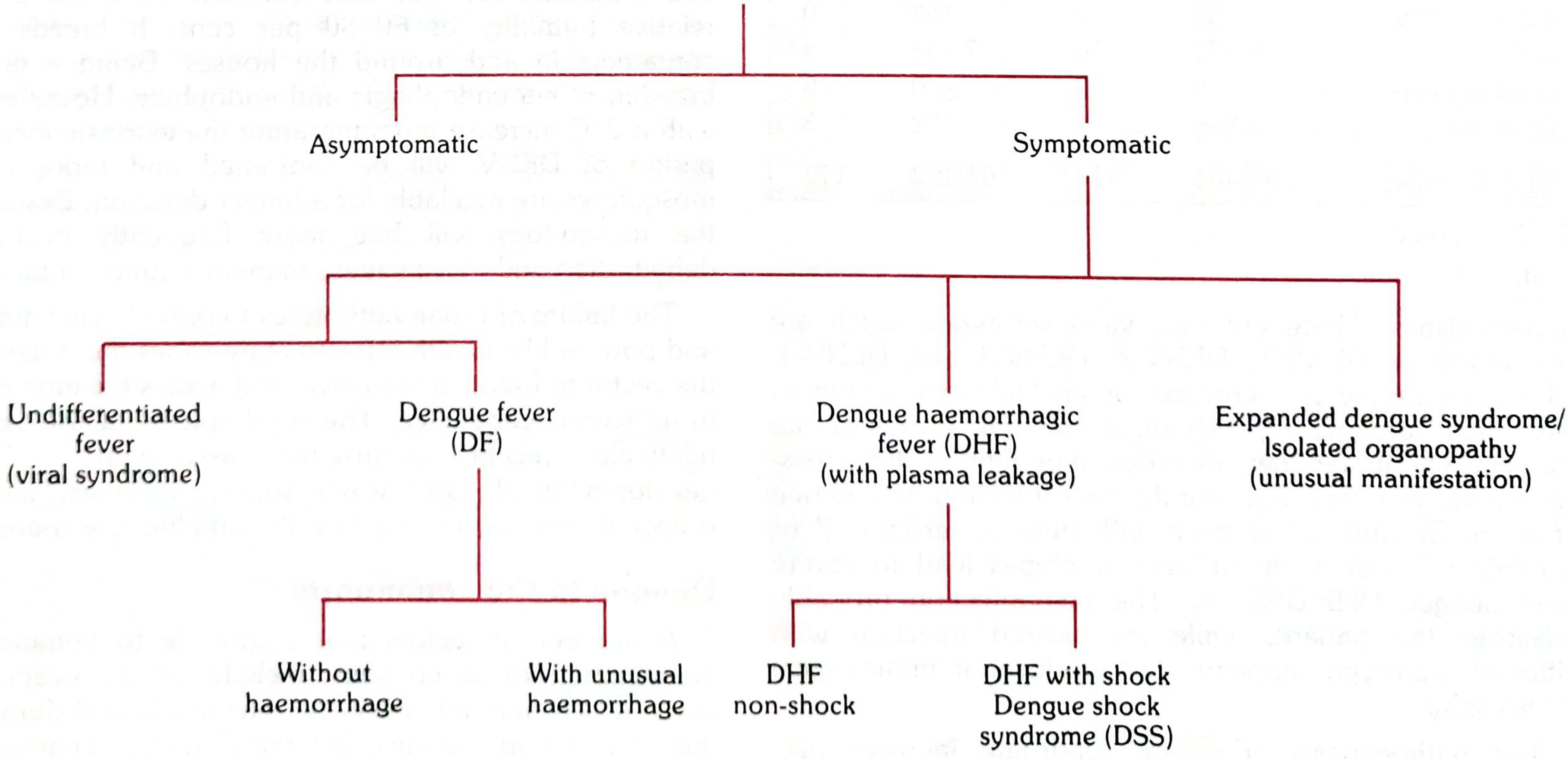
Type 1: Undifferentiated Fever (Viral Syndrome)
- Mildest form; especially in young children
- Non-specific febrile illness with no clear clinical diagnosis
- Only identifiable retrospectively by serology
- No hemorrhage, no plasma leakage
Type 2: Dengue Fever (DF)
- The "classic" dengue - self-limiting, rarely fatal
- High fever, severe myalgia, arthralgia ("breakbone fever"), retro-orbital pain, headache
- Rash (biphasic): flushing early, then maculopapular rash on trunk spreading outward
- May have unusual hemorrhage (petechiae) or positive tourniquet test
- No significant plasma leakage
- Sub-types: DF without hemorrhage; DF with unusual hemorrhage
Type 3: Dengue Hemorrhagic Fever (DHF)
- Severe form with plasma leakage - the life-threatening form
- All four WHO criteria must be met for diagnosis:
- Fever lasting 2-7 days
- Hemorrhagic manifestations (positive tourniquet test, petechiae, ecchymoses, mucosal bleeding)
- Thrombocytopenia: platelets ≤ 100,000/mm³
- Objective plasma leakage: hematocrit rise ≥20%, pleural effusion, ascites, hypoalbuminemia
- Graded I-IV by severity
- Typically occurs in second infections with a different serotype
| Grade | Features |
|---|---|
| Grade I | Fever + non-specific constitutional symptoms + positive tourniquet test only |
| Grade II | Grade I + spontaneous bleeding (skin, gum, nose, GI) |
| Grade III | Grade II + circulatory failure (rapid/weak pulse, narrow pulse pressure ≤20 mmHg, hypotension) = Dengue Shock Syndrome (DSS) |
| Grade IV | Profound shock (undetectable BP and pulse) |
Grades I-II = DHF without shock; Grades III-IV = Dengue Shock Syndrome (DSS)
Type 4: Expanded Dengue Syndrome / Isolated Organopathy
- Unusual manifestations: dengue encephalitis, dengue myocarditis, dengue hepatitis (severe), dengue nephropathy, dengue retinopathy
- Occurs without obvious plasma leakage or shock
- Increasingly recognized; not rare
4. PATHOPHYSIOLOGY
Course of Dengue Illness (from Park's Textbook)
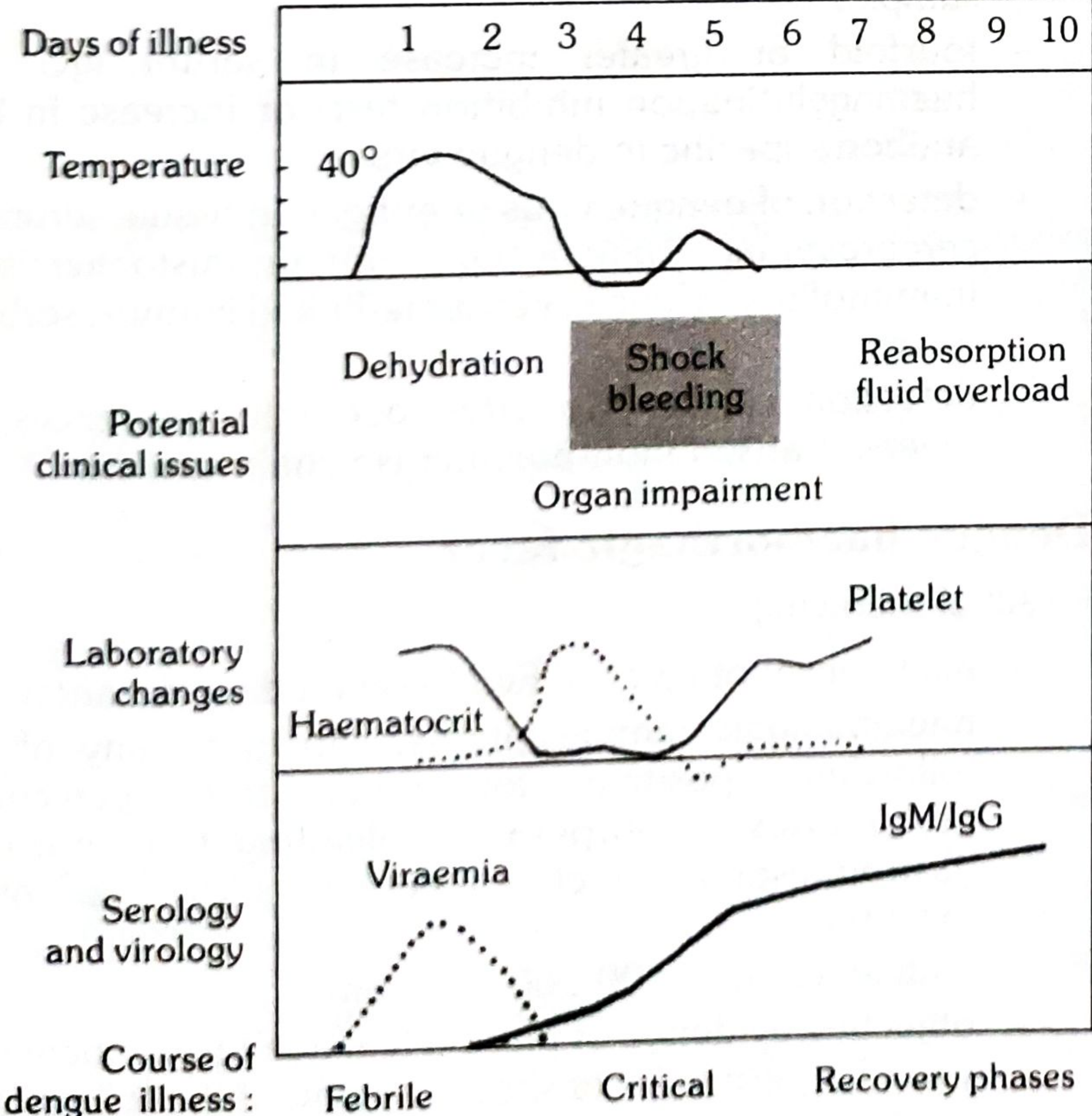
Step-by-Step Pathophysiology Flowchart
Infected Aedes aegypti mosquito bites human
↓
DENGUE VIRUS (DENV 1-4) injected into skin
→ Virus infects skin dendritic cells (Langerhans cells) and macrophages
→ Travels via lymphatics to regional lymph nodes
→ PRIMARY VIREMIA: virus enters bloodstream
↓
VIRUS INFECTS MONOCYTES/MACROPHAGES (the main target cells)
→ Replicates within mononuclear phagocytes
→ Secondary viremia: widespread dissemination to liver, spleen, lymph nodes, bone marrow
→ INCUBATION PERIOD: 4-7 days (range 3-10 days; clinically silent)
↓
FEBRILE PHASE begins (Days 1-3):
→ Viral PAMPs trigger innate immunity → cytokine release: IFN-α, TNF-α, IL-1, IL-6
→ HIGH FEVER (39-40°C), sudden onset
→ Severe myalgia and arthralgia: cytokines cause muscle/joint inflammation
→ Headache: cytokines trigger prostaglandin-mediated neuroinflammation
→ Retro-orbital pain: congestion of periorbital vessels
→ Early rash: facial flushing from vasodilation
↓
[In MOST patients - simple dengue fever]:
Defervescence around Day 3-5
→ Immune clearance of virus
→ Recovery over 7-10 days
↓
[In SECOND INFECTION WITH A DIFFERENT SEROTYPE - Severe Dengue]:
ANTIBODY-DEPENDENT ENHANCEMENT (ADE):
→ Pre-existing non-neutralizing antibodies from previous infection bind new serotype
→ Virus-antibody complex binds Fc receptors on monocytes/macrophages
→ INCREASES viral entry and replication in MORE macrophages
→ Far greater viral load than primary infection
↓
MASSIVE CYTOKINE STORM:
→ Infected macrophages + CD4+ and CD8+ T cells secrete: IFN-γ, TNF-α, IL-2, IL-8
→ Complement activation via circulating immune complexes (C3a, C5a)
→ These mediators act on VASCULAR ENDOTHELIUM
↓
INCREASED VASCULAR PERMEABILITY (the central event in DHF):
→ Cytokines damage tight junctions between endothelial cells
→ Plasma (water + proteins) leaks OUT of blood vessels into:
→ Pleural cavity (pleural effusion - predominantly RIGHT-SIDED)
→ Peritoneal cavity (ascites)
→ Retroperitoneum
→ Interstitial tissues (edema)
→ HEMATOCRIT RISES (hemoconcentration from plasma loss)
→ Blood volume FALLS → SHOCK (Dengue Shock Syndrome)
↓
THROMBOCYTOPENIA (parallel mechanism):
→ Dengue virus directly infects megakaryocytes in bone marrow → suppressed platelet production
→ Activated complement + antibodies destroy circulating platelets (immune-mediated)
→ Platelets consumed at sites of endothelial damage
→ PLATELET COUNT FALLS rapidly in critical phase
→ Combined with capillary fragility: HEMORRHAGE
↓
CRITICAL PHASE (Days 3-7 - period of defervescence):
→ Temperature drops (looks like improvement - DANGEROUS TRAP)
→ Plasma leakage peaks over 24-48 hours
→ Shock can develop RAPIDLY during this "apparent improvement"
↓
RECOVERY PHASE (Days 7-10):
→ Capillary integrity restored
→ Leaked plasma RE-ABSORBED back into circulation
→ Hematocrit falls (dilutional effect of fluid reabsorption)
→ Platelet count recovers
→ RISK: Fluid overload/pulmonary edema if too much IV fluid was given during critical phase
Why Second Infections Are More Dangerous (ADE - Exam Cornerstone)
FIRST INFECTION (DENV-1) → IgG antibodies against DENV-1 formed
↓
SECOND INFECTION (DENV-2):
Pre-existing DENV-1 antibodies recognize DENV-2 partially but CANNOT NEUTRALIZE it
→ Instead, antibody-virus complex binds Fc receptors on monocytes
→ TROJAN HORSE effect: virus gets DIRECTLY DELIVERED into the cell
→ 100× more macrophages infected compared to primary infection
→ Cytokine storm 100× more intense → DHF/DSS
5. CLINICAL FEATURES
Three-Phase Clinical Course
PHASE 1: FEBRILE PHASE (Days 1-3)
↓ High fever (39-40°C), sudden onset
↓ Severe frontal headache
↓ Retro-orbital pain (especially on eye movement or eye pressure)
↓ Severe myalgia + arthralgia = "BREAKBONE FEVER" (pain so severe it feels like bones breaking)
↓ Facial flushing
↓ Nausea and vomiting
↓ Anorexia
↓ Early rash: diffuse facial flushing or mottling
PHASE 2: CRITICAL PHASE (Days 3-7)
↓ Fever begins to FALL (defervescence)
↓ "WARNING SIGNS" appear (see below)
↓ Plasma leakage → pleural effusion, ascites, periorbital edema
↓ Hematocrit RISES ≥20%
↓ Platelets FALL rapidly
↓ Hemorrhagic manifestations appear
↓ If severe: SHOCK, organ impairment
PHASE 3: RECOVERY PHASE (Days 7-10)
↓ General well-being returns
↓ Appetite returns
↓ Second rash: "Isles of white in a sea of red" - characteristic convalescent rash
↓ Diuresis (reabsorption of leaked plasma)
↓ Bradycardia common
↓ Platelet count recovers (later than WBC)
↓ RISK: Fluid overload if excessive IV fluids were given during critical phase
Symptoms
| Symptom | Timing | Notes |
|---|---|---|
| High fever (39-40°C) | Day 1 onwards | Sudden onset; may be biphasic (saddle-back pattern in classic dengue: drops then rises again briefly) |
| Severe frontal headache | Day 1-3 | Often severe; prominent from onset |
| Retro-orbital pain | Day 1-2 | Pain behind the eyes, especially on moving eyes or pressing on eyeballs; characteristic of dengue |
| Severe myalgia | Day 1-4 | "Breakbone fever" - excruciating muscle and joint pain |
| Arthralgia | Day 1-4 | Joint pain; less prominent than in chikungunya |
| Nausea and vomiting | Day 1-4 | Common; anorexia profound |
| Abdominal pain | Day 3-5 | Especially epigastric and right upper quadrant (hepatic tenderness); WARNING SIGN if persistent |
| Sore throat, cough | Early | Mild; leads to misdiagnosis as URTI |
| Altered taste sensation | Variable | Metallic taste reported by many patients |
Signs
| Sign | Timing | Notes |
|---|---|---|
| Facial flushing | Days 1-2 | Bright red face from cutaneous vasodilation |
| Biphasic rash (saddleback) | - Early rash Day 1-2: diffuse flushing, mottling on face/chest. - Late rash Day 3-5: maculopapular/scarlatiniform on trunk spreading to extremities | The classic "isles of white in a sea of red" = sparing of small islands of normal skin within the erythematous rash. Itching and hyperaesthesia common. Lasts 2 hours to several days. May have desquamation |
| Positive tourniquet test (Rumpel-Leede test) | From Day 1 onwards | BP cuff inflated to midpoint between systolic and diastolic × 5 minutes. Positive = ≥10 petechiae per 2.5×2.5 cm area. DHF threshold: ≥20 petechiae. Tests capillary fragility |
| Petechiae | Day 3 onwards | Small red dots from capillary rupture; skin, conjunctivae, palate |
| Hepatomegaly | Day 2-5 | Mild; tender; >2 cm below costal margin = WARNING SIGN |
| Relative bradycardia | Recovery phase | Bradycardia during convalescence is a characteristic dengue finding |
| Conjunctival injection | Variable | Mild; not always present |
| Lymphadenopathy | Absent/minimal | Absence of lymphadenopathy differentiates dengue from rubella, measles, and West Nile fever |
| Narrow pulse pressure / hypotension | Critical phase (DHF Grade III-IV) | Pulse pressure ≤20 mmHg = early shock sign in DSS |
Why Important Signs Occur
| Sign | Mechanism |
|---|---|
| Breakbone pain | Cytokines (TNF-α, IL-1, IL-6) act on muscle and joint nociceptors + prostaglandin-mediated inflammation in periosteum |
| Rash (biphasic) | Phase 1 (flushing): intense cytokine-mediated vasodilation. Phase 2 (maculopapular): immune complex deposition in dermal vessels + platelet-endothelium interactions |
| Thrombocytopenia | Virus infects bone marrow megakaryocytes (suppressed production) + immune-mediated platelet destruction (antibody + complement) + consumption at damaged endothelial sites |
| Hematocrit rise | Plasma leaks out of capillaries into third spaces → blood becomes more concentrated → hematocrit rises (hemoconcentration) |
| Pleural effusion (right > left) | Right-sided predominance because right lung drains to the retroperitoneum and liver hilum where lymphatic channels are affected; permeability changes affect right pleural first |
| Shock in DSS | Critical volume of plasma lost through leaky capillaries → intravascular volume collapses → compensated then decompensated shock |
| Retro-orbital pain | Congestion and edema of periorbital tissues + congestion of intraocular vessels from increased vascular permeability |
| "Isles of white in sea of red" | Sparing of areas where skin is compressed against underlying structures + irregular distribution of immune complex deposition in dermis |
6. KEY SYMPTOMS THAT SUGGEST THE DIAGNOSIS
Hallmark Symptom
Sudden high fever + severe "breakbone" myalgia + retro-orbital pain + thrombocytopenia in a patient from or returning from a tropical/subtropical area - especially if accompanied by a biphasic rash or positive tourniquet test.
Classic Clinical Clues
| Clue | What It Suggests |
|---|---|
| Pain behind the eyeballs (retro-orbital pain, worse on eye movement/pressure) | Near-pathognomonic for dengue; ask specifically - patients rarely volunteer it |
| Fever of sudden onset reaching 39-40°C within hours | Dengue's explosive viremic onset |
| Myalgia so severe the patient "can't move" | Breakbone fever - distinguishes dengue from most other viral fevers |
| Thrombocytopenia ≤100,000/mm³ + leukopenia + fever | Classic dengue CBC triad |
| Biphasic ("saddle-back") fever | Fever → brief defervescence → second fever peak; classic for dengue, though not universal |
| Positive tourniquet test (≥10 petechiae/inch²) | Demonstrates increased capillary fragility from thrombocytopenia + plasma leakage |
| Rising hematocrit (≥20% above baseline) as fever falls | Plasma leakage = dengue entering critical phase |
| No lymphadenopathy in a febrile patient with rash | Distinguishes dengue from rubella, infectious mononucleosis, West Nile fever |
| Travel to Southeast Asia, South Asia, Latin America, Caribbean | High-transmission zones for all 4 serotypes |
| Day-biting mosquito exposure (no bed net protection) | Aedes aegypti bites in daytime; standard malaria nets don't protect |
| Second febrile illness worse than first in a patient from endemic area | Suggests second-serotype infection → ADE → higher risk of DHF |
| Pleural effusion (right-sided) + ascites during defervescence | Critical phase plasma leakage = severe dengue |
Warning Signs (RED FLAG - Critical Phase Is Beginning)
- 🚨 Abdominal pain or tenderness (persistent, worsening)
- 🚨 Persistent vomiting (≥3 vomits in 24 hours)
- 🚨 Clinical fluid accumulation (pleural effusion, ascites, facial/leg edema)
- 🚨 Mucosal bleeding (gum bleeding, epistaxis, hematemesis, melena)
- 🚨 Lethargy or restlessness (altered mental status)
- 🚨 Liver enlargement >2 cm below costal margin
- 🚨 Rapid rise in hematocrit with rapid drop in platelet count
The most dangerous clinical trap in dengue: When fever falls (defervescence around Day 3-5), it looks like the patient is getting better. This is actually the START of the critical phase when plasma leakage peaks. Patients (and sometimes doctors) go home or relax monitoring at precisely the moment closest monitoring is needed. Any patient who "improves" from dengue fever but then develops abdominal pain, persistent vomiting, or hypotension has entered Dengue Shock Syndrome.
Red Flag Findings - Severe Dengue Criteria (WHO 2009)
- 🚨 Severe plasma leakage → dengue shock (pulse pressure ≤20 mmHg; narrow pulse + rapid heart rate = DSS)
- 🚨 Severe bleeding → hematemesis, melena, massive hematuria, vaginal bleeding
- 🚨 Severe organ impairment: - Liver: AST/ALT ≥1000 IU/L (severe hepatitis) - CNS: altered consciousness, seizures (dengue encephalitis) - Heart: tachycardia out of proportion, poor cardiac function (dengue myocarditis) - Kidneys: oliguria, rising creatinine (dengue nephropathy)
Exam memory hook - "DENGUE" warning signs mnemonic:
- D - Drop in platelets with rise in hematocrit
- E - Extreme/persistent vomiting
- N - No urine (oliguria)
- G - Gut pain (abdominal tenderness)
- U - Unease (restlessness/lethargy)
- E - Edema/fluid accumulation (pleural effusion, ascites)
Act as a World-Class Professor of General Medicine, Clinical Pharmacology, Pathology, and Medical Education. Teach me Dengue disease in a simple, exam-oriented, and clinically practical manner. For every disease, follow this format: 1. Definition - One-line exam definition - Simple explanation 2. Causes (Etiology) - Common causes - Important risk factors 3. Types / Classification - Brief explanation of each type 4. Pathophysiology - Step-by-step flowchart with arrows - Easy to understand 5. Clinical Features - Symptoms - Signs - Explain why important symptoms occur 6. Key Symptoms That Suggest the Diagnosis - Hallmark symptom - Classic clinical clues - Red flag findings
🦟 DENGUE FEVER - Complete Exam-Oriented Clinical Guide
1. DEFINITION
2. CAUSES (ETIOLOGY)
The Virus
| Feature | Detail |
|---|---|
| Virus | Dengue virus (DENV) - single-stranded positive-sense RNA virus |
| Family | Flaviviridae |
| Genus | Flavivirus (same genus as Zika, Yellow fever, West Nile, Japanese encephalitis) |
| Serotypes | 4 serotypes: DENV-1, DENV-2, DENV-3, DENV-4 |
| Key point | Infection with one serotype = lifelong immunity to THAT serotype only; cross-immunity to others is partial and short-lived (6-12 months) |
Vectors
| Vector | Notes |
|---|---|
| Primary: Aedes aegypti | Day-biting (peak: 2 hours after sunrise and before sunset); breeds in clean, stagnant, man-made containers (flower pots, tires, water tanks, coolers); urban mosquito |
| Secondary: Aedes albopictus | Tiger mosquito; more widely distributed; less competent vector; rural/suburban |
Exam pearl: Aedes mosquitoes bite during the DAY - not at night like Anopheles (malaria). Standard bed nets do NOT protect against dengue.
Important Risk Factors
| Risk Factor | Reason |
|---|---|
| Living in or traveling to tropical/subtropical areas (Southeast Asia, Indian subcontinent, Latin America, Caribbean, Pacific Islands) | Highest Aedes density and DENV circulation |
| Urban/semi-urban environments | Aedes aegypti is an urban mosquito |
| Prior infection with a DIFFERENT dengue serotype | Most important risk factor for severe dengue - antibody-dependent enhancement (ADE) |
| Infants of immune mothers | Maternal anti-dengue IgG causes ADE in the infant |
| Children <15 years in endemic areas | High exposure; lower baseline immunity |
| Rainy season | Increased mosquito breeding sites |
| Poor vector control | Stagnant water collections near homes |
| Genetic factors | Certain HLA types and TNF-α polymorphisms increase severity |
3. TYPES / CLASSIFICATION
WHO 2009 Classification (Current Standard)
Type 1: Undifferentiated Fever (Viral Syndrome)
- Mildest form, especially in young children
- Non-specific febrile illness with no clear clinical diagnosis
- Only identifiable retrospectively by serology
- No hemorrhage, no plasma leakage
Type 2: Dengue Fever (DF)
- Classic dengue - self-limiting, rarely fatal
- Sudden high fever, severe myalgia/arthralgia ("breakbone fever"), retro-orbital pain, headache, rash
- May have unusual hemorrhage (petechiae) or positive tourniquet test
- No significant plasma leakage
- Sub-types: DF without hemorrhage; DF with unusual hemorrhage
Type 3: Dengue Hemorrhagic Fever (DHF)
- Severe form with plasma leakage - the life-threatening presentation
- All four WHO criteria must be met:
- Fever lasting 2-7 days
- Hemorrhagic manifestations (positive tourniquet test, petechiae, ecchymoses, mucosal bleeding)
- Thrombocytopenia: platelets ≤100,000/mm³
- Objective plasma leakage: hematocrit rise ≥20%, pleural effusion, ascites, or hypoalbuminemia
| Grade | Features |
|---|---|
| Grade I | Fever + constitutional symptoms + positive tourniquet test only |
| Grade II | Grade I + spontaneous bleeding (skin, gum, nose, GI) |
| Grade III | Grade II + circulatory failure (rapid/weak pulse, narrow pulse pressure ≤20 mmHg, hypotension) = Dengue Shock Syndrome (DSS) |
| Grade IV | Profound shock with undetectable BP and pulse |
Grades I-II = DHF without shock; Grades III-IV = Dengue Shock Syndrome (DSS)
Type 4: Expanded Dengue Syndrome / Isolated Organopathy
- Unusual manifestations without classic plasma leakage
- Includes: dengue encephalitis, dengue myocarditis, dengue hepatitis (severe), dengue nephropathy, dengue retinopathy
- Increasingly recognized; requires high clinical suspicion
4. PATHOPHYSIOLOGY
Course of Dengue Illness
Step-by-Step Pathophysiology Flowchart
Infected Aedes aegypti mosquito bites human
↓
DENGUE VIRUS injected into skin
→ Infects dendritic cells (Langerhans cells) and skin macrophages
→ Travels via lymphatics to regional lymph nodes
→ PRIMARY VIREMIA: virus enters bloodstream
↓
VIRUS INFECTS MONOCYTES/MACROPHAGES (the main target cells)
→ Replicates within mononuclear phagocytes
→ Secondary viremia: disseminates to liver, spleen, lymph nodes, bone marrow
→ INCUBATION PERIOD: 4-7 days (range 3-10 days; clinically silent)
↓
FEBRILE PHASE (Days 1-3):
→ Viral PAMPs trigger innate immunity → cytokine release: IFN-α, TNF-α, IL-1, IL-6
→ SUDDEN HIGH FEVER (39-40°C)
→ Severe myalgia/arthralgia: cytokines inflame muscle and joints
→ Headache: prostaglandin-mediated neuroinflammation
→ Retro-orbital pain: periorbital vascular congestion
→ Early rash: cytokine-driven vasodilation → facial flushing
↓
[In MOST patients → simple dengue fever]:
Defervescence around Day 3-5 → immune clearance → full recovery in 7-10 days
↓
[In SECOND INFECTION WITH A DIFFERENT SEROTYPE → Severe Dengue]:
ANTIBODY-DEPENDENT ENHANCEMENT (ADE) - THE KEY MECHANISM:
→ Pre-existing non-neutralizing IgG from previous infection binds new serotype
→ Cannot neutralize virus but virus-antibody complex binds Fc receptors on macrophages
→ TROJAN HORSE: virus delivered DIRECTLY into far more macrophages
→ Viral load 100× higher than primary infection
→ MASSIVE CYTOKINE STORM: IFN-γ, TNF-α, IL-2, IL-8 from infected macrophages
+ activated CD4+ and CD8+ T cells
→ Complement activation via circulating immune complexes (C3a, C5a)
↓
INCREASED VASCULAR PERMEABILITY (the central event in severe dengue):
→ Cytokines disrupt endothelial tight junctions
→ Plasma (water + proteins) leaks OUT of blood vessels into:
→ Pleural cavity (pleural effusion - predominantly RIGHT-SIDED)
→ Peritoneal cavity (ascites)
→ Interstitial tissues (edema)
→ HEMATOCRIT RISES ≥20% (hemoconcentration from plasma loss)
→ Intravascular volume FALLS → SHOCK (Dengue Shock Syndrome)
↓
THROMBOCYTOPENIA (parallel mechanism):
→ Virus directly infects bone marrow megakaryocytes → suppressed production
→ Antibody + complement-mediated destruction of circulating platelets
→ Platelet consumption at damaged endothelial sites
→ + Capillary fragility → HEMORRHAGE
↓
CRITICAL PHASE (Days 3-7 - period of defervescence):
→ Temperature DROPS (appears like improvement - THE MOST DANGEROUS TRAP)
→ Plasma leakage PEAKS over 24-48 hours
→ Shock develops RAPIDLY during apparent improvement
↓
RECOVERY PHASE (Days 7-10):
→ Capillary integrity restored
→ Leaked plasma RE-ABSORBED back into circulation
→ Hematocrit falls; platelets recover
→ RISK: Fluid overload/pulmonary edema if too much IV fluid given during critical phase
The ADE Mechanism (The Exam Cornerstone)
1st Infection (DENV-1) → IgG antibodies against DENV-1 formed → recovery
↓
2nd Infection (DENV-2):
Pre-existing DENV-1 antibodies recognize DENV-2 but CANNOT neutralize it
→ Antibody-virus complex binds Fc receptors on monocytes/macrophages
→ TROJAN HORSE: virus gets directly delivered into cells
→ Far more macrophages infected → cytokine storm → DHF/DSS
5. CLINICAL FEATURES
Three-Phase Clinical Course
FEBRILE PHASE (Days 1-3):
→ Sudden high fever 39-40°C
→ Severe frontal headache
→ RETRO-ORBITAL PAIN (pain behind eyes on movement/pressure)
→ BREAKBONE MYALGIA + arthralgia
→ Facial flushing
→ Nausea, vomiting, anorexia
→ Early rash: diffuse facial flushing/mottling
CRITICAL PHASE (Days 3-7):
→ Fever FALLS (defervescence - looks like improvement)
→ WARNING SIGNS appear (see below)
→ Plasma leakage → pleural effusion (right > left), ascites
→ Hematocrit RISES ≥20%
→ Platelet count FALLS rapidly (may reach <20,000)
→ Hemorrhagic manifestations: petechiae, gum bleeding, hematemesis
→ If severe: DENGUE SHOCK SYNDROME
RECOVERY PHASE (Days 7-10):
→ General well-being returns
→ Appetite returns
→ Second rash: "Isles of white in a sea of red" (convalescent rash)
→ Diuresis (plasma reabsorption)
→ Bradycardia (characteristic finding in recovery)
→ RISK: Fluid overload from reabsorbed plasma + IV fluids given
Symptoms
| Symptom | Timing | Notes |
|---|---|---|
| High fever (39-40°C) | Day 1 onwards | Sudden onset; may be biphasic "saddle-back" pattern |
| Severe frontal headache | Day 1-3 | Prominent from onset |
| Retro-orbital pain | Day 1-2 | Behind the eyes; worse on eye movement or direct pressure; near-characteristic of dengue |
| Severe myalgia | Day 1-4 | "Breakbone fever" - excruciating; patient cannot move comfortably |
| Arthralgia | Day 1-4 | Joint pain; less severe than chikungunya |
| Nausea, vomiting | Day 1-4 | Common; anorexia profound |
| Abdominal pain | Day 3-5 | Epigastric/RUQ; if persistent = WARNING SIGN |
| Altered taste | Variable | Metallic taste described |
Signs
| Sign | Timing | Notes |
|---|---|---|
| Facial flushing | Day 1-2 | Bright red face from intense vasodilation |
| Biphasic rash | Phase 1 (Day 1-2): flushing/mottling on face/neck/chest. Phase 2 (Day 3-5): maculopapular/scarlatiniform on trunk spreading to extremities | Classic "isles of white in a sea of red" in convalescent phase. Itching common |
| Positive tourniquet test | From Day 1 | BP cuff at midpoint × 5 min; positive = ≥10 petechiae per 2.5×2.5 cm; DHF threshold = ≥20 petechiae |
| Petechiae | Day 3 onwards | Skin, conjunctivae, palate |
| Hepatomegaly | Day 2-5 | Mild, tender; >2 cm below costal margin = WARNING SIGN |
| Relative bradycardia | Recovery phase | Characteristic bradycardia during convalescence |
| No lymphadenopathy | Throughout | Absence distinguishes dengue from rubella, EBV, West Nile fever |
| Narrow pulse pressure | Critical phase | ≤20 mmHg = early dengue shock |
Why Important Signs Occur
| Sign | Mechanism |
|---|---|
| Breakbone pain | TNF-α, IL-1, IL-6 act on muscle/joint nociceptors + prostaglandin-mediated periosteal inflammation |
| Retro-orbital pain | Congestion and edema of periorbital vessels from increased vascular permeability |
| Thrombocytopenia | Virus infects megakaryocytes (↓ production) + immune-mediated platelet destruction (antibody + complement) + consumption at damaged endothelial sites |
| Hematocrit rise | Plasma leaks into third spaces → blood becomes concentrated → hematocrit rises (hemoconcentration) |
| Right-sided pleural effusion | Right pleural drainage connects to hepatic hilum and retroperitoneum, most affected by cytokine-driven permeability changes |
| Shock in DSS | Critical plasma volume lost through leaky capillaries → intravascular collapse |
| "Isles of white in sea of red" | Irregular distribution of immune complex-mediated dermal inflammation sparing compressed skin areas |
| Bradycardia in recovery | Vagal predominance as autonomic system stabilizes after cytokine storm; possible direct myocardial effect |
6. KEY SYMPTOMS THAT SUGGEST THE DIAGNOSIS
Hallmark Symptom
Sudden high fever + severe "breakbone" myalgia + retro-orbital pain + thrombocytopenia in a patient from or returning from a tropical area - especially with a biphasic rash and positive tourniquet test.
Classic Clinical Clues
| Clue | What It Suggests |
|---|---|
| Pain specifically behind the eyes (retro-orbital), worse on eye movement or pressure | Near-pathognomonic for dengue; always ask for it specifically |
| Fever of explosive sudden onset reaching 39-40°C within hours | Dengue's viremic burst; typhoid rises gradually by contrast |
| Myalgia so severe the patient cannot move | Breakbone fever - distinguishes dengue from most other viral fevers |
| Thrombocytopenia + leukopenia + fever | The dengue CBC triad |
| Saddle-back (biphasic) fever pattern | Fever → brief dip → second fever peak; classic but not universal |
| Positive tourniquet test (≥10 petechiae/inch²) | Capillary fragility from thrombocytopenia + plasma leakage |
| Rising hematocrit ≥20% as fever falls | Plasma leakage = critical phase beginning |
| Absence of lymphadenopathy in febrile patient with rash | Distinguishes dengue from rubella, EBV/mononucleosis, West Nile fever |
| Travel to Southeast Asia, South Asia, Latin America, Caribbean | All four serotypes co-circulate in these zones |
| Day-biting mosquito exposure; no bed net use | Aedes aegypti is a daytime biter - bed nets don't protect |
| Second dengue illness worse than first | Suggests ADE from second-serotype infection → higher DHF risk |
| Right-sided pleural effusion + ascites during defervescence | Critical phase plasma leakage = severe dengue |
Warning Signs - RED FLAGS (Critical Phase Onset)
- 🚨 Abdominal pain or tenderness (persistent, worsening)
- 🚨 Persistent vomiting (≥3 episodes in 24 hours)
- 🚨 Clinical fluid accumulation (pleural effusion, ascites, periorbital/limb edema)
- 🚨 Mucosal bleeding (gum, epistaxis, hematemesis, melena, menorrhagia)
- 🚨 Lethargy or restlessness (reduced brain perfusion)
- 🚨 Liver enlargement >2 cm below costal margin
- 🚨 Rapid rise in hematocrit with rapid fall in platelet count
Red Flags for Severe Dengue
- 🚨 Severe plasma leakage → DSS: narrow pulse pressure ≤20 mmHg (e.g., 100/80 mmHg); cold extremities; capillary refill >2 seconds
- 🚨 Severe bleeding: hematemesis, melena, massive hemorrhage
- 🚨 Severe organ impairment: AST/ALT ≥1000 (hepatitis); altered consciousness (encephalitis); severe cardiac dysfunction (myocarditis); oliguria (nephropathy)
The most dangerous clinical trap in dengue: When fever falls around Day 3-5, it looks like the patient is improving. This is actually the START of the critical phase when plasma leakage is at its worst. Patients (and clinicians) relax at the exact moment maximum vigilance is needed. Never discharge a dengue patient just because the fever has broken.
Exam memory hook - "DENGUE" warning signs:
- D - Drop in platelets / rise in hematocrit
- E - Excessive vomiting (persistent)
- N - No urine (oliguria)
- G - Gut pain (persistent abdominal tenderness)
- U - Unease (restlessness or lethargy)
- E - Edema/fluid accumulation (pleural effusion, ascites)
7. Laboratory Investigations of dengue disease Create a table: Investigation | Expected Finding | Clinical Significance Include: - CBC - Biochemistry - Disease-specific tests - Imaging findings if relevant 8. Differential Diagnosis Create a comparison table: Disease | Similar Features | Differentiating Features 9. Short Clinical Case Example format: "A 55-year-old male presents with chest pain radiating to the left arm, sweating, and breathlessness. ECG shows ST elevation in leads II, III, and aVF. Troponin-I is elevated." Diagnosis: Reasoning: Provide only 1–2 short high-yield cases.
7. LABORATORY INVESTIGATIONS OF DENGUE FEVER
COMPLETE BLOOD COUNT (CBC)
| Investigation | Expected Finding | Clinical Significance |
|---|---|---|
| Total Leukocyte Count (TLC/WBC) | Leukopenia (WBC <5,000/mm³) from Day 2 onwards; may drop to 2,000-3,000/mm³ | Cytokine-mediated bone marrow suppression + viral destruction of leukocyte precursors. Leukopenia is the earliest and most consistent CBC finding in dengue. Progressive leukopenia just before defervescence signals the approaching critical phase |
| Neutrophil count | Neutropenia; relative lymphocytosis | Bone marrow suppression preferentially affects neutrophil line |
| Atypical lymphocytes | Present on peripheral blood smear | Activated T cells responding to dengue antigen; similar to infectious mononucleosis appearance |
| Platelet count | Thrombocytopenia - falls progressively from Day 3; may reach <20,000/mm³ at nadir in severe dengue | Direct bone marrow megakaryocyte infection + immune-mediated platelet destruction + consumption at damaged endothelial sites. Platelet <100,000 = DHF diagnostic criterion. Rate of fall matters more than absolute value - rapid fall = critical phase |
| Hematocrit (PCV) | Rising hematocrit ≥20% above baseline during critical phase | Hemoconcentration from plasma leakage into third spaces. This is the single most important parameter to monitor in dengue. A rise in hematocrit while fever falls = plasma leakage is occurring. In recovery phase, hematocrit falls as plasma reabsorbs |
| Hemoglobin | Initially normal; may fall in recovery (dilutional) or if significant hemorrhage | During recovery, plasma reabsorption dilutes the blood → hematocrit falls; does NOT mean re-bleeding |
Exam-critical rule: Monitor hematocrit every 4-6 hours in dengue with warning signs. A rapidly decreasing platelet count in parallel with a rising hematocrit = transition to critical phase. This combination is the most clinically important CBC pattern in dengue.
BIOCHEMISTRY
| Investigation | Expected Finding | Clinical Significance |
|---|---|---|
| Serum AST (SGOT) and ALT (SGPT) | Elevated - typically 2-5× normal in uncomplicated dengue; may rise to >1000 IU/L in severe dengue hepatitis | Dengue virus directly infects hepatocytes (Kupffer cells) + cytokine-mediated hepatocellular injury. AST:ALT ratio typically >2 (unlike viral hepatitis where ALT > AST). Severe hepatitis (AST/ALT ≥1000) = severe dengue criterion requiring hospitalization |
| Serum albumin | Low (hypoalbuminemia) in DHF | Plasma proteins including albumin leak through damaged capillaries into third spaces. Hypoalbuminemia = confirms plasma leakage = DHF diagnostic criterion |
| Serum sodium | Hyponatremia common | From fluid shifts + inappropriate ADH secretion from viral inflammation |
| Coagulation profile (PT, aPTT) | Prolonged in severe dengue | Liver dysfunction impairs clotting factor synthesis; consumption coagulopathy (DIC) in severe DHF/DSS; fibrinogen may be low, D-dimer elevated |
| Serum urea and creatinine | Elevated in dengue-associated AKI | Mechanisms: hypovolemia (pre-renal), direct viral tubular injury, myoglobinuria from rhabdomyolysis, hemoglobinuria, immune complex glomerulonephritis. AKI is seen in ~5% of hospitalized dengue cases |
| Urine protein | Proteinuria (mild to moderate; rarely nephrotic) | From glomerular injury (immune complex deposition, anti-GBM antibodies via molecular mimicry) |
| Serum CRP | Mildly elevated or normal | Lower than expected for the severity of fever; distinguishes dengue from bacterial infections where CRP is markedly elevated. Low CRP in dengue vs. high CRP in bacterial sepsis is a useful distinguishing point |
| Blood glucose | Hypoglycemia in severe cases | Poor oral intake + hepatic dysfunction impairs gluconeogenesis |
| Serum lactate | Elevated in DSS | Tissue hypoperfusion and anaerobic metabolism during shock |
| Serum electrolytes (K+) | Hypokalemia common | From vomiting, poor intake, GI losses during febrile phase |
DISEASE-SPECIFIC TESTS
| Investigation | Expected Finding | Clinical Significance |
|---|---|---|
| NS1 Antigen (Non-Structural Protein 1) ELISA / RDT | Positive from Day 1 to Day 5-6 of illness | NS1 is a dengue-specific viral glycoprotein secreted early during infection. First test to order in a febrile patient presenting within 5 days of symptom onset. Detects all 4 serotypes but cannot differentiate between them. Sensitivity ~80-90% in primary infection; slightly lower in secondary. Negative NS1 after Day 5 does NOT rule out dengue |
| Dengue IgM antibody (MAC-ELISA) | Positive from Day 5 onwards (primary infection); Day 4 (secondary infection); peaks at 2 weeks; persists 2-3 months | Test of choice for patients presenting after Day 5. IgM = current/recent infection. Cannot distinguish serotypes |
| Dengue IgG antibody (ELISA) | In primary infection: rises slowly, detectable from Day 7-10. In secondary infection: IgG rises rapidly and dramatically even from Day 1-2 (anamnestic response) | Primary vs. Secondary infection differentiation: Low/absent IgG = primary; high IgG from early in illness = secondary. IgM/IgG ratio: high IgM:IgG ratio = primary infection; low ratio = secondary infection. Secondary infection carries higher DHF/DSS risk |
| RT-PCR (Reverse Transcriptase PCR) | Positive in Days 1-5 (during viremia); can identify specific serotype | Most sensitive and specific early test; identifies all 4 serotypes; confirms active viral replication. Not available in most routine labs; used in reference/research labs and outbreak surveillance |
| Virus isolation | Positive in Days 1-6 from serum, plasma, buffy coat | Gold standard for serotype confirmation; complex, slow (5-7 days), not for clinical management; used for epidemiological surveillance and vaccine research |
| Haemagglutination Inhibition Assay (HIA) | ≥4-fold rise in paired sera or titre ≥1:1280 | Traditional serological test; detects anti-dengue antibodies; cross-reacts with other flaviviruses (Zika, yellow fever, West Nile); requires paired acute and convalescent samples 2 weeks apart; not useful for rapid clinical diagnosis |
| Tourniquet test (Rumpel-Leede / capillary fragility test) | Positive ≥10 petechiae per 2.5×2.5 cm (1 inch²); DHF: ≥20 petechiae | Bedside test requiring only a BP cuff. Inflates to midpoint between systolic and diastolic pressure × 5 minutes. Tests capillary fragility - positive in thrombocytopenia and plasma leakage. The single most important bedside physical examination test in dengue |
Timing of Diagnostic Tests - Critical Summary
| Day of Illness | Best Test(s) to Order | Why |
|---|---|---|
| Day 1-5 (Febrile phase, early) | NS1 Antigen RDT/ELISA + CBC + hematocrit | Viremia is at peak; NS1 is highly positive; CBC shows early leukopenia/thrombocytopenia |
| Day 5 onwards (Defervescence, critical phase) | NS1 + dengue IgM ELISA + CBC + hematocrit every 4-6h | IgM rises from Day 5; NS1 may still be positive; hematocrit monitoring is paramount for plasma leakage detection |
| After Day 7 (Recovery phase) | Dengue IgM + IgG ELISA | NS1 usually negative; serology is the primary diagnostic method; IgM persists 2-3 months |
| Any phase in reference lab | RT-PCR (Days 1-5) or Virus isolation (Days 1-6) | Serotype identification; outbreak surveillance; research |
IMAGING FINDINGS
| Investigation | Expected Finding | Clinical Significance |
|---|---|---|
| Chest X-ray (CXR) - erect | Right-sided pleural effusion (most common); bilateral effusions in severe disease; perihilar haziness/pulmonary edema in fluid overload | Right-sided pleural effusion on CXR is one of the key indicators of plasma leakage (DHF criterion). Appears as blunting of right costophrenic angle. Also used to monitor fluid overload during recovery - bilateral opacities = too much IV fluid given |
| Ultrasound abdomen | Gallbladder wall edema (thickening >3 mm - an early sign of plasma leakage); ascites (free fluid in peritoneal cavity); pleural effusion (especially right); hepatomegaly; splenomegaly | Gallbladder wall edema is an early ultrasound marker of plasma leakage - it appears BEFORE clinical ascites becomes detectable. Abdominal ultrasound is more sensitive than CXR for detecting early plasma leakage. Also monitors for splenic rupture (rare complication of dengue splenomegaly) |
| CT scan | Pleural effusions, ascites, periorbital edema, hepatomegaly, rarely cerebral edema in dengue encephalitis | Reserved for complicated dengue - suspected encephalitis, hepatic failure, or atypical presentations |
| Echocardiography | Pericardial effusion; reduced ventricular function in dengue myocarditis | Indicated if new arrhythmia, cardiac symptoms, or hypotension out of proportion to plasma leakage |
8. DIFFERENTIAL DIAGNOSIS
| Disease | Similar Features to Dengue | Differentiating Features |
|---|---|---|
| Malaria | Fever, thrombocytopenia, headache, myalgia, leukopenia, travel to tropics | Cyclic paroxysmal fever (48h or 72h) with rigors and sweating vs. dengue's continuous high fever with saddle-back pattern; no retro-orbital pain; splenomegaly more prominent; positive blood smear/RDT for Plasmodium; can have very high WBC in severe malaria; no NS1 positivity |
| Chikungunya | Fever, myalgia, rash, thrombocytopenia (mild), travel to tropics, same Aedes vector | Dominant ARTHRITIS - severe, symmetrical, polyarthritis affecting small joints (fingers, wrists, ankles) that may persist for months or years vs. dengue's predominantly myalgia; rash more maculopapular and prominent; thrombocytopenia less severe; plasma leakage and shock do NOT occur; positive Chikungunya IgM/PCR; self-limiting |
| Typhoid fever | Fever, headache, leukopenia, thrombocytopenia, hepatomegaly, travel to endemic area | Step-ladder fever (gradually rising over days) vs. dengue's sudden explosive onset; relative bradycardia (pulse-temperature dissociation); no retro-orbital pain; no plasma leakage or DHF; constipation more common than diarrhea; rose spots (vs dengue's flushing/maculopapular rash); positive blood culture for S. Typhi; no NS1 |
| Leptospirosis | Fever, myalgia, headache, thrombocytopenia, hepatomegaly, leukopenia, jaundice, renal failure, travel to tropics | Biphasic fever (leptospiremic + immune phase); prominent calf muscle tenderness (almost pathognomonic); conjunctival suffusion (bright red eyes without discharge); exposure to water/animals; more severe jaundice and AKI; uveitis; positive MAT or IgM ELISA for Leptospira; no NS1 |
| Influenza | Sudden high fever, headache, severe myalgia, dry cough, malaise | Less severe myalgia than dengue; no retro-orbital pain; no thrombocytopenia (unless severe); no plasma leakage; seasonal clustering; positive rapid influenza test/PCR; rhinorrhea more common; predominantly respiratory symptoms |
| Rubella | Fever, maculopapular rash, thrombocytopenia, young patients | Prominent cervical and post-auricular lymphadenopathy (vs. NO lymphadenopathy in dengue - a key distinguishing feature); milder fever; no myalgia; no retro-orbital pain; Koplik's spots absent (those are in measles); positive rubella IgM |
| Measles | Fever, rash, conjunctival injection, myalgia | Koplik's spots (white spots on buccal mucosa) - pathognomonic; rash starts on face/behind ears and descends (vs dengue rash starting on trunk); cough + coryza + conjunctivitis (the 3 Cs); lymphadenopathy present; positive measles IgM; no thrombocytopenia early |
| Zika virus | Fever, rash, myalgia, headache, same Aedes vector | Fever is usually MILD and short; conjunctivitis (non-purulent) prominent; arthralgia more than myalgia; associated with microcephaly (in congenital infection) and Guillain-Barré syndrome; NO significant thrombocytopenia; NO plasma leakage/DHF; positive Zika PCR/IgM |
| Scrub typhus | Fever, headache, myalgia, rash, thrombocytopenia, travel to Asia | Classic eschar at mite bite site (pathognomonic - found in groin, axilla, behind ears); cervical lymphadenopathy; positive Weil-Felix (OX-K) or Scrub typhus IgM; responds dramatically to doxycycline; no plasma leakage |
| Viral Hemorrhagic Fevers (Ebola, Marburg, Yellow Fever) | Fever, thrombocytopenia, hemorrhage, shock | Different geographic exposures (Ebola = Central/West Africa; Yellow fever = Africa/South America); Yellow fever: jaundice is prominent early; Faget's sign; Ebola/Marburg: contact with infected bodily fluids/bushmeat; specific PCR/serology required; different clinical trajectories |
Clinical exam principle: Among all differentials, Chikungunya is the hardest to distinguish from dengue because it uses the SAME vector. The key distinction: arthralgia in chikungunya vs. myalgia in dengue; chikungunya arthritis can persist for months while dengue arthralgia is brief.
9. SHORT CLINICAL CASES
Case 1 - Classic Dengue Fever with Warning Signs
- Travel to Thailand (highly endemic for all 4 DENV serotypes) + sudden explosive onset = classic dengue epidemiology and presentation
- Retro-orbital pain (pain behind eyes on movement) - near-pathognomonic for dengue; always ask for it
- Breakbone myalgia so severe he cannot walk - characteristic of dengue's cytokine-driven myalgia
- NS1 antigen positive on Day 3 - confirms dengue during the febrile/viremic phase (Days 1-5)
- Leukopenia (2,800) + thrombocytopenia (68,000) = classic dengue CBC pattern
- The most dangerous moment: fever partially breaks (appears like improvement) but hematocrit has risen from 38% to 47% = a 23.7% rise = plasma leakage beginning - this is crossing from febrile to critical phase
- Warning signs are now all present: persistent vomiting + abdominal pain + dizziness (orthostatic hypotension) = imminent DHF/shock
- Next step: Immediate hospitalization; IV isotonic fluid resuscitation; hematocrit monitoring every 4-6 hours; platelet monitoring; avoid NSAIDs/aspirin; do NOT discharge despite apparent "improvement" of fever
Case 2 - Dengue Shock Syndrome (Severe Dengue)
- Age 9, endemic area (Chennai) = classic demographic for severe dengue
- Day 5 - the fever has just broken = this IS the critical phase (Days 3-7 of defervescence)
- Pulse pressure of only 10 mmHg (88/78) = the classic early shock sign of DSS (WHO criterion: pulse pressure ≤20 mmHg). The diastolic rises as peripheral vasoconstriction compensates while systolic falls - the narrowing pulse pressure indicates falling cardiac output and hypovolemia
- Cold extremities + weak thready pulse + capillary refill 4 sec + sleepiness (reduced brain perfusion) = compensated shock rapidly becoming decompensated
- Hematocrit 56% vs. baseline 37% = a 51% rise - massive plasma leakage from vascular space
- Right-sided pleural effusion (dullness at right base) + ascites = plasma in third spaces confirms DHF criteria
- Petechiae + platelets 22,000 = hemorrhagic manifestations
- All 4 DHF criteria met + signs of shock = DHF Grade III (Dengue Shock Syndrome)
- Next step: IMMEDIATE IV isotonic crystalloid bolus (10-20 mL/kg over 15-30 minutes); repeat and reassess; switch to colloid if no improvement; continuous monitoring of vital signs, urine output, and hematocrit; avoid over-hydration (risk of pulmonary edema in recovery phase). Mortality without treatment: >10%; with prompt fluid resuscitation: <1%
Exam trap illustrated by Case 2: The child's fever broke yesterday - at this point many caregivers and even some physicians think the worst is over and the child is recovering. In dengue, this is the most dangerous assumption. The falling temperature signals the START of plasma leakage, not resolution. Any child (or adult) becoming quiet, lethargic, or complaining of abdominal pain after dengue fever breaks must be assessed urgently for DSS.
10. Treatment Of dengue A. Non-Pharmacological Treatment B. Pharmacological Treatment Create a table: Drug | Drug Group | Mechanism of Action | Indication | Dose | Major Adverse Effects For every disease mention: - Drug of Choice (DOC) - First-line drugs - Alternative drugs - Emergency drugs (if applicable) 11. Clinical Pearls - Important bedside clues - High-yield exam facts - Viva points 12. Common Diagnostic Mistakes - Frequently missed findings - Common confusion with other diseases - How to avoid errors
10. TREATMENT OF DENGUE FEVER
A. NON-PHARMACOLOGICAL TREATMENT
| Intervention | Details | Rationale |
|---|---|---|
| Triage and risk stratification | Classify all dengue patients into: (1) Dengue without warning signs - home management; (2) Dengue with warning signs - hospitalize; (3) Severe dengue (DHF/DSS) - ICU/high-dependency care | The single most important management decision. Appropriate triage prevents both under-treatment (missing DSS) and over-treatment (unnecessary hospitalization) |
| Oral hydration (uncomplicated DF) | ORS, fruit juices, coconut water, electrolyte solutions, soups - 2-3 litres/day minimum. Fluids with electrolytes preferred over plain water. Caution: glucose/sugar-containing fluids may worsen hyperglycemia in diabetics | Replaces losses from fever (insensible), sweating, and vomiting; maintains circulating volume; adequate oral intake reduces hospitalization rates |
| IV fluid resuscitation (DHF/DSS) | Start with isotonic crystalloid (normal saline 0.9% or Ringer's Lactate); initial bolus 5-10 mL/kg over 1 hour for DHF with warning signs; 10-20 mL/kg over 15-30 minutes for DSS; reassess every 15-30 minutes; if no improvement after 1000 mL crystalloid → switch to colloid (Dextran 40 or hydroxyethyl starch/Haemaccel) | Plasma has leaked OUT of blood vessels; IV fluids temporarily restore intravascular volume. Crystalloids are first choice - they fill the vascular space. Colloids are given when crystalloids fail because they have larger molecules that remain in the intravascular space longer. Over-infusion is as dangerous as under-infusion |
| Fluid monitoring - hematocrit-guided | Serial hematocrit every 4-6 hours during critical phase; urine output monitored hourly (target: 0.5-1 mL/kg/hour); weigh patient daily | Hematocrit rise = plasma still leaking → more fluid needed. Hematocrit fall + stable BP = plasma reabsorbing → reduce IV fluids immediately to prevent overload. Urine output is the best bedside indicator of organ perfusion |
| Strict bed rest | During the entire febrile and critical phase | Reduces metabolic demand; prevents injury from orthostatic hypotension; reduces risk of traumatic bleeding in thrombocytopenic patients |
| Tepid sponging | Lukewarm water sponging for fever >39°C | Physical cooling reduces fever without the GI/anticoagulant risks of NSAIDs |
| Blood transfusion | Packed RBCs: for overt blood loss ≥10% of blood volume OR refractory shock with falling hematocrit. Fresh whole blood 10 mL/kg/hour. Fresh frozen plasma (FFP) if significant coagulopathy/DIC | Overt hemorrhage causes falling hematocrit (opposite of the expected rising HCT from plasma leakage) - this must be recognized. FFP replaces consumed clotting factors |
| Oxygen therapy | All patients in shock (DSS Grade III/IV) | Tissue hypoperfusion → anaerobic metabolism; oxygen delivery improves cellular function and reduces lactate acidosis |
| Mosquito protection (source control) | Isolate patient under mosquito net during viremic phase (Days 1-5); Aedes aegypti avoidance for household contacts | Prevents further mosquito bites while patient is viremic → prevents transmission to other people via a mosquito → limits outbreak spread |
| Avoid invasive procedures when platelets <20,000 | No IM injections, no unnecessary venepuncture, no arterial lines unless essential | Thrombocytopenia + capillary fragility → bleeding from puncture sites; minimize trauma |
| Nutrition support | Small frequent meals; high-calorie, easily digestible foods; avoid spicy/acidic foods | Anorexia and vomiting are severe; adequate calories reduce catabolism; soft foods minimize GI mucosal irritation in thrombocytopenic patients |
| Recovery phase fluid management | Reduce IV fluids at first sign of hematocrit falling (plasma reabsorption); monitor for signs of fluid overload (rising BP, bounding pulse, respiratory distress) | Leaked plasma begins returning to circulation from Day 7-10. If IV fluids continue at the same rate → dangerously high intravascular volume → pulmonary edema and cardiac failure. This is the most preventable cause of death in the recovery phase |
B. PHARMACOLOGICAL TREATMENT
Core principle for dengue pharmacology: There is NO antiviral drug for dengue. Treatment is entirely supportive. Dengue pharmacotherapy consists of what you SHOULD use (paracetamol, IV fluids, colloids) and - critically - what you must NEVER use (NSAIDs, aspirin, corticosteroids, prophylactic platelet transfusions, antibiotics unless secondary bacterial infection is proven).
PRIORITY LEGEND
- 🥇 DOC / Essential first-line
- 🚨 Emergency/critical phase drug
- ⛔ CONTRAINDICATED - do NOT use
| Drug | Drug Group | Mechanism of Action | Indication | Dose | Major Adverse Effects |
|---|---|---|---|---|---|
| 🥇 Paracetamol (Acetaminophen) | Non-opioid analgesic / Antipyretic | Inhibits COX-2 centrally (in CNS/hypothalamus) → reduces prostaglandin E₂ synthesis → lowers temperature set-point. Does NOT inhibit platelet COX-1 (unlike NSAIDs) → no antiplatelet effect | DOC for fever and pain in ALL forms of dengue. The only recommended analgesic/antipyretic. Keeps temperature <39°C | Adults: 500-1000 mg PO every 6 hours (maximum 4 g/day). Children: 10-15 mg/kg every 6 hours | Hepatotoxicity at overdose (>10 g/day in adults; lower in liver disease). Safe in thrombocytopenic dengue at standard doses |
| 🚨 Isotonic Crystalloids - Normal Saline 0.9% (NaCl) OR Ringer's Lactate (RL) | IV crystalloid solution | Isotonic fluid replaces lost intravascular volume from plasma leakage; maintains oncotic pressure temporarily | First-line IV fluid for DHF/DSS. Given when: patient cannot tolerate oral fluids OR warning signs are present OR hematocrit rising ≥20% | DHF with warning signs: 5-7 mL/kg/hour × 1-2h, then reduce to 3-5 mL/kg/hour once improving. DSS/shock: 10-20 mL/kg bolus over 15-30 min; reassess; repeat as needed | Fluid overload (pulmonary edema, hyponatremia with excess NS) if given in recovery phase; Ringer's Lactate preferred over NS in most guidelines due to lower risk of hyperchloremic acidosis |
| 🚨 Colloids - Dextran-40 OR Hydroxyethyl starch (HES) OR Gelatin-based (Haemaccel/Gelofusine) | Plasma expanders (colloids) | Large molecular weight particles remain in the intravascular space → sustain oncotic pressure → draw fluid back into vessels → maintain circulatory volume for longer than crystalloids | Second-line (escalation) fluid for DSS when crystalloids fail to stabilize hemodynamics; also when hematocrit fails to fall adequately with isotonic fluids | 10-20 mL/kg of colloid after approximately 1000 mL crystalloid has been given without adequate response; titrated to hematocrit and vital signs | Anaphylaxis (rare but serious with dextran); coagulopathy with excess HES; renal impairment with high-dose HES; use minimum effective volume |
| 🚨 Fresh Whole Blood / Packed Red Blood Cells (pRBCs) | Blood component therapy | Restores oxygen-carrying capacity and circulating volume | Overt hemorrhage with blood loss ≥10% total blood volume OR refractory shock with declining hematocrit (indicates internal hemorrhage, not just plasma leakage) | Whole blood: 10 mL/kg/hour. pRBCs: 5-10 mL/kg transfusion; monitor hematocrit response | Transfusion reactions; fluid overload; TRALI; CMV/hepatitis transmission; must always check group and crossmatch |
| 🚨 Fresh Frozen Plasma (FFP) | Blood component therapy | Replenishes all coagulation factors including V and VIII; also contains fibrinogen | Significant coagulopathy / DIC with active severe bleeding + prolonged PT/aPTT; NOT routine | 10-15 mL/kg; guided by coagulation profile | Volume overload; allergic reactions; alloimmunization |
| 🚨 Platelet concentrate transfusion | Blood component therapy | Temporarily raises platelet count to reduce spontaneous bleeding | NOT indicated prophylactically. Only indicated for: active significant hemorrhage WITH platelets <20,000/mm³ OR pre-operatively (e.g., emergency surgery for perforation) | Single-donor platelet unit or pooled concentrate; dose guided by clinical response | Refractoriness with repeated transfusions; alloimmunization; key exam point: prophylactic platelet transfusions do NOT prevent bleeding in dengue and are NOT recommended by WHO |
| ⛔ Aspirin (Acetylsalicylic acid) | NSAID / Antiplatelet | Irreversibly inhibits COX-1 AND COX-2 → inhibits thromboxane A₂ → impairs platelet aggregation permanently for the platelet's lifespan (7-10 days) | ABSOLUTELY CONTRAINDICATED in dengue | - | Worsens thrombocytopenia-induced bleeding; gastric mucosal damage → GI hemorrhage; Reye's syndrome in children (<12 yrs) - fatal hepatic failure + encephalopathy; irreversible platelet dysfunction |
| ⛔ Ibuprofen / Diclofenac / other NSAIDs | Non-selective COX inhibitors | Inhibit COX-1 and COX-2 → reduce platelet thromboxane synthesis → impair clotting → worsen GI mucosa integrity | CONTRAINDICATED in dengue | - | Platelet dysfunction → hemorrhage in already thrombocytopenic patients; gastric erosion and GI bleeding; may mask fever and impair monitoring of disease progress |
| ⛔ Corticosteroids (Dexamethasone, Prednisolone) | Anti-inflammatory steroids | Suppress cytokine production and immune response | NOT indicated in dengue - do NOT use | - | Multiple randomized clinical trials have shown NO mortality benefit from corticosteroids in dengue. They worsen hyperglycemia, increase secondary infection risk, impair immune clearance of virus, and may worsen GI bleeding. This is a critical "do NOT do" exam point |
| ⛔ Antibiotics (empirical/prophylactic) | Antimicrobials | Kill bacteria | NOT indicated unless secondary bacterial infection is confirmed or strongly suspected | - | No role in viral dengue; promotes antibiotic resistance; unnecessary risk of C. difficile; fever will not improve. Only use if culture-proven bacterial co-infection develops |
Treatment Decision Flowchart by Severity
ALL DENGUE PATIENTS
→ Stop all NSAIDs/aspirin immediately
→ Start paracetamol only for fever/pain
→ Assess for warning signs
↓
NO WARNING SIGNS (Simple DF):
→ Oral hydration 2-3 L/day (ORS/fruit juice/coconut water)
→ Paracetamol for fever
→ Daily CBC + hematocrit monitoring
→ Return IMMEDIATELY if warning signs develop
→ Discharge when afebrile ≥24h, no warning signs, stable HCT
↓
WARNING SIGNS PRESENT (Dengue with warning signs):
→ ADMIT to hospital
→ IV isotonic crystalloid (RL or NS): 5-7 mL/kg/hour
→ Reassess every 1-2 hours; adjust fluid rate by HCT and clinical response
→ Urine output monitoring (target 0.5 mL/kg/hour)
→ Serial HCT every 4-6 hours
↓
SEVERE DENGUE / DSS (DHF Grade III-IV):
→ ICU/high-dependency care
→ IV crystalloid BOLUS: 10-20 mL/kg over 15-30 min
→ Reassess after each bolus: BP, pulse pressure, capillary refill, HCT
→ If no improvement after ~1000 mL crystalloid → SWITCH TO COLLOID
→ Oxygen for all shock patients
→ Blood transfusion if overt hemorrhage/refractory shock + falling HCT
→ FFP if DIC with active bleeding
→ Platelet transfusion ONLY for active hemorrhage + platelets <20,000
↓
RECOVERY PHASE (Day 7-10):
→ REDUCE IV FLUIDS immediately when HCT falls (plasma reabsorbing)
→ Monitor for fluid overload: rising BP, bradycardia, respiratory distress
→ Switch to oral fluids as soon as possible
→ Continue monitoring for pulmonary edema
Dengue Vaccines (Prevention Pharmacology)
| Vaccine | Type | Efficacy | Notes |
|---|---|---|---|
| Dengvaxia (CYD-TDV) by Sanofi Pasteur | Live attenuated tetravalent chimeric yellow fever-dengue vaccine (covers all 4 serotypes) | ~60% overall efficacy against dengue disease; ~93% efficacy against severe dengue in seropositive individuals | FDA-approved only for individuals 9-16 years with confirmed PRIOR dengue infection. In dengue-naive (seronegative) individuals, vaccination acts like a "first infection" → subsequent natural infection triggers ADE → increased risk of severe dengue. Mandatory pre-vaccination serological testing (anti-dengue IgG) required |
| TAK-003 (Qdenga) by Takeda | Live attenuated tetravalent dengue vaccine (DENV-2 backbone) | ~80% efficacy against all dengue illness; ~84% against hospitalized dengue | Approved in several countries (EU, Brazil, Indonesia, others) for ages 4-60 years regardless of prior exposure; more favorable safety profile than Dengvaxia for seronegative individuals |
11. CLINICAL PEARLS
Bedside Clues
- Always ask specifically about retro-orbital pain - pain behind the eyes on movement or pressure. Patients rarely volunteer it unless asked. It is near-pathognomonic for dengue and quickly separates dengue from influenza, typhoid, and most other febrile illnesses.
- Measure the pulse pressure at every examination in any dengue patient during defervescence. Pulse pressure = systolic BP minus diastolic BP. Normal is >40 mmHg. Pulse pressure narrowing to ≤20 mmHg (e.g., 100/80) = early DSS. The diastolic RISES before the systolic falls - this is the first hemodynamic sign of shock in dengue and is easily missed if you only measure systolic BP.
- Watch the hematocrit, not just the platelet count. Platelet count decline is expected and by itself does not indicate DSS. It is the COMBINATION of rising hematocrit + falling platelets that signals critical phase plasma leakage - the hematocrit is the more actionable number.
- The recovery phase is as dangerous as the critical phase. When plasma reabsorbs (Days 7-10), patients receiving IV fluids can develop acute pulmonary edema. A sudden rise in blood pressure, bounding pulse, or breathing difficulty in a recovering dengue patient = fluid overload. Reduce IV fluids immediately.
- Gallbladder wall edema on ultrasound = earliest sign of plasma leakage. It appears before clinical ascites or pleural effusion are detectable. In any febrile patient with dengue suspicion, an ultrasound abdomen showing gallbladder wall thickening >3 mm is an early alarm.
High-Yield Exam Facts
- Four serotypes (DENV 1-4). Infection with one confers lifelong immunity to THAT serotype only; second infection with a DIFFERENT serotype = highest risk of DHF/DSS via Antibody-Dependent Enhancement (ADE).
- Aedes aegypti bites during the DAY (not at night). Standard malaria bed nets do NOT protect against dengue.
- NS1 antigen positive: Days 1-5. Dengue IgM positive: Day 5 onwards. This is the most tested diagnostic sequence in exams.
- DHF four criteria (all four must be present): (1) Fever 2-7 days, (2) Hemorrhagic manifestations, (3) Platelets ≤100,000/mm³, (4) Evidence of plasma leakage (HCT rise ≥20%, pleural effusion, ascites, hypoalbuminemia).
- DSS diagnostic criteria: DHF + narrow pulse pressure ≤20 mmHg OR frank hypotension.
- Aspirin and NSAIDs are absolutely contraindicated - worsen bleeding in thrombocytopenic patients + cause Reye's syndrome in children.
- Corticosteroids do NOT help in dengue - multiple RCTs showed no benefit; this is opposite to cerebral malaria where dexamethasone is contraindicated, AND opposite to typhoid shock where dexamethasone helps.
- Prophylactic platelet transfusion is NOT recommended even with very low platelet counts (20,000-50,000) unless the patient has active significant hemorrhage. Platelets recover spontaneously once the infection resolves.
- CRP is typically LOW in dengue despite high fever - because dengue is a viral illness; bacterial infections raise CRP much higher. This is a useful bedside triage tool.
- Bradycardia during the recovery phase is a characteristic dengue finding - do not mistake it for cardiac pathology. It is a vagal response during convalescence and resolves spontaneously.
- "Isles of white in a sea of red" = the convalescent rash of dengue. Sparing of normal skin islands within the widespread erythematous rash during recovery. Pathognomonic appearance.
- Right-sided pleural effusion is more common than left (or bilateral) in dengue - the right pleural space is preferentially affected due to its drainage connections to the hepatic and retroperitoneal lymphatics affected by plasma leakage.
High-Yield Viva Points
- Q: Why does a second dengue infection cause worse disease than the first? A: Antibody-Dependent Enhancement (ADE). Non-neutralizing IgG from the first infection binds to the new serotype's virus particle. This complex binds Fc receptors on monocytes/macrophages, dramatically increasing viral entry and replication. The result is 100× more infected macrophages, a massive cytokine storm (TNF-α, IL-8, IFN-γ), complement activation, and endothelial permeability - leading to DHF/DSS.
- Q: Why is hematocrit rising if the patient has plasma leakage? A: Plasma (the liquid part of blood) leaks OUT of capillaries, but red blood cells (which are too large to cross damaged endothelium) stay behind. The red cells now occupy a larger proportion of a smaller blood volume → hematocrit (% of blood that is red cells) RISES = hemoconcentration. It is a direct measure of plasma loss.
- Q: Why is aspirin specifically banned in dengue in children? A: Two reasons - (1) Antiplatelet effect worsens bleeding in thrombocytopenic dengue; (2) Reye's syndrome - aspirin given to children with viral illnesses can cause fatal hepatic failure + encephalopathy. This is specifically called out in WHO dengue guidelines.
- Q: Why must IV fluids be reduced in the recovery phase? A: Leaked plasma reabsorbs back from third spaces (pleural cavity, ascites) into the intravascular space. If IV fluids continue at the same infusion rate, the circulating volume suddenly becomes excessive → pulmonary edema and acute cardiac failure. The hematocrit FALLS as plasma re-enters (dilutional) - this fall is the signal to reduce fluid rate.
- Q: What is the Dengvaxia controversy? A: Dengvaxia was initially given to school children in the Philippines. In dengue-naive (seronegative) children, the vaccine acted as a "first infection" - leaving them with non-neutralizing antibodies. When they subsequently encountered natural dengue, ADE occurred and they developed severe dengue at higher rates than unvaccinated children. Mandatory pre-vaccination serology testing is now required.
12. COMMON DIAGNOSTIC MISTAKES
| Mistake | Why It Happens | How to Avoid It |
|---|---|---|
| Diagnosing "influenza" or "viral fever" and discharging patient without CBC | Dengue Week 1 is clinically similar to flu - fever, headache, myalgia, dry cough | Any febrile patient from a dengue-endemic area or with relevant travel must have a CBC + hematocrit done. If the WBC is <5,000/mm³ (leukopenia), dengue is the top diagnosis. Do NOT discharge without at least one CBC |
| Missing retro-orbital pain because it wasn't asked | Patients describe headache and body pain but don't volunteer retro-orbital pain unless specifically asked | Always ask: "Do you have pain behind your eyes? Is it worse when you move your eyes or when I press gently on your closed eyelids?" A "yes" strongly points to dengue |
| Reassuring the patient when fever breaks (Days 3-5) | Temperature falling = appearing to improve; families are relieved; clinicians reduce monitoring | Defervescence = START of critical phase, not resolution. Any patient with dengue fever who becomes afebrile must be watched MOST closely for the next 24-48 hours. Hematocrit must be checked at this point |
| Missing narrow pulse pressure in DSS | Routinely measuring only systolic BP; diastolic BP not carefully noted | Always calculate pulse pressure (systolic - diastolic). A reading of 100/80 mmHg looks "not too bad" on the systolic, but the pulse pressure of 10 mmHg is a DSS criterion. Train yourself to note both |
| Confusing dengue with chikungunya | Both use the same Aedes vector, same geography, both cause fever + rash + thrombocytopenia | Joint arthritis vs myalgia is the key distinguisher. Chikungunya causes severe symmetrical ARTHRITIS that persists for weeks-months; dengue causes MYALGIA (muscle pain). If joints are swollen and stiff for weeks, it's chikungunya. Test for both simultaneously with PCR/serology in endemic areas |
| Giving aspirin or ibuprofen for fever/pain | These are the most commonly used over-the-counter antipyretics worldwide; patients often self-medicate before presenting | The first question to ask every dengue patient: "Have you taken any aspirin, ibuprofen, diclofenac, or Brufen?" If yes, document it - it explains unexpected bleeding and thrombocytopenia severity. Educate every patient and family: only paracetamol for fever in dengue |
| Giving prophylactic platelet transfusions for low platelet count | A platelet count of 20,000-30,000 looks alarming; the instinct is to transfuse | Prophylactic platelet transfusion has NO proven benefit in dengue and can cause fluid overload, transfusion reactions, and alloimmunization. Platelets will recover spontaneously. Only transfuse for active significant hemorrhage with platelets <20,000 |
| Over-infusing IV fluids and not reducing them in recovery | Critical phase fluid given appropriately; recovery phase not recognized | Monitor hematocrit serially. When HCT starts falling AND BP normalizes/rises AND urine output is good → plasma is reabsorbing → reduce IV fluid rate immediately and switch to oral fluids |
| Misdiagnosing dengue hemorrhagic fever as a primary bleeding disorder | Thrombocytopenia + purpura/petechiae in a febrile child → misdiagnosed as ITP or other hematological disorder | In any tropical/endemic area: fever + thrombocytopenia + hemorrhage = dengue first. Check NS1 antigen. ITP is usually afebrile and without systemic symptoms. Send dengue serology before hematology workup |
| Not repeating blood tests during critical phase | A single CBC showing platelets 80,000 on Day 3 may falsely reassure | Dengue platelet counts can drop dramatically within hours. A patient with platelets 80,000 in the morning can have 15,000 by evening on Day 4-5. Serial monitoring every 4-6 hours during the critical phase is mandatory |
| Missing dengue in non-tropical travelers after return | Dengue is thought of as a "tropical disease"; clinicians in non-endemic areas don't think of it | Any febrile returning traveler from Southeast Asia, South Asia, Latin America, or the Caribbean = dengue until proven otherwise. The incubation period is 4-7 days, so fever within 2 weeks of return should always trigger dengue workup |