
Crhd
- CRHD - Chronic Rheumatic Heart Disease?
- A medication, condition, or abbreviation in another context?
Yes
"rheumatic heart disease"[MeSH Terms] AND management
chronic rheumatic heart disease mitral valve stenosis echocardiogram
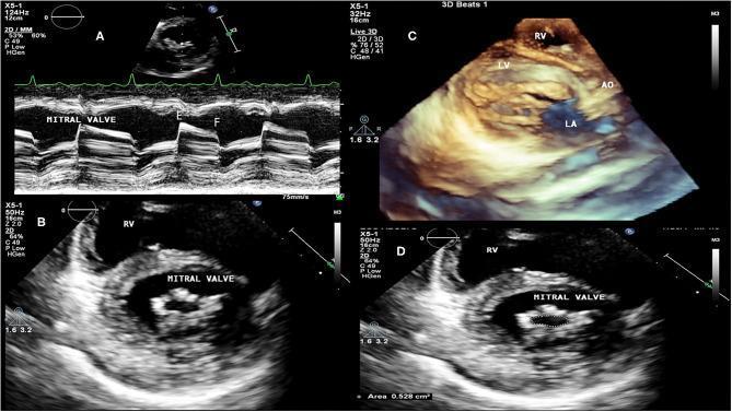
A multi-panel echocardiographic series demonstrating chronic rheumatic heart disease with severe mitral valve stenosis. Panel A shows an M-mode transthoracic echocardiogram of the mitral valve, highlighting a classic rectification (flattening) of the E to F slope, indicating a slow diastolic filling rate. Panel B and D provide 2D transthoracic views in the short-axis/transversal plane, revealing significant thickening and calcification of the mitral leaflets and a restricted, 'buttonhole' or 'fish-mouth' shaped orifice; panel D includes planimetry measuring a critical mitral valve area of 0.528 cm². Panel C utilizes 3D transthoracic echocardiography to visualize the complex subvalvular apparatus, showing chordae fusion and anatomical relationships between the right ventricle (RV), left ventricle (LV), aorta (AO), and left atrium (LA). The collection illustrates diagnostic hallmarks of rheumatic valvular aggression, including commissural fusion and restricted diastolic opening, used for clinical staging and procedural planning.
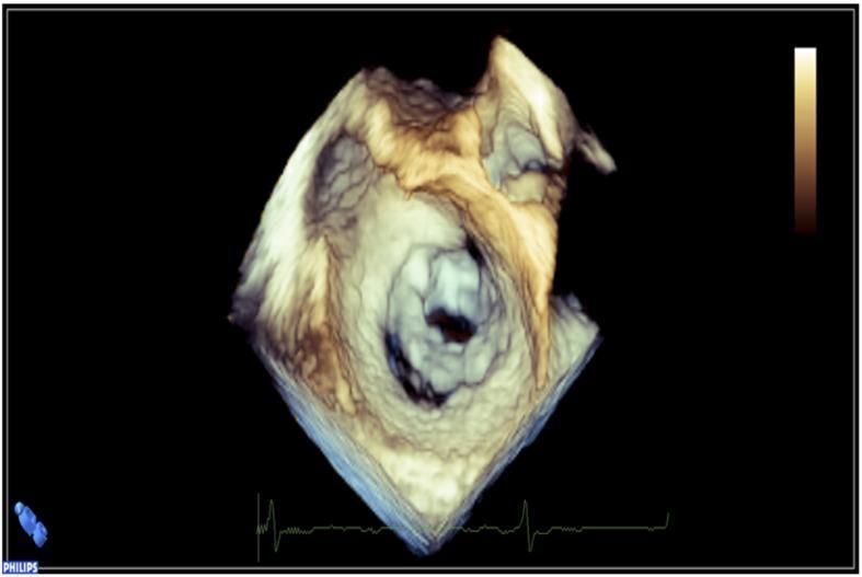
This diagnostic image is a 3D echocardiogram reconstruction of the heart, specifically providing an 'en face' view of the mitral valve. The image illustrates severe mitral valve stenosis, likely secondary to rheumatic heart disease. Key visual features include significant thickening and calcification of the valve leaflets, which appear rigid and echo-dense. There is evidence of commissural fusion, resulting in a markedly reduced and restricted mitral valve orifice (the 'fish-mouth' appearance). The surrounding valvular anatomy shows distorted morphology and increased tissue density, indicative of chronic fibrotic changes. This visual is characteristic of advanced valvular disease requiring clinical intervention such as mitral commissurotomy. The echocardiogram also includes a simultaneous EKG tracing at the bottom of the frame for temporal correlation within the cardiac cycle.
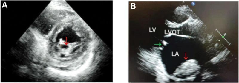
Two-panel transthoracic echocardiogram (TTE) demonstrating classic cardiac complications of Rheumatic Heart Disease (RHD). Panel A shows a short-axis view at the level of the mitral valve, where a red arrow highlights a stenotic mitral valve orifice. The valve leaflets appear significantly thickened with increased echogenicity, characteristic of fibrotic or calcific changes. Panel B displays a parasternal long-axis view showing severe left atrial (LA) enlargement, a common sequela of mitral stenosis. Within the dilated LA, a red arrow identifies a large, echogenic, and heterogeneous mass consistent with an intracavitary thrombus. A green arrow indicates the mitral valve apparatus between the LA and the left ventricle (LV). The image provides a clinical demonstration of the structural remodeling and thromboembolic risks associated with chronic rheumatic mitral valve disease, particularly in the context of atrial fibrillation.
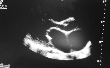
This diagnostic image is a transthoracic echocardiogram in a parasternal long-axis (PLAX) view, illustrating classic findings of rheumatic mitral stenosis. The visual focus is on the mitral valve apparatus, where the anterior mitral valve leaflet demonstrates characteristic 'doming' during diastole, often referred to as a 'hockey-stick' deformity. Both the anterior and posterior leaflets appear significantly thickened with increased echogenicity, suggesting fibrosis and possible calcification. There is evidence of subvalvular involvement, including thickening of the chordae tendineae. The left atrium appears dilated, a common secondary hemodynamic consequence of restricted mitral inflow and chronic pressure overload. In contrast, the left ventricle chamber size remains within relatively normal proportions in this view. The image serves as an educational example of valvular heart disease, specifically highlighting the morphological changes associated with chronic rheumatic carditis in a clinical cardiology setting.
Chronic Rheumatic Heart Disease (CRHD)
Definition
Epidemiology
- Remains the leading cause of valvular heart disease worldwide, particularly in developing countries and low-income populations with limited healthcare access
- In endemic regions, it is a leading cause of bacterial endocarditis and cardiovascular morbidity and mortality
- Degenerative valve disease now predominates in developed countries; RHD is decreasing but still common in underdeveloped regions
- Approximately 35-72% of patients with ARF develop clinical carditis; another ~18% have subclinical carditis detectable only on echocardiogram
- Goldman-Cecil Medicine, p. 3044
Pathogenesis
Mechanism
- GAS pharyngitis triggers an immune response in susceptible individuals
- Cross-reactive antibodies are generated: antibodies against streptococcal N-acetyl-glucosamine (a carbohydrate antigen) also recognize cardiac myosin, tropomyosin, and laminin
- These antibodies bind to valvular endothelium, upregulating VCAM-1 and other adhesion molecules
- This facilitates CD4+ T lymphocyte infiltration into valve tissue via very late activation antigen-4 (VLA-4) interactions
- Infiltrating T cells react against light meromyosin (LMM) - cardiac myosin epitopes; ~63% of intralesional T cell clones in RHD patients recognized LMM peptides
- Th1/Th17 cytokine dominance: large numbers of IFN-γ, TNF, IL-17, and IL-23 producing cells are found in valvular tissue; low IL-4 (a regulatory cytokine) contributes to progressive valve damage
- Reduced T regulatory cell (Treg) activity (CD4+CD25+FoxP3+) allows ongoing autoimmunity
- Firestein & Kelley's Textbook of Rheumatology, p. 2579
Valvular Pathology
| Valve | Involvement | Type of Lesion |
|---|---|---|
| Mitral | Nearly 100% | MR (most common initially), then MS (chronic scarring) |
| Aortic | 20-30% | AR > AS |
| Tricuspid | 15-40% (histologic) | Usually not clinically relevant |
| Pulmonic | Rare | Rarely involved |
- Mitral stenosis: CRHD is the most common cause of MS worldwide
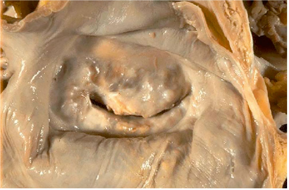
- Pathognomonic lesion: "fish-mouth" or "buttonhole" funnel valve appearance from commissural fusion, leaflet thickening, and chordae tendineae fusion and shortening
Clinical Features
- Progressive disease - severe valvular disease often appears in the 3rd or 4th decade of life
- Symptoms of MS: exertional dyspnea, orthopnea, PND, hemoptysis, AF, systemic embolism
- Symptoms of MR: fatigue, palpitations, progressive HF symptoms
- Classic murmurs: opening snap + mid-diastolic rumble (MS); holosystolic murmur at apex radiating to axilla (MR)
- Complications: atrial fibrillation, LA thrombus and systemic embolism, pulmonary hypertension, infective endocarditis, heart failure
Echocardiographic Findings
- Leaflet thickening and calcification
- Commissural fusion
- Restricted leaflet mobility
- Subvalvular apparatus fusion (chordae thickening and shortening)
- LA enlargement (in MS)
- M-mode: loss of E-F slope (flattening) in MS
- Planimetry for mitral valve area
Diagnosis
- Evidence of recent GAS infection (elevated ASOT/anti-DNase B, positive throat culture)
- Throat swab: gold standard for GAS pharyngitis (note: ~75% are negative by the time ARF symptoms appear)
- Echocardiography for valvular assessment (subclinical carditis detection)
Management
1. Secondary Prophylaxis (Most Important)
- Intramuscular benzathine penicillin G every 3-4 weeks - first-line, preferred over oral therapy (better compliance)
- Prevents recurrent ARF episodes which worsen valvular damage
- Duration depends on age, number of prior attacks, and presence/severity of CRHD
- Recent Cochrane review (2024) confirmed long-term prophylaxis is effective at preventing recurrence and progression - Bray et al., Cochrane 2024
- A 2025 systematic review (PMID 40333929) quantified severe adverse reactions to benzathine penicillin G in RHD - real but rare, and the benefit-risk ratio still strongly favors prophylaxis
2. Symptomatic Medical Management
- Rate control for AF (beta-blockers, digoxin)
- Anticoagulation for AF or LA thrombus (warfarin for mechanical valves or rheumatic MS with AF)
- Diuretics for pulmonary congestion
- Vasodilators/ACE inhibitors in MR with LV dysfunction
3. Interventional/Surgical Management
- Percutaneous mitral balloon commissurotomy (PMBC): procedure of choice for suitable MS (pliable, non-calcified valves, no significant MR, no LA thrombus)
- Surgical mitral valve repair: preferred over replacement when feasible (commissurotomy, chordal repair)
- Mitral valve replacement (MVR): for severe, calcified, or heavily subvalvular disease not amenable to repair; mechanical valves require lifelong anticoagulation
- Aortic valve surgery if significant AR/AS present
- A 2025 meta-analysis (PMID 40516887) compared survival after mitral valve repair vs replacement by etiology; RHD-specific outcomes are being actively studied
4. Echocardiographic Screening
- In endemic regions, population-based echocardiographic screening can detect latent RHD and guide earlier initiation of secondary prophylaxis
- Goldman-Cecil Medicine, p. 3044
Summary Table
| Feature | Detail |
|---|---|
| Cause | Repeated GAS pharyngitis → ARF → valvular damage |
| Mechanism | Molecular mimicry, cross-reactive antibodies, T-cell mediated inflammation |
| Most common valve | Mitral (100%), then aortic (20-30%) |
| Classic MS lesion | "Fish-mouth" valve with commissural fusion |
| Most common cause of MS | RHD (worldwide) |
| Key investigation | Echocardiography |
| Secondary prevention | IM benzathine penicillin G |
| Intervention for MS | PMBC (suitable cases) or MVR |