
4 type of necrosis flowchart and sharp points
A clean educational flowchart titled "4 Types of Necrosis" for medical students. The flowchart starts at the top with a box labeled "NECROSIS - Pathologic cell death with membrane destruction, enzyme leakage, and inflammation". Below it, four branches flow down to four main type boxes, each with sub-boxes showing key features: 1. COAGULATIVE NECROSIS (blue) - sub-boxes: Cause: Ischemia/infarction (except brain), Gross: Firm, pale tissue, Micro: Architecture preserved, anucleate ghost cells, eosinophilic; Key: Most common type, all solid organs except brain 2. LIQUEFACTIVE NECROSIS (green) - sub-boxes: Cause: Bacterial/fungal infection OR brain ischemia, Gross: Creamy yellow pus / liquid, Micro: Complete digestion of dead cells, viscous liquid; Key: CNS infarcts + abscesses 3. CASEOUS NECROSIS (orange) - sub-boxes: Cause: Tuberculosis (and some fungi), Gross: Cheese-like, friable, yellow-white, Micro: Amorphous granular pink debris, architecture obliterated; Key: Surrounded by granuloma 4. FAT NECROSIS (purple) - sub-boxes: Cause: Acute pancreatitis / abdominal trauma, Gross: Chalky white lesions (calcium soap), Micro: Shadowy fat cell outlines, basophilic Ca2+ deposits; Key: Fatty acids + Ca2+ = saponification At the bottom, a horizontal comparison bar showing: Architecture preserved? Yes=Coagulative, No=all others. Inflammation? All types yes. Specific cause clue arrows connecting each type to its trigger.
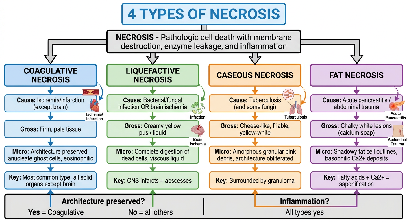
4 Types of Necrosis - Flowchart & Sharp Points
Master Framework
NECROSIS
├── Membrane destroyed
├── Enzymes leak → digest cell
├── Inflammation always present
└── Nuclear changes: pyknosis → karyorrhexis → karyolysis
         ↓
    4 MAIN PATTERNS
    ┌──────────────┬─────────────────┬──────────────┬──────────────┐
    │ Coagulative  │ Liquefactive    │   Caseous    │     Fat      │
    └──────────────┴─────────────────┴──────────────┴──────────────┘
1. Coagulative Necrosis
| Feature | Detail |
|---|---|
| Cause | Ischemia / infarction |
| Where | All solid organs except brain |
| Gross | Firm, pale, "mummified" tissue |
| Micro | Architecture preserved (ghost cells), anucleate, eosinophilic cells |
| Mechanism | Injury denatures structural proteins AND enzymes → proteolysis blocked |
| Timeline | Cells persist days to weeks before leukocytes clear them |
- Architecture preserved = coagulative. This is the single most tested distinguishing feature.
- "Ghost cells" = anucleate eosinophilic cell outlines - the classic histology term.
- Brain is the exception - brain ischemia gives liquefactive, not coagulative.
- All myocardial infarcts are coagulative necrosis.
2. Liquefactive Necrosis
| Feature | Detail |
|---|---|
| Cause | Bacterial/fungal infection OR CNS ischemia |
| Where | Brain (ischemia), anywhere with abscess |
| Gross | Viscous liquid, creamy-yellow pus |
| Micro | Dead cells completely digested - no architecture remains |
| Mechanism | Leukocyte enzymes "liquefy" the tissue |
| Localized collection | = Abscess (pus = creamy yellow liquid) |
- The brain uniquely undergoes liquefactive necrosis with ischemia - reason is "obscure" per Robbins.
- Pus = liquefactive necrosis caused by acute bacterial inflammation.
- Neutrophils are the key effector cells (their lysosomal enzymes digest tissue).
- Wet gangrene = coagulative + superimposed bacterial infection → becomes liquefactive.
3. Caseous Necrosis
| Feature | Detail |
|---|---|
| Cause | Tuberculosis (most common), some fungi (Histoplasma, Coccidioides) |
| Where | Lung (TB focus), lymph nodes, any organ with TB |
| Gross | Cheese-like, friable, yellow-white appearance |
| Micro | Amorphous granular pink debris, architecture obliterated, cellular outlines absent |
| Surrounding cells | Granuloma = macrophages + epithelioid cells + Langhans giant cells + lymphocytes |
| Special feature | Enclosed within a granuloma |
- "Caseous" = Latin for cheese (caseus). Cheese appearance = TB until proven otherwise.
- Architecture obliterated = distinguishes it from coagulative necrosis.
- A granuloma without caseous necrosis can be sarcoidosis; with caseous necrosis = TB.
- Ghon complex = primary TB = caseous necrosis in lung + hilar lymph node.
4. Fat Necrosis
| Feature | Detail |
|---|---|
| Cause | Acute pancreatitis or abdominal trauma |
| Where | Peritoneal fat, omentum, breast (traumatic fat necrosis) |
| Gross | Chalky white opaque lesions ("calcium soap") |
| Micro | Shadowy outlines of necrotic fat cells + basophilic Ca²⁺ deposits + inflammatory reaction |
| Mechanism | Pancreatic lipases digest triglycerides → fatty acids + saponification with Ca²⁺ |
- Saponification = fatty acids + calcium = chalky white deposits = classic gross finding.
- Elevated serum lipase in pancreatitis is the enzyme responsible.
- Breast fat necrosis (traumatic): no pancreas involved - caused by direct trauma releasing fat.
- Ca²⁺ deposits = basophilic on H&E = dystrophic calcification.
Quick Comparison Table
| Type | Architecture Preserved? | Cause Clue | Color/Consistency | Unique Marker |
|---|---|---|---|---|
| Coagulative | YES (ghost cells) | Ischemia | Pale, firm | All organs except brain |
| Liquefactive | NO (digested away) | Bacteria / brain | Yellow liquid / pus | CNS + abscesses |
| Caseous | NO (obliterated) | TB / fungi | Yellow-white, friable | Granuloma surrounds it |
| Fat | Partial (shadowy outlines) | Pancreatitis / trauma | Chalky white | Ca²⁺ saponification |
Nuclear Changes in Necrosis (Any Type)
Normal nucleus
    ↓
Pyknosis → nuclear shrinkage + increased basophilia (dark, dense mass)
    ↓
Karyorrhexis → pyknotic nucleus fragments
    ↓
Karyolysis → basophilia fades (DNase digests DNA) → nucleus dissolves in 1-2 days
Note on Fibrinoid Necrosis (5th type - bonus)
- Seen in immune vasculitis and severe hypertension
- Immune complexes + leaked plasma proteins deposit in vessel walls
- Bright pink, amorphous appearance on H&E (fibrin-like)
- Only detectable microscopically - no gross appearance