
Bilirubin metabolism and jaundice
bilirubin metabolism pathway diagram jaundice
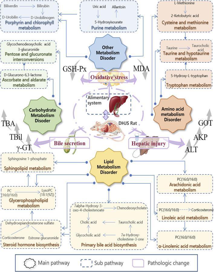
A metabolic network diagram illustrating the pathophysiological pathways of the Dampness-Heat Jaundice Syndrome (DHJS) model. The illustration is centered on a 'DHJS Rat' and the 'Alimentary system' (liver and gallbladder), with four octagonal 'Main pathway' boxes representing Carbohydrate, Lipid, Amino Acid, and Other Metabolism Disorders. Radiating from these nodes are numerous 'Sub-pathway' dashed boxes containing specific KEGG-based metabolic pathways, including Steroid Hormone Biosynthesis, Primary Bile Acid Biosynthesis, and Glycerophospholipid Metabolism. Key metabolites mentioned include bilirubin, cholic acid, corticosterone, and LysoPC. The diagram highlights 'Pathologic changes' such as Oxidative Stress and Hepatic Injury, indicated by thick purple arrows. Clinical biomarkers including TBil, TBA, ̳̳γ-GT, ALT, and MDA are shown with upward gray arrows, signifying elevation in the diseased state. This integrative map demonstrates the complex biochemical dysregulation across multiple systems associated with hepatic injury and bile secretion disorders.
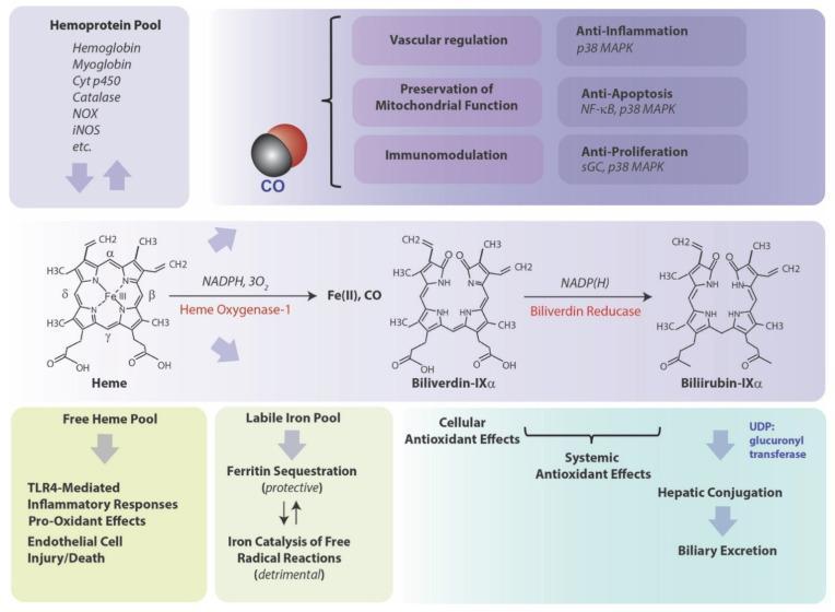
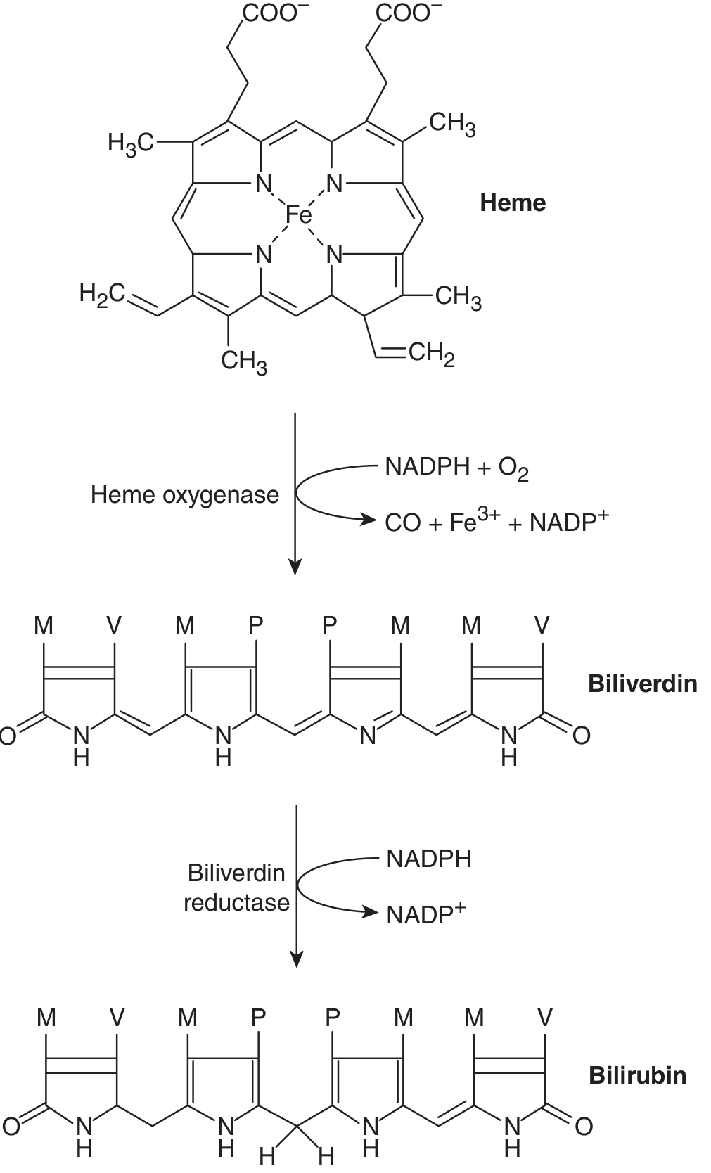
A detailed pathophysiology diagram illustrating the metabolic pathway of heme degradation and its systemic physiological implications. The central panel displays the biochemical conversion of Heme to Biliverdin-IXα by the rate-limiting enzyme Heme Oxygenase-1 (requiring NADPH and 3O2), which releases carbon monoxide (CO) and ferrous iron (Fe II). Subsequently, Biliverdin-IXα is reduced to Bilirubin-IXα by Biliverdin Reductase using NADP(H). The diagram maps the downstream outcomes of these byproducts: CO is linked to vascular regulation, mitochondrial preservation, and immunomodulation through signaling molecules like p38 MAPK, NF-κB, and sGC. The 'Labile Iron Pool' is shown bifurcating into protective Ferritin sequestration or detrimental free radical catalysis. Bilirubin is associated with cellular and systemic antioxidant effects before undergoing hepatic conjugation via UDP-glucuronyl transferase for biliary excretion. Additionally, a 'Free Heme Pool' is noted for its role in TLR4-mediated inflammatory responses and endothelial injury. This clinical illustration serves as a comprehensive guide for medical students and researchers studying oxidative stress, heme metabolism, and cytoprotective mechanisms.
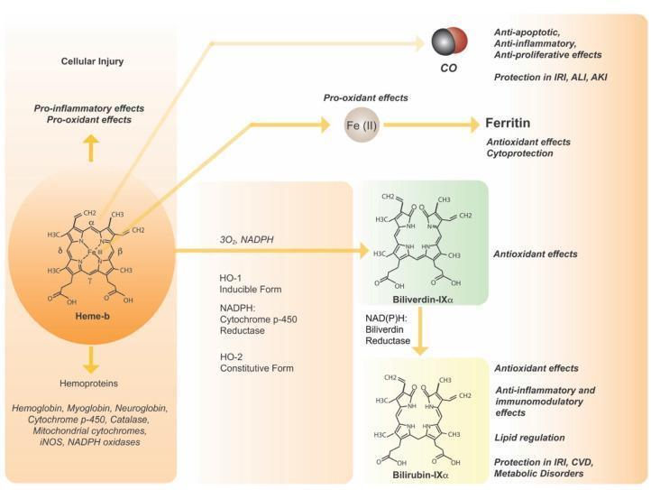
A detailed metabolic signaling pathway diagram illustrating the enzymatic catabolism of heme and its clinical implications. The diagram begins with Heme-b (central iron coordinated in a tetrapyrrole ring), which can contribute to hemoproteins or induce pro-inflammatory/pro-oxidant effects during cellular injury. The primary pathway shows heme oxygenase (inducible HO-1 and constitutive HO-2) catalyzing the conversion of heme to Biliverdin-IX̑ in the presence of 3O2, NADPH, and NADPH: Cytochrome p-450 Reductase. This reaction releases Carbon Monoxide (CO), which exerts anti-apoptotic and anti-inflammatory effects (protecting against IRI, ALI, and AKI), and Ferrous Iron (Fe-II), which can act as a pro-oxidant or trigger Ferritin for cytoprotection. Biliverdin-IX̑ is subsequently reduced to Bilirubin-IX̑ via NAD(P)H: Biliverdin Reductase. Both biliverdin and bilirubin are shown with their molecular structures and are associated with antioxidant, immunomodulatory, and lipid-regulating effects, offering protection in cardiovascular and metabolic disorders. This infographic serves as a comprehensive visual for cellular biochemistry and the cytoprotective roles of the HO system.
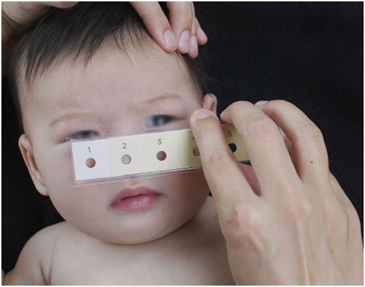
Clinical photograph of a neonate undergoing a non-invasive screening for neonatal jaundice (hyperbilirubinemia) using a Bili-ruler. The image shows a healthcare provider holding a rectangular, transparent-sleeved Bili-ruler against the infant's face. The ruler features a standardized color progression strip with six numbered patches (1–6). Each color patch increases in yellow intensity and contains a central circular aperture (hole). These apertures allow the clinician to compare the underlying skin tone directly against the calibrated reference colors to estimate bilirubin levels. The neonate demonstrates visible icterus (yellowish discoloration) of the skin and sclera. This diagnostic tool is designed as a low-cost, point-of-care alternative to electronic transcutaneous bilirubinometers (TcB) and serum testing, following the principle of cephalocaudal progression of jaundice. It is used in clinical settings to determine the need for further laboratory testing, phototherapy, or management of physiological and pathological jaundice.
Bilirubin Metabolism and Jaundice
1. Overview
2. Bilirubin Production (Prehepatic Phase)
3. Hepatic Handling (Intrahepatic Phase)
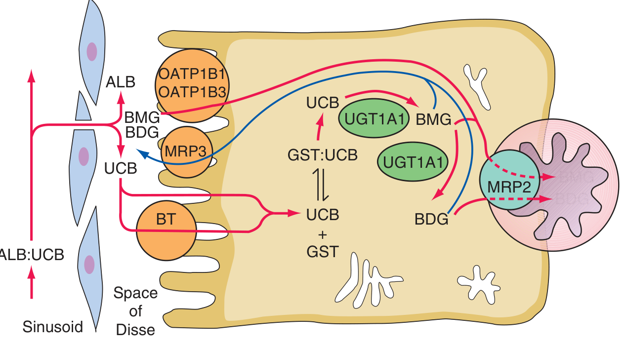
Step 1: Hepatocellular Uptake
Step 2: Intracellular Binding
Step 3: Conjugation
- Conjugation disrupts the internal hydrogen bonds, making bilirubin highly water-soluble - this is called conjugated (direct) bilirubin
- UGT1A1 is encoded by exon A1 of the UGT1 gene complex on chromosome 2
Step 4: Biliary Excretion
4. Intestinal Fate and Enterohepatic Circulation
- Conjugated bilirubin passes into the duodenum and down the GI tract without reabsorption (intestinal mucosa is impermeable to conjugated bilirubin)
- Gut bacteria convert conjugated bilirubin into urobilinogen (colorless) and further into urobilin (orange-yellow - gives urine its color) and stercobilin (brown - gives stool its color)
- ~10-20% of urobilinogen is reabsorbed into the portal circulation (enterohepatic circulation), most is re-excreted by the liver; a small amount spills into systemic circulation and is excreted in urine
- Stool urobilinogen/stercobilin is responsible for normal fecal pigmentation
5. Jaundice - Classification by Phase
A. Prehepatic (Hemolytic) Jaundice
- Mechanism: Excessive heme breakdown overwhelms the liver's conjugating capacity → unconjugated hyperbilirubinemia
- Bilirubin type: Unconjugated (indirect) ↑↑
- Urine bilirubin: Absent (unconjugated bilirubin is albumin-bound, not filtered by kidney)
- Urine urobilinogen: Increased (more bilirubin reaches the gut → more urobilinogen formed and absorbed)
- Stool color: Dark (increased stercobilin)
- Causes: Hereditary hemolytic anemias (sickle cell, thalassemia, G6PD deficiency, hereditary spherocytosis); acquired hemolytic anemias (autoimmune - positive Coombs; non-immune - drugs, toxins, mechanical, microangiopathic); hypoalbuminemia (burns, malnutrition - impaired transport)
B. Intrahepatic Jaundice
- Mechanism: Defective hepatocellular uptake, conjugation, or excretion
- Caused by hepatocellular disease (hepatitis, cirrhosis, drugs), or inherited enzyme defects
| Syndrome | UGT1A1 Activity | Bilirubin Level | Features |
|---|---|---|---|
| Gilbert's syndrome | ~30% of normal | Mild ↑ (<3 mg/dL) | Benign; fasting/stress triggers; ~4-7% of population |
| Crigler-Najjar Type I | Absent | 20-45 mg/dL | Neonatal onset; kernicterus risk; no response to phenobarbital |
| Crigler-Najjar Type II | Markedly reduced | 6-25 mg/dL | Less severe; responds to phenobarbital |
| Syndrome | Defect | Hallmark |
|---|---|---|
| Dubin-Johnson syndrome | MRP2 mutation (canalicular transport) | Black liver pigment; conjugated hyperbilirubinemia; benign |
| Rotor syndrome | OATP1B1/1B3 absent (reuptake defect) | Conjugated hyperbilirubinemia; no liver pigment |
- Hepatitis (viral, alcoholic, drug-induced), cirrhosis, ischemic hepatitis: Mixed picture with both unconjugated and conjugated bilirubin elevated; AST/ALT elevated
C. Posthepatic (Obstructive/Cholestatic) Jaundice
- Mechanism: Obstruction of bile flow (intra- or extrahepatic) → back-pressure → conjugated bilirubin regurgitates into blood
- Bilirubin type: Conjugated (direct) ↑↑
- Urine bilirubin: Present (conjugated bilirubin is water-soluble, passes the glomerular filter → dark "tea-colored" urine)
- Urine urobilinogen: Absent or decreased (no bilirubin reaching the gut)
- Stool color: Pale/clay-colored (no stercobilin reaching the gut)
- Causes:
- Intrahepatic cholestasis: Primary biliary cholangitis (PBC), primary sclerosing cholangitis (PSC), drug-induced cholestasis, intrahepatic cholestasis of pregnancy
- Extrahepatic obstruction: Choledocholithiasis (gallstones in CBD), pancreatic cancer (head of pancreas - classically painless jaundice), cholangiocarcinoma, biliary strictures, pancreatitis
6. Key Lab Differentiators
| Feature | Prehepatic | Hepatocellular | Obstructive |
|---|---|---|---|
| Total bilirubin | ↑ | ↑↑ | ↑↑ |
| Direct (conjugated) | Normal | ↑ | ↑↑ |
| Indirect (unconjugated) | ↑↑ | ↑ | Normal/mild ↑ |
| AST/ALT | Normal | ↑↑↑ | Mildly ↑ |
| ALP/GGT | Normal | Mild ↑ | ↑↑↑ |
| Urine bilirubin | Absent | Present | Present (dark urine) |
| Urine urobilinogen | ↑↑ | Variable | ↓ or absent |
| Stool color | Dark | Variable | Pale/clay |
7. Special Situations
Neonatal Jaundice (Physiological)
- Immature UGT1A1 + increased hemolysis of fetal hemoglobin → unconjugated hyperbilirubinemia peaking days 2-4
- Usually resolves within 2 weeks as enzyme matures
- Danger: Unconjugated bilirubin >~20 mg/dL (340 µmol/L) crosses the immature blood-brain barrier → deposits in basal ganglia = kernicterus (bilirubin encephalopathy), ranging from neurologic deficits to death
- Treatment: Phototherapy (converts bilirubin to water-soluble photoisomers excreted directly in bile) or exchange transfusion
- Breast milk jaundice: Fatty acids in breast milk inhibit UGT1A1; also increased enterohepatic circulation
Drugs Causing Conjugation Defects
8. Summary Pathway
Heme (from RBCs, myoglobin, cytochromes)
     ↓  Heme oxygenase (NADPH + O₂ → CO + Fe³⁺)
Biliverdin
     ↓  Biliverdin reductase (NADPH)
Unconjugated bilirubin (lipid-soluble)
     ↓  Bound to albumin in plasma
Hepatocyte uptake (OATP1B1/1B3)
     ↓  Bound to glutathione-S-transferase
Conjugation with glucuronic acid (UGT1A1)
     ↓  → Bilirubin mono/diglucuronide (water-soluble)
Biliary excretion via MRP2 (canalicular)
     ↓
Intestine → Urobilinogen (bacteria) → Urobilin (urine) / Stercobilin (feces)
              ↑ 10-20% reabsorbed → enterohepatic circulation