
The efferent pathway of the lower urinary tract is controlled by three components of the peripheral nervous system: the parasym pathetic, sympathetic, and somatic nervous systems. (Fig. 1) The parasympathetic nerve pathway (green) originates from the sacrum (S2–S4) and runs through the pelvic nerves, the ganglion cells of the pelvic plexus, and into the terminal nerve ganglia of the blad der wall and urethra. It releases acetylcholine at nerve endings and acts mainly in response to M3 muscarinic receptors to contract the bladder. The sympathetic nerve pathway (blue) originates from the thoracolumbar spine (T11–L2) and passes through the intermes enteric plexus and hypogastric nerves into the pelvic plexus and genitourinary organs. Iexplain
Efferent Innervation of the Lower Urinary Tract (LUT)
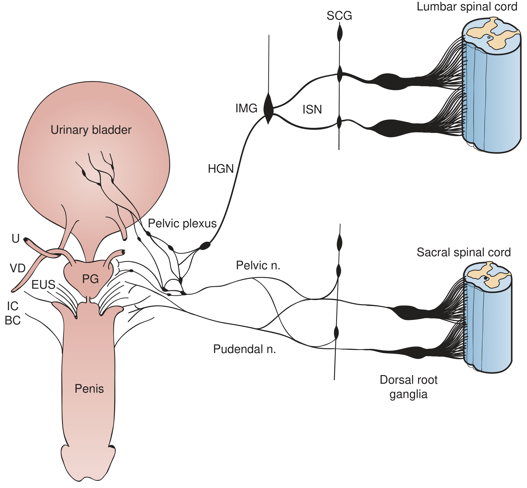
1. Parasympathetic Pathway (Green) - "The Voiding Nerve"
| Feature | Detail |
|---|---|
| Origin | Sacral parasympathetic nucleus (S2-S4) in the lateral intermediate gray matter |
| Preganglionic route | Ventral roots → pelvic nerves (nervi erigentes) |
| Synapse site | Pelvic plexus ganglia, vesical ganglia (on bladder surface), and intramural ganglia within the bladder and urethral walls |
| Postganglionic transmitter | Acetylcholine (ACh) |
| Receptor | M3 muscarinic receptors on detrusor smooth muscle |
| Effect | Detrusor contraction + urethral smooth muscle relaxation → voiding |
- The preganglionic transmitter at the ganglion acts on nicotinic receptors (cholinergic ganglionic transmission), and the postganglionic terminal releases ACh onto M3 receptors to contract the detrusor.
- A small atropine-resistant (NANC) component exists, mediated mainly by ATP (purinergic) signaling, though it contributes minimally to normal human detrusor contraction.
- Because postganglionic neurons are located within the bladder wall itself, even a cauda equina or pelvic plexus injury does not cause complete denervation - residual intramural ganglionic activity may persist.
2. Sympathetic Pathway (Blue) - "The Storage Nerve"
| Feature | Detail |
|---|---|
| Origin | Thoracolumbar spinal cord (T11-L2/L3) |
| Preganglionic route | Sympathetic chain ganglia → inferior splanchnic nerves (ISN) → inferior mesenteric ganglia (IMG) |
| Postganglionic route | Hypogastric nerves (HGN) → pelvic plexus → bladder and urethra |
| Postganglionic transmitter | Noradrenaline (norepinephrine) |
| Receptors & effects | β3-adrenoceptors on detrusor → relaxation; α1-adrenoceptors at bladder neck/urethra → contraction |
| Net effect | Bladder body relaxes + bladder outlet/urethra contracts → urine storage |
- The sympathetic pathway is activated during the storage phase via a vesicospinovesical reflex: bladder distension activates Aδ afferents in the pelvic nerve → spinal cord → lumbar sympathetic efferents are triggered.
- At the ganglionic level, sympathetic firing reduces excitatory parasympathetic input to the bladder (ganglionic inhibition).
- The sympathetic innervation of the human detrusor is limited; it acts predominantly at the outlet region (bladder neck and proximal urethra) to maintain continence.
- During voiding, supraspinal mechanisms strongly inhibit this sympathetic storage reflex, allowing the bladder to contract and the urethra to relax.
3. Somatic Pathway (Red) - "The Sphincter Nerve"
| Feature | Detail |
|---|---|
| Origin | Onuf's nucleus (Onufrowicz's nucleus) in the sacral ventral horn (S2-S4) |
| Nerve | Pudendal nerve |
| Target | External urethral sphincter (EUS) - rhabdosphincter (striated muscle) |
| Transmitter | ACh acting on nicotinic cholinergic receptors |
| Effect | EUS contraction → maintains continence |
- This pathway is tonically active during urine storage to keep the rhabdosphincter contracted.
- During a sudden rise in intra-abdominal pressure (cough, sneeze, laugh), the guarding/continence reflex (pelvic-to-pudendal reflex) rapidly activates Onuf's nucleus to dynamically contract the EUS.
- During micturition, this somatic reflex is inhibited by spinal and supraspinal (pontine) mechanisms, allowing sphincter relaxation.
- The pontine micturition center (PMC) projects directly to Onuf's nucleus, providing voluntary control of the rhabdosphincter.
Coordinated Storage vs. Voiding
| Phase | Parasympathetic | Sympathetic | Somatic |
|---|---|---|---|
| Storage | Inactive (inhibited) | Active - relaxes detrusor, contracts outlet | Tonically active - contracts EUS |
| Voiding | Active - contracts detrusor, relaxes urethra | Inhibited (supraspinal) | Inhibited - relaxes EUS |