
Explain about three lead ECG, why it is done, its diagnosis, its method of preparation and what are the places all these leads are placed in the body.
Reading File
Finding Sources
Finding Sources
Searching Images
diagram placement of electrodes for three lead ECG right arm left arm left leg torso monitoring
Searching PubMed
electrocardiography monitoring
Searching the Web
3 lead ECG electrode placement right arm left arm left leg monitoring lead I II III
Reading File
Reading File
Reading File
Reading Web Page
https://pmc.ncbi.nlm.nih.gov/articles/PMC5067828/
What is a 3-lead ECG?
A 3-lead ECG is a simple, continuous heart-monitoring system. It detects the heart’s electrical activity through three skin electrodes and produces the three standard bipolar limb-lead views:
| ECG lead | Negative electrode | Positive electrode | Main view |
|---|---|---|---|
| Lead I | Right arm (RA) | Left arm (LA) | Left lateral view |
| Lead II | Right arm (RA) | Left leg (LL) | Inferior view, excellent for P waves |
| Lead III | Left arm (LA) | Left leg (LL) | Inferior view |
These electrodes form an imaginary triangle around the heart called Einthoven’s triangle. Guyton and Hall Textbook of Medical Physiology, p. 146.
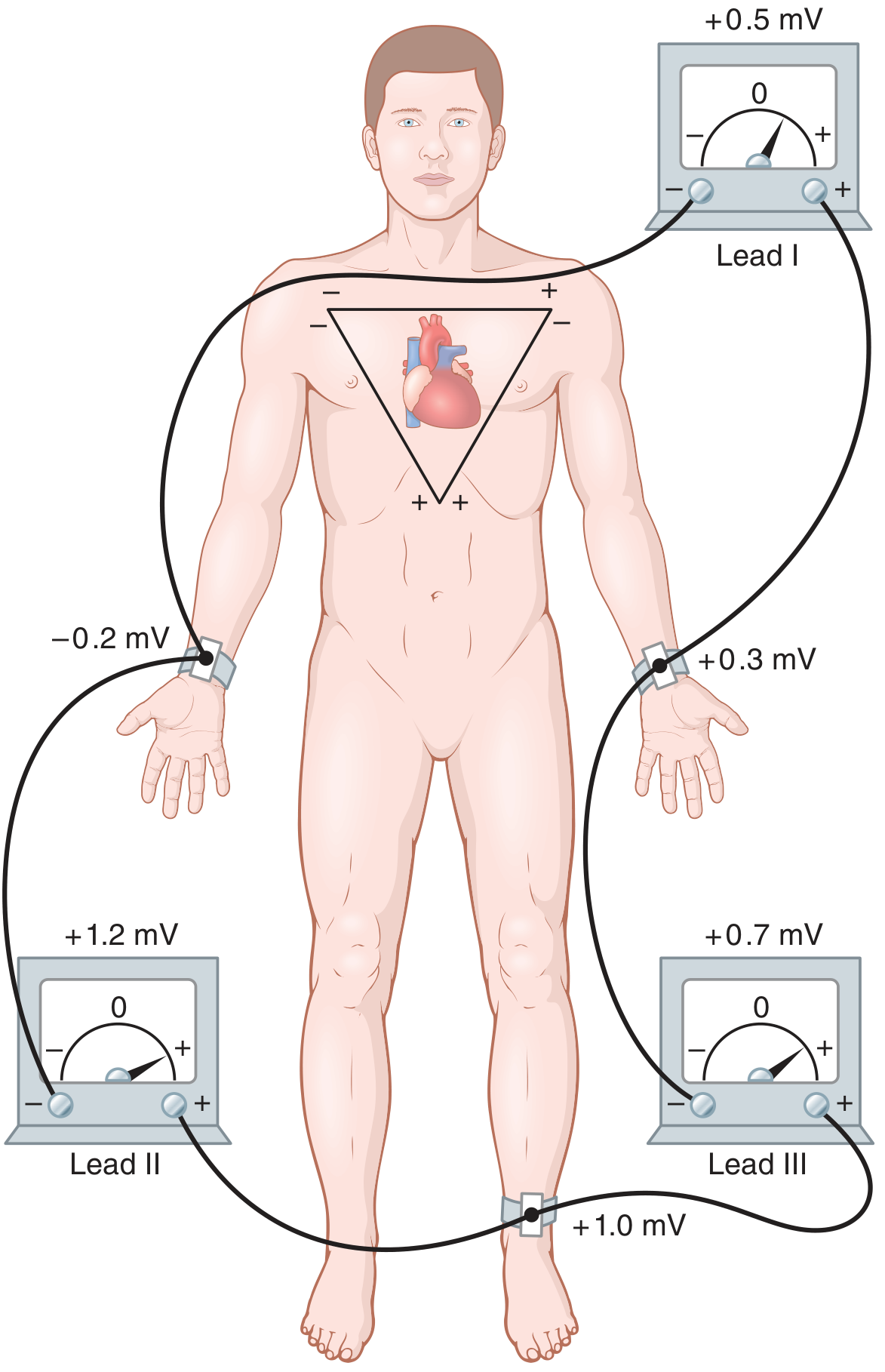
Why it is done
A 3-lead ECG is mainly used for continuous cardiac monitoring, including in hospital wards, ICU, operating rooms, ambulances, and during procedures. It helps staff monitor:
- Heart rate
- Heart rhythm
- P waves and AV conduction, especially in Lead II
- Sudden rhythm changes
- Response to medicines, anesthesia, pacing, or defibrillation
- Alarm-triggering rhythms such as marked bradycardia or tachycardia
It is quick, painless, noninvasive, and can run continuously. ECG electrodes measure electrical voltages on the body surface, not the mechanical pumping strength of the heart. Schwartz’s Principles of Surgery, section “Electrocardiographic Monitoring.”
What can a 3-lead ECG diagnose or suggest?
It is particularly useful to identify or monitor:
- Sinus bradycardia and sinus tachycardia
- Atrial fibrillation and atrial flutter
- Supraventricular tachycardia
- Ventricular tachycardia or ventricular fibrillation
- Premature atrial contractions and premature ventricular contractions
- AV blocks and other conduction disturbances
- Pacemaker rhythm or possible pacemaker malfunction
- Possible electrolyte-related rhythm changes, such as very tall T waves with hyperkalemia
Lead II is commonly selected because its electrical direction is close to normal atrial depolarization, so P waves are often clearer. It is useful for rhythm assessment. Morgan and Mikhail’s Clinical Anesthesiology, pp. 170-172.
Important limitation
A 3-lead monitor is not a replacement for a diagnostic 12-lead ECG. It has limited ability to diagnose:
- Myocardial infarction or acute coronary syndrome
- Exact location of ischemia
- Hypertrophy
- Detailed axis abnormalities
- Many subtle conduction or repolarization changes
If chest pain, suspected heart attack, persistent ST changes, or a new serious rhythm abnormality occurs, obtain a 12-lead ECG and assess the patient urgently. Monitoring filters and movement artifact can distort the ST segment. Morgan and Mikhail’s Clinical Anesthesiology, p. 172.
Where are the electrodes placed?
A. Standard anatomical limb positions
For a conventional recording, place the three electrodes on:
- RA, right arm: right wrist or lower right forearm
- LA, left arm: left wrist or lower left forearm
- LL, left leg: left ankle or lower left leg
These locations create Leads I, II, and III.
B. Usual monitoring positions on the torso
For continuous monitoring, electrodes are commonly moved to the torso to reduce motion artifact and avoid wires on the limbs:
| Electrode | Typical torso placement |
|---|---|
| RA | Right upper chest, just below the right clavicle |
| LA | Left upper chest, just below the left clavicle |
| LL | Left lower chest or left upper abdomen, below the left rib margin |
Keep all electrodes on a similar horizontal plane where possible, and avoid placing them directly over bone, large muscle masses, wounds, breast tissue, implanted devices, or very hairy skin.
A three-electrode monitor records a bipolar signal between two selected electrodes. Depending on the monitor setting, it can show Lead I, II, or III. For rhythm monitoring, Lead II is commonly used. The ECG monitoring review notes that torso placement is often used to reduce movement artifact during continuous monitoring.
Note about the right-leg electrode
A true 3-electrode system uses RA, LA, and LL. Some ECG machines use a fourth electrode, usually on the right leg (RL) or lower right abdomen, as a reference or ground electrode. That is often called a 3-lead ECG with a ground electrode, even though four adhesive pads may be seen.
Do not depend only on electrode color because colors vary by country and manufacturer. Always follow the labels printed on the cables: RA, LA, LL, and RL.
Preparation and method
- Explain the procedure and provide privacy.
- Check the monitor, cables, electrodes, and patient identity.
- Position the patient comfortably, usually supine or semi-recumbent, and ask them to remain still.
- Expose the required areas while maintaining dignity.
- Inspect the skin. Avoid broken, irritated, burned, infected, or heavily scarred areas where possible.
- Prepare the skin:
- Clean with soap and water or alcohol wipe, then allow it to dry.
- Clip excessive hair if needed. Do not unnecessarily shave fragile skin.
- Light skin abrasion may be used according to local policy to improve contact.
- Attach fresh gel electrodes to the prepared skin.
- Connect each lead wire to the correctly labelled electrode:
- RA to right upper chest/right arm region
- LA to left upper chest/left arm region
- LL to left lower chest/left leg region
- Turn on monitoring and select the required displayed lead, commonly Lead II.
- Confirm that the tracing is clear and that the displayed heart rate agrees with the patient’s pulse.
- Correct artifact by checking loose electrodes, dry gel, cable movement, patient shivering, and electrical interference.
- Document the time, lead selected, rhythm, heart rate, relevant symptoms, and any abnormal findings.
Conductive gel and clean skin lower skin resistance and improve ECG signal quality. Morgan and Mikhail’s Clinical Anesthesiology, p. 172.
Common problems
- Loose or dry electrode: wandering baseline or intermittent tracing
- Patient movement, shivering, tremor: can mimic arrhythmia
- Wrong electrode placement: can invert or alter the ECG appearance
- Electrical interference: regular fine artifact, often from nearby equipment
- Monitor artifact misread as a rhythm: always check the patient and palpate a pulse before treating an alarming rhythm strip
A simple safety rule: treat the patient, not only the monitor.
This is a shared conversation. Sign in to Orris to start your own chat.