
So final patient clinical picture is in between eczema and psoriasis As there are erythematous plaques but no scaling with severe itch on dorsum of foot and toes but sole is free. 2 or 3 crackes seen Patient complains severe itch
chronic eczema dorsum foot erythematous plaques no scaling
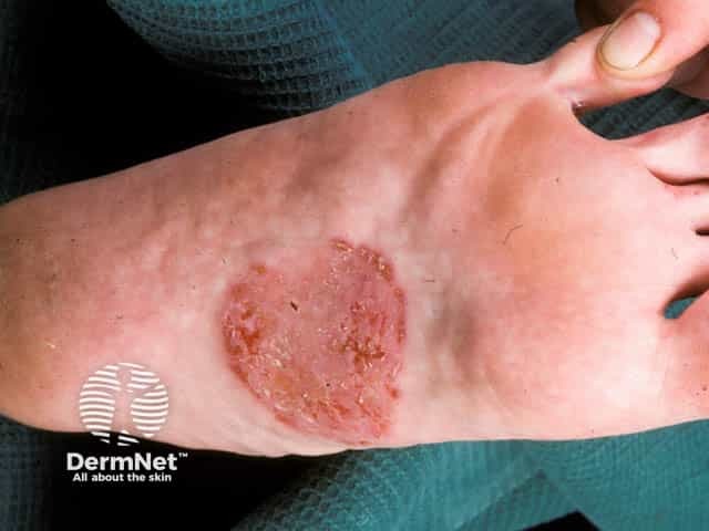
Imaging modality: Clinical photography of a cutaneous lesion on the foot. Anatomic location: dorsum/instep of the foot. The image shows a single, well-demarcated coin-shaped plaque with a erythematous, scaly surface and central crust, located on the instep near the midfoot. The lesion is circular to oval, appearing slightly raised at the edges with a flatter, desquamative center. The surrounding skin appears mildly inflamed without obvious secondary infection. This presentation is characteristic of discoid eczema (nummular dermatitis), a chronic inflammatory dermatosis that produces circumscribed plaques that itch and erode. The plaque displays typical features: polygonal to circular configuration, sharply defined border, erythema, scaling, and crusting from scratching or secondary infection. Differential diagnoses include tinea pedis/ corporis, psoriasis, contact dermatitis, asteatotic eczema, and other papulosquamous or infectious dermatoses. The image highlights the need for correlation with pruritus history, chronic dryness, and potential irritant or allergic exposures. In clinical practice, recognition of discoid eczema guides topical therapy (emollients, medium-potency topical corticosteroids), evaluation for secondary infection, and consideration of phototherapy or systemic therapy in refractory cases. This photograph is relevant for educational demonstration of classic nummular dermatitis morphology on the foot instep and for differential diagnosis training. Correlation with history strengthens diagnostic confidence significantly.
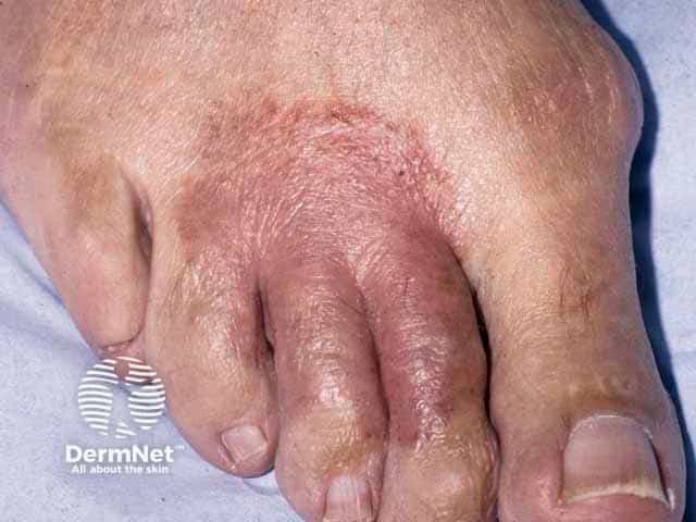
This is a high-resolution clinical photograph of the dorsal aspect of the foot in a patient with a suspected atopic dermatitis presenting as erythematous, dry, and scaly plaques. Visible features include xerosis of the dorsal skin, mild edema, and fissuring around the toe bases with subtle lichenification on chronic lesions. The distribution appears localized to the dorsum of the foot with interdigital involvement; there is no obvious vesiculation. The image emphasizes the differential diagnostic challenge of eczema versus fungal infection, specifically tinea pedis, which can mimic or coexist with atopic dermatitis in the toes and interdigital spaces. For diagnostic workup, clinicians should consider noninvasive tests such as potassium hydroxide (KOH) preparation and fungal culture, and refer for PAS staining or biopsy if atypical features persist. Clinically, adult atopic dermatitis manifests with pruritus, chronic relapsing inflammation, thickened plaques (lichenification), and variable erythema, which may respond to emollients and topical anti-inflammatory therapy. The photograph is valuable for dermatology education, clinical decision support, and research into foot dermatoses, aiding pattern recognition of dorsal-foot eczema versus dermatophytosis, documenting baseline morphology, monitoring therapeutic response, and teaching differential diagnosis. In summary, the image depicts classic eczema features while underscoring the essential step of excluding fungal etiology to guide treatment.
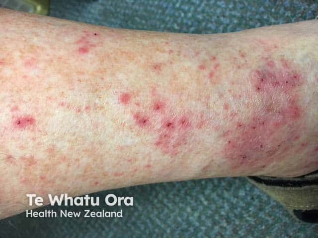
This image is a high-resolution clinical photograph of the left ankle region showing discoid eczema (nummular dermatitis). The skin exhibits multiple coin-shaped erythematous plaques, approximately 0.5–2 cm in diameter, with dry scaling, peripheral crusting, and mild surface excoriation from pruritus. Lesions are well-demarcated and arranged singly and in small clusters along the lateral malleolar area and distal lower leg. Perilesional erythema and xerosis are evident; surrounding skin appears dry without overt infection. The pattern is localized to the ankle region, a common site for discoid eczema, though plaques may appear on dorsum of hands or extensor surfaces in other contexts. Differential considerations include tinea corporis, contact dermatitis, psoriasis, and seborrheic dermatitis. Clinically, discoid eczema is associated with atopy and chronic itch, and management focuses on skin hydration, regular emollients, and anti-inflammatory therapy (topical corticosteroids and calcineurin inhibitors) with avoidance of irritants. This image serves as an educational reference for annular plaque dermatoses, differential diagnosis, and treatment response assessment in chronic eczematous skin disease.
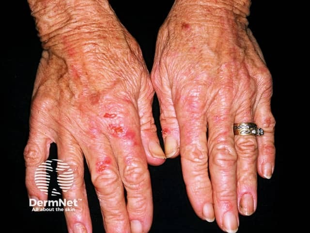
This clinical photograph shows bilateral dorsal surfaces of the hands in an elderly patient. The skin demonstrates chronic, erythematous plaques with fine scaling and superficial erosions greatest over the dorsum of the fingers and metacarpophalangeal joints. The plaques are irregular in shape, with a xerotic, dry appearance and areas of mild lichenification suggesting chronic rubbing or scratching. Subtle edema is present around the knuckles, and the overall skin surface appears coarse and wrinkled consistent with aging. No obvious vesicles, purpura, or open ulcers are seen. The ring and fingernails are normal in morphology apart from minor cuticular shedding; no prior surgery is evident. The color balance is accurate for a skin-tone photograph; lighting is even with minimal shadowing, enabling evaluation of surface texture, scale, and lesion distribution. This pattern, bilateral and symmetric involvement of the dorsal hands with erythematous, scaly patches, fits chronic dermatitis etiologies such as irritant contact dermatitis, allergic contact dermatitis, or atopic dermatitis (eczema), with dyshidrotic eczema as a less likely differential in this location. Clinical correlation should include history of irritant exposure, cosmetic or cleansing products, and pruritus. Management implications include emollient therapy, barrier protection, topical corticosteroids, and consideration of patch testing to identify triggers today.
shoe contact dermatitis foot dorsum sole spared
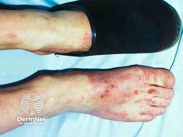
Clinical photography of bilateral feet demonstrating erythematous, vesiculopapular, and crusted dermatitis concentrated where the shoe upper contacts the skin. The dorsum and toes show patchy erythema with adjacent edema, scaling, and excoriations, more pronounced along the medial and dorsal foot margins. The distribution is consistent with a contact irritant/allergic dermatitis pattern influenced by footwear, with a linear to patchy arrangement at the boundary of the shoe. The appearance suggests allergic contact dermatitis to a component of the shoe upper rather than a fungal infection or venous stasis changes. Potential allergens include leather tanning chromates (chromium VI), rubber accelerators, dyes, adhesives, or synthetic polymers used in the upper. Patch testing is recommended to identify the precise allergen; management includes avoidance of the offending material, protective footwear, and skin barrier care. Acute flares respond to topical corticosteroids (e.g., medium-potency to high-potency depending on body area), plus saline or bland emollients and antipruritic measures. Educate patient on product labeling, alternative footwear materials, and routine follow-up to monitor improvement. This image is useful for dermatology training, clinical case discussions, and educational resources on footwear-related dermatitis, allergen exposure, and the value of diagnostic patch testing in suspected allergic contact dermatitis.
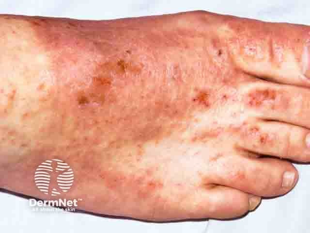
This clinical dermatology photograph depicts the dorsal aspect of the foot with evidence of allergic contact dermatitis consistent with chromate exposure from leather shoe uppers. The image highlights erythematous, pruritic plaques distributed over the dorsum of the foot and between toes, with fine scale, crusting, and secondary fissuring in some regions. The changes are most pronounced along the shoe-closure margins and toe web spaces, where direct skin contact with chromate-containing leather is likely. Inset details include diffuse edema, multiple coalescent patches, and a variable pigmentary change that may reflect postinflammatory hyperpigmentation. This presentation aligns with chromium allergy, a common cause of delayed-type hypersensitivity dermatitis in patients wearing leather footwear; hexavalent chromium (Cr(VI)) is used in leather tanning and can transfer to the skin. Differential diagnoses include irritant dermatitis, atopic dermatitis, and fungal infection such as tinea pedis, but the discrete distribution to footwear contact supports an occupational or recreational exposure history. Diagnostic significance emphasizes the importance of patch testing to confirm chromium sensitivity and to guide avoidance strategies. Clinically, management focuses on topical corticosteroids, barrier protection, and chromium-free footwear, with avoidance of leather with chromate pretreatment. Patch test results would refine future shoe choices and prevent recurrence.
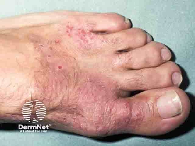
Clinical photograph of the dorsal aspect of the foot demonstrating an acute to subacute eczematous eruption with erythema, vesiculation, and surrounding scale and excoriations, highly suggestive of an allergic contact dermatitis to a shoe upper constituent. The lesions are localized to the dorsum and toe regions, with mild edema and moist, crusted patches adjacent to interdigital spaces. This imaging modality, clinical photography, provides a color-accurate representation of cutaneous inflammation without invasive procedures. Anatomic involvement includes the skin of the foot (epidermis and superficial dermis) in the dorsal surface and toe webs, consistent with a contact-related dermatitis pattern rather than a diffuse systemic eruption. Pathognomonic features are difficult to confirm radiographically; the clinical morphology points toward an allergen exposure, particularly footwear materials such as leather dyes, chromium salts, rubber accelerators, adhesives, or finishing agents. Diagnostic significance lies in supporting a diagnosis of allergic contact dermatitis and guiding allergen identification via patch testing. Differential considerations include irritant contact dermatitis from footwear, tinea pedis, dyshidrotic eczema, psoriasis, and candidal intertrigo in web spaces. Clinical correlation should focus on recent shoe purchases or changes, in-use footwear, and exposure history; management includes allergen avoidance, topical corticosteroids, emollients, and follow-up; education on prevention is essential for optimal outcomes.
Diagnosis: Shoe Dermatitis (Allergic Contact Dermatitis to Footwear)
How the Features Map
| Feature | Your Patient | Shoe Dermatitis |
|---|---|---|
| Erythematous plaques | ✅ | Classic |
| Scaling | Absent / minimal | Variable - often absent in early/subacute phase |
| Sole spared | ✅ | Classic pattern - dorsum involved, sole often spared |
| Dorsum of foot + toes | ✅ | Classic distribution |
| Severe itch | ✅ | Hallmark |
| Fissures (2-3) | ✅ | Seen in severe cases |
Why NOT Psoriasis
- Psoriasis has well-defined silvery-white scaling (not absent)
- Psoriasis on the foot typically involves the sole prominently (palmoplantar psoriasis)
- Itch in psoriasis is usually mild to moderate, not severe
- No Auspitz sign (pinpoint bleeding on scale removal) mentioned
- No nail changes mentioned
Why NOT Simple Eczema (Atopic/Endogenous)
- Atopic eczema on feet in adults is much less common
- The sole-sparing + dorsum involvement pattern is the hallmark of a contact etiology, not endogenous eczema
- If purely atopic, you'd expect toe webs and soles to also be involved
Most Likely Causative Allergens in Shoe Dermatitis
- Chromium (from chromate leather tanning) - most common
- Rubber accelerators - mercaptobenzothiazole, thiuram, carba mix
- Adhesives - p-tert-butylphenol formaldehyde resin
- Shoe dyes - disperse dyes, azo dyes
- Biocides in insoles
What to Do Next
- Patch testing - essential to identify the specific allergen (chromate series, rubber series, adhesive series)
- Ask about shoe type - leather shoes are the most common culprit; synthetic rubber soles/uppers also common
- Ask about new shoes recently purchased - onset often correlates with new footwear
- Rule out tinea pedis with KOH prep (interdigital space involvement would point toward fungal cause; sole involvement with scaling would strongly suggest tinea)
Treatment
- Stop wearing the offending footwear immediately
- Topical corticosteroids - medium to high potency (e.g., betamethasone valerate or clobetasol propionate) twice daily
- Emollients liberally for barrier repair
- Once allergen identified on patch test: wear chromate-free or allergen-free footwear (100% cotton lining, no rubber soles with accelerators)
- Oral antihistamines for itch (e.g., hydroxyzine at night for sedative effect, cetirizine during day)
- If severe: short course of oral prednisolone
Clinical picture is changed as pstient intermittently usinf clobetasol plus saly 3% cream
steroid modified psoriasis no scale erythema
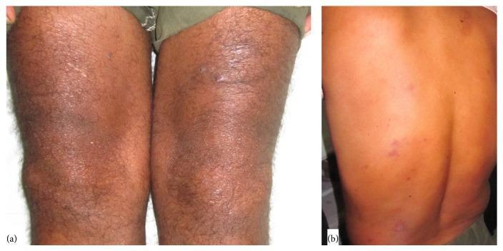
Clinical photographs showing dermatological manifestations of borderline lepromatous leprosy. Panel (a) displays the anterior lower extremities, highlighting a shiny, atrophic plaque on the left knee with overlying xerosis and mild desquamation, which had been previously misdiagnosed as steroid-modified psoriasis. Panel (b) shows the patient's posterior trunk (back) featuring multiple discrete, erythematous plaque-like lesions. These lesions represent cutaneous involvement in Hansen's disease, mimicking clinical psoriasis. The presentation is significant for medical education as it demonstrates the 'great imitator' quality of leprosy, where skin plaques and symmetric polyarthritis can lead to a misdiagnosis of psoriatic arthritis or seronegative rheumatoid arthritis. The morphological features include erythema, induration, and surface changes that are characteristic of type 2 lepra reactions (Erythema Nodosum Leprosum) in the context of chronic polyarthritis.
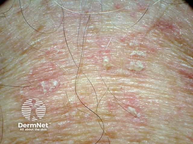
Imaging modality: Clinical photography of the anterior chest. Technique: Macroscopic, high-resolution photograph of the presternal region to document superficial skin changes; no magnification; color balance preserved to show erythema and scale. Anatomical location: presternal chest wall; skin of the anterior chest; midline region; integumentary system. Visual features: well-demarcated erythematous plaques with greasy, white-yellow scale adherent to the surface; mild edema; no vesiculation; slight pruritus typically reported. These findings are characteristic of seborrheic dermatitis in sebaceous-rich areas and may co-localize with scalp and facial involvement. The dermatosis presents with erythema, scale, and occasional crusting; margins are variable; surface is shiny and slightly oily; scales may be greasy and pale; regional distribution is limited here but commonly involves the chest, scalp, eyebrows, nasolabial folds. Pathology: not assessable from image; however, such lesions correlate with seborrheic dermatitis histology showing mild spongiosis, parakeratosis, and inflammatory infiltrate. Diagnostic significance: visual documentation supports clinical diagnosis of seborrheic dermatitis; helps differentiate from tinea corporis (annular scaling with central clearing) and psoriasis (thicker plaques with silvery scales). Potential use: dermatology education, telemedicine triage, treatment monitoring, antifungal/steroid therapy guidance. Clinical relevance includes dermoscopy planning, patient education, and documenting response to antifungal and anti-inflammatory therapy during follow-up.
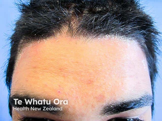
Clinical photography of facial skin demonstrating plaque-type psoriasis along the anterior hairline. Modality: Dermatologic clinical photography; frontal view of the forehead and frontal scalp margin under standardized lighting. The image depicts erythematous, well-demarcated plaques with mild to moderate scaling confined to the anterior hairline and forehead, extending along the temporal margins in places. The surrounding skin shows mild erythema without active oozing. The plaques appear slightly infiltrated with silvery scales, consistent with plaque psoriasis. No swelling of underlying subcutaneous tissues is evident. The hair follicles appear preserved; no ulceration or crusting. The distribution is focal to the hairline region, a common site for facial psoriasis, which may resemble seborrheic dermatitis but differs by the extent and silvery scale. Clinically, this finding supports a diagnosis of psoriasis vulgaris with facial involvement; it may correlate with psoriatic plaques at other sites. This image is useful for dermatology education, patient counseling, and longitudinal assessment of treatment response to topical corticosteroids or vitamin D analogs. Potential differential diagnoses include seborrheic dermatitis, eczema, contact dermatitis, and less likely fungal infection. Clinical correlation with patient history and examination of palms, elbows, knees, and nails is recommended for comprehensive assessment. Useful for therapy planning and ongoing monitoring.
Why Clobetasol + Salicylic Acid 3% Changes the Clinical Picture
The "Double Masking" Effect
| Agent | What it Does to the Skin | Effect on Clinical Picture |
|---|---|---|
| Clobetasol propionate (Class I super-potent steroid) | Powerfully suppresses inflammation, vasoconstriction | Erythema reduced, scale suppressed, itch temporarily reduced |
| Salicylic acid 3% (keratolytic) | Physically dissolves and removes scale/keratin | Any residual scale gets chemically removed |
| Combined | Both acting together | Plaques WITHOUT scale = exactly what you're seeing |
- Scale disappears (salicylic acid removes it physically + clobetasol suppresses keratinocyte turnover)
- Erythema partially persists (because it's a deep inflammatory process that steroid can't fully suppress with intermittent use)
- Itch persists (because the underlying cause - contact allergen or psoriasis - is still there)
What This Means for Diagnosis
- Salicylic acid + clobetasol is actually the standard combination used TO TREAT psoriasis
- Clobetasol clears 68-89% of psoriasis patients - [Textbook of Family Medicine 9e]
- Intermittent use = partial suppression = erythematous plaques remain but scale is gone
- The fissures can be seen in palmoplantar psoriasis
- Sole-sparing with dorsal involvement is less typical but palmoplantar variants exist
- The sole-sparing + dorsum + toe pattern remains highly characteristic
- Severe itch is more typical of eczema/contact dermatitis than psoriasis
- Clobetasol + salicylic acid would similarly mask the eczema morphology
- This is a critical one: applying potent topical steroids to a fungal infection causes tinea incognito
- Tinea incognito = atypical, modified fungal infection where scale is suppressed, borders become irregular, and the classic ring pattern disappears
- Itch can be severe
- Fissures occur in tinea pedis too
- KOH microscopy is now MANDATORY before any further steroid use
Immediate Action Required
- STOP clobetasol for at least 1-2 weeks (steroid washout period)
- KOH scraping from the scales/edge of lesion - rule out tinea pedis
- Fungal culture if KOH negative but suspicion remains
- After washout: re-examine the lesions - scale pattern, border characteristics, and Auspitz sign will return
- Patch testing to confirm/exclude shoe contact dermatitis
Why This Matters Clinically
| Diagnosis | If Clobetasol Continued |
|---|---|
| Psoriasis | Acceptable short-term, but intermittent use causes rebound flares; tachyphylaxis develops |
| Contact dermatitis | Temporarily suppresses but does not cure; allergen must be identified |
| Tinea pedis | DANGEROUS - promotes fungal spread, destroys diagnostic morphology, can cause extensive tinea incognito |
| Tinea incognito | Spreading, atypical, difficult-to-treat fungal infection |
Summary
I gave short course wysolone 20mg 5days taper to 10mg 5days with plain topucal clobetasol
Assessment of Your Treatment Approach
What You Prescribed
- Wysolone (prednisolone) 20mg x 5 days → 10mg x 5 days (total 10-day taper)
- Plain clobetasol propionate topically
Evaluation by Likely Diagnosis
If the Diagnosis is Contact Dermatitis / Eczema ✅ Appropriate
- Oral prednisolone for systemic anti-inflammatory effect
- Topical clobetasol for local barrier suppression
- ...is a well-accepted stepdown strategy
If the Diagnosis is Psoriasis ⚠️ CAUTION - Significant Risk
- Rebound flare on withdrawal - lesions come back worse than before
- Pustular psoriasis - the most feared complication, where plaques convert to widespread sterile pustules (von Zumbusch pattern) upon steroid withdrawal
- Erythrodermic psoriasis - generalized erythroderma on tapering
If the Diagnosis is Tinea Pedis / Tinea Incognito ❌ Harmful
- Further suppress inflammatory signs
- Promote fungal proliferation
- Expand the area of infection
- Make the tinea even harder to diagnose and treat
Key Question Now: How Did the Patient Respond?
| Response | Suggests |
|---|---|
| Dramatic improvement, itch gone, lesions clearing well | Contact dermatitis or eczema - steroids are working appropriately |
| Partial improvement but itch persisting | Mixed picture - underlying allergen still present, or psoriasis partially suppressed |
| No improvement or worsening | Tinea incognito, or diagnosis still incorrect |
| Flare on tapering / new pustules appearing | Psoriasis - steroid withdrawal reaction |
What to Do Now
- Watch carefully for rebound at day 8-10 as you step down to 10mg and stop - if lesions come back briskly, psoriasis is more likely
- Do KOH scraping NOW - you still need to rule out fungal infection. Steroids will make it harder to identify but a KOH can still be attempted
- After completing the course and allowing 2-week washout: Re-examine the lesions in their "natural" state
- Patch testing - essential for shoe contact dermatitis confirmation
- Do NOT repeat oral steroids if psoriasis remains on the differential - pivot to safer alternatives (topical vitamin D analogs, narrowband UVB, methotrexate if severe)