
Act as a DM Neonatology faculty teaching neonatal neurology. Teach the topic Intraventricular Hemorrhage (IVH) in preterm infants in a crisp, clinically relevant manner suitable for a neonatology fellow. Avoid textbook theory and focus only on points that change bedside management and counselling. Cover the following: 1. Why IVH occurs in preterm infants Germinal matrix anatomy Why extremely preterm infants are at highest risk Major risk factors (antenatal, perinatal, postnatal) 2. Papile grading (I–IV) Ultrasound features of each grade Easy memory tricks Prognosis associated with each grade Common misconceptions (especially Grade IV = periventricular hemorrhagic infarction, not simply blood entering brain tissue) 3. Neurosonogram interpretation Teach a stepwise approach: What structures to examine first Ventricular size Echogenicity Clots Ventricular dilatation Midline shift Periventricular changes Cysts Doppler if relevant Red flags that require urgent discussion 4. Cyst formation after IVH Explain in depth: Why cysts form after hemorrhage Difference between clot liquefaction, porencephalic cyst, post-hemorrhagic cyst and periventricular leukomalacia (PVL) Which cysts represent healing and which indicate irreversible brain injury How serial neurosonograms help differentiate them Typical timeline of cyst evolution Specifically answer this scenario: 'An extremely preterm infant had Grade III IVH on Day 2. On Day 10 the ultrasound shows resolving intraventricular blood with cyst formation.' Explain: Is cyst formation reassuring or concerning? What additional ultrasound findings determine prognosis? When should I reassure parents? When should I be worried? What follow-up scans or MRI are indicated? 5. How prognosis changes with ultrasound evolution Explain prognosis if: Blood decreases without ventricular enlargement Blood decreases but ventricles enlarge Small localized cyst develops Large porencephalic cyst develops Diffuse cystic PVL develops Progressive hydrocephalus develops 6. Counselling according to IVH grade For each grade (I–IV), provide: What to tell parents today Short-term risks Long-term neurodevelopmental risks Chance of cerebral palsy Risk of cognitive impairment Risk of epilepsy Whether prognosis can still improve on serial imaging Example counselling sentences suitable for family discussions 7. Post-hemorrhagic ventricular dilatation (PHVD) How to monitor Ventricular measurements (VI, AHW, TOD) Thresholds for intervention When to involve neurosurgery Lumbar puncture vs ventricular reservoir vs VP shunt 8. Clinical correlation Teach how ultrasound findings should be interpreted together with: Gestational age Neurological examination Head circumference trend Seizures Fontanelle findings MRI findings Developmental follow-up 9. High-yield viva questions Include 20 consultant-level questions with ideal answers. 10. Take-home messages End with the 15 most important bedside learning points every neonatologist should remember about IVH. Base the teaching on current evidence and recent neonatal guidelines (2024–2026). Present the content using headings, tables, algorithms and clinical pearls. Emphasize interpretation of serial neurosonography rather than isolated ultrasound findings."
"intraventricular hemorrhage" AND "preterm" AND management
"intraventricular hemorrhage" AND "preterm" AND "post-hemorrhagic ventricular dilatation"
PMID: 39044109
PMID: 41916837
PMID: 41896077
"periventricular hemorrhagic infarction" AND "preterm" AND prognosis
intraventricular hemorrhage preterm infant cranial ultrasound grading
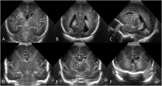
This diagnostic image series consists of six cranial ultrasound (cUS) scans (A-F) in preterm infants, illustrating Grade II germinal matrix hemorrhage-intraventricular hemorrhage (GMH-IVH). Images A-C demonstrate a common pattern in a 26-week gestational age infant. (A) Coronal view shows an echogenic clot involving the caudate nucleus without ventricular dilatation. (B) Coronal view at the trigone shows dependent layering of intraventricular clot. (C) Parasagittal view identifies the clot at the caudothalamic notch and within the occipital horn, distinct from the choroid plexus. Images D-F show a delayed hemorrhage pattern in a 27-week infant. (D) Initial coronal scan at 2 days post-birth appears normal. (E) Follow-up at 6 days reveals a new echogenic clot in the left lateral ventricle and normal choroid plexus in the third ventricle roof. (F) Coronal view anterior to the foramen of Monro confirms intraventricular hemorrhage in the frontal horns. These images serve as educational examples for identifying echogenicity (hemorrhage) within ventricular structures and differentiating it from the choroid plexus for accurate GMH-IVH grading.
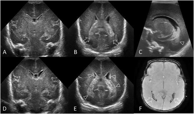
A multi-panel medical image illustrating the radiological evolution and retrospective diagnosis of intraventricular hemorrhage (IVH) in a preterm infant (GA 24 weeks). Panels (A) and (D) are coronal cranial ultrasound (cUS) scans at the level of the frontal horns showing initially normal lateral ventricles with no germinal matrix hemorrhage (GMH). Panel (B), a coronal scan at the level of the trigone, demonstrates small echogenic clots (arrows) within the posterior lateral ventricles. Panel (C) provides a parasagittal view confirming this small-volume IVH (arrowhead). Panel (E), a follow-up coronal scan 14 days later, reveals pathognomonic hyperechogenic ependyma (arrowheads) outlining the lateral ventricles, serving as an indirect sign of subacute IVH. Panel (F) is an axial T2* gradient-echo MRI scan taken 3 months post-birth, showing prominent hypointense susceptibility effects (arrows) in the occipital horns, indicating hemosiderin deposition from the previous hemorrhage. This series highlights the utility of serial ultrasound for identifying subacute changes like echogenic ependyma and MRI for confirming retrospective neonatal intracranial injury.
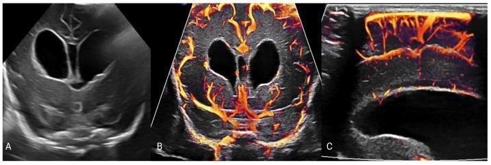
This composite of neonatal cranial ultrasound images illustrates post-hemorrhagic hydrocephalus in a 22-day-old preterm infant. Image A is a grayscale coronal view showing marked symmetric dilatation of the lateral ventricles (hydrocephalus) with internal echogenic material representing layering blood products from a prior intraventricular hemorrhage. Images B and C utilize advanced microvascular Doppler imaging to map slow-flow cerebral vessels. Image B (coronal) and Image C (sagittal) display these microvessels using a velocity-coded color scale, where yellow indicates relatively higher flow velocities and orange-to-red indicates slower flow. The images demonstrate the spatial distribution of the cerebral microvasculature in relation to the enlarged ventricles, providing a visual assessment of cerebral perfusion. This educational material highlights the diagnostic utility of combining conventional grayscale ultrasound for structural evaluation with microvascular imaging to monitor hemodynamic changes associated with increased intracranial pressure in neonatal hydrocephalus.
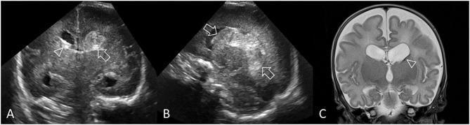
This composite figure illustrates a case of Grade III germinal matrix hemorrhage-intraventricular hemorrhage (GMH-IVH) in a preterm infant through cranial ultrasound (cUS) and follow-up MRI. Panel A is a coronal cUS at the level of the lateral ventricular bodies, showing a large, hyperechoic left-sided hemorrhage (arrow) causing acute distension and ventriculomegaly. A smaller amount of hyperechoic blood is visible in the right lateral ventricle (arrowhead), which exhibits 'ballooning' of the frontal horn. Panel B is a parasagittal cUS demonstrating that the echogenic clot occupies more than 50% of the distended left lateral ventricle, consistent with Grade III IVH. Panel C shows a coronal T2-weighted MR image taken 3 months post-birth, revealing sequelae of the hemorrhage. Notable findings include subtle hypointense staining at the left caudothalamic notch (arrowhead), indicating hemosiderin deposition. There is persistent, asymmetrical ventriculomegaly and widened subarachnoid spaces, reflecting ex-vacuo changes secondary to cerebral tissue volume loss.
periventricular leukomalacia cystic PVL ultrasound preterm brain
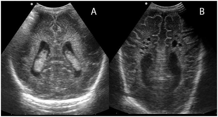
This diagnostic image consists of two coronal cranial ultrasound panels (A and B) demonstrating different grades of periventricular leukomalacia (PVL) in a neonatal brain. Panel A illustrates Grade 1 PVL, characterized by increased, relatively homogeneous echogenicity (echodensities) in the periventricular white matter surrounding the lateral ventricles. These bright regions are bilateral and lack cystic changes. Panel B demonstrates a more severe, cystic form of PVL (Grade 2 or higher). It reveals multiple discrete, small, anechoic (dark) fluid-filled cystic lesions within the periventricular regions where white matter necrosis has occurred. The lateral ventricles in Panel B appear irregular, and the surrounding brain parenchyma shows disrupted architecture compared to the solid echodensities in Panel A. These images are clinically significant for evaluating hypoxic-ischemic injury in preterm infants, showing the progression from transient echogenicity to permanent parenchymal loss and cavitation.
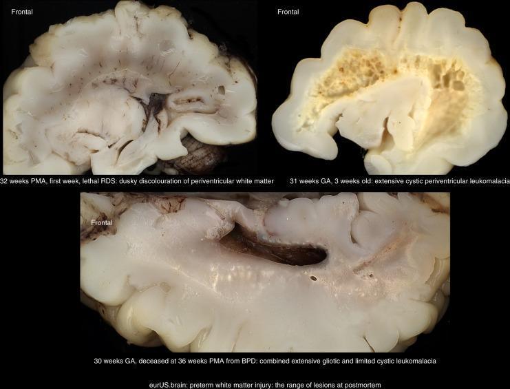
This clinical photograph displays three postmortem frontal brain sections from preterm infants, illustrating the spectrum of preterm white matter injury (WMI). The top-left specimen (32 weeks PMA) demonstrates mild injury characterized by dusky discoloration of the periventricular white matter without focal necrosis. The top-right specimen (31 weeks GA) shows severe cystic periventricular leukomalacia (PVL), featuring extensive, well-defined cystic formations and a spongy texture that disrupts the normal tissue architecture. The bottom specimen (30 weeks GA, deceased at 36 weeks PMA) displays a chronic stage of injury with combined extensive gliotic changes (glial scarring) and limited cystic leukomalacia, indicating both focal and diffuse white matter damage. These specimens serve as pathological correlates for imaging findings such as periventricular hyperechogenicity on cranial ultrasound (CUS) and signal abnormalities on MRI. The image highlights the morphological differences between non-cystic WMI, characterized by gliosis, and cystic PVL, which is associated with more severe clinical outcomes like spastic bilateral cerebral palsy.
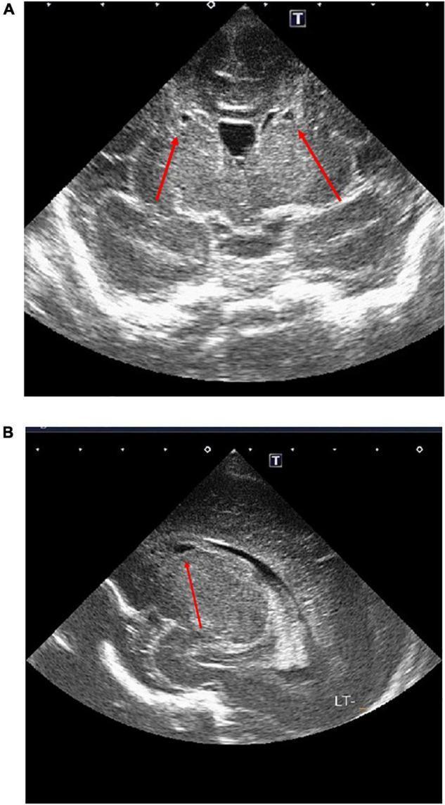
This diagnostic image displays two panels (A and B) of a neonatal head ultrasound (cranial ultrasound) illustrating cystic changes in the periventricular white matter. Panel A shows a coronal view where red arrows indicate bilateral, small, anechoic (fluid-filled) cystic lesions positioned symmetrically adjacent to the anterior horns of the lateral ventricles. Panel B provides a sagittal or parasagittal view (marked 'LT-' for left side) showing a similar discrete cystic abnormality within the periventricular region near the anterior horn. These findings are characteristic of periventricular leukomalacia (PVL), likely Grade 2 based on the localized punctate cystic morphology. The ultrasound serves as a critical bedside diagnostic tool for assessing white matter injury in preterm or high-risk neonates, providing evidence of potential hypoxic-ischemic damage or inflammatory response in the brain's germinal matrix and white matter zones.
post hemorrhagic ventricular dilatation hydrocephalus neonatal
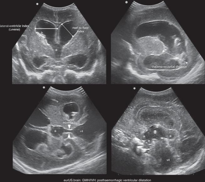
This composite of four neonatal cranial ultrasound images demonstrates post-hemorrhagic ventricular dilatation (PHVD) following germinal matrix-intraventricular hemorrhage (GMH/IVH). The top left coronal scan shows significantly enlarged lateral ventricles with labeled measurements for the 'Lateralventricle index (Levene)' and the 'roof to floor index.' The top right parasagittal scan highlights a dilated lateral ventricle and occipital horn, measured via the 'thalamo-occipital distance.' The bottom left image, a pseudo-axial view, identifies a dilated aqueduct (marked with arrowheads) and fourth ventricle (v4), alongside the third ventricle (*), suggesting tetraventricular hydrocephalus. The bottom right image shows a further view of the dilated ventricular system, including the third ventricle with extended protrusions (*) and a prominent fourth ventricle. The images illustrate the clinical progression and standardized sonographic metrics used to assess the severity of ventricular distension and the level of CSF flow obstruction in preterm infants.
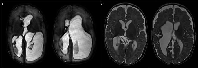
This diagnostic image displays four axial MRI brain scans of infants at term-equivalent age, categorized into two clinical scenarios (a and b) to evaluate post-hemorrhagic ventricular dilatation (PHVD). Panel (a) shows two T2-weighted axial slices demonstrating severe, asymmetric ventricular enlargement (hydrocephalus). The lateral ventricles are markedly dilated and hyperintense, causing significant compression and effacement of the surrounding brain parenchyma and cortical sulci. This appearance indicates failed management by a temporizing ventriculosubgaleal shunt (VSGS), necessitating permanent ventriculoperitoneal (VP) shunt insertion. Panel (b) shows two axial slices from a different infant where the ventricles are well-controlled and significantly smaller in volume. There is better preservation of cerebral mantle thickness and visible cortical folding (gyri and sulci), indicating successful management where the VSGS could be safely removed without permanent shunting. The comparison illustrates the use of MRI in neonatal neurosurgery to assess ventricular volume, brain tissue organization, and the clinical need for permanent cerebrospinal fluid diversion.
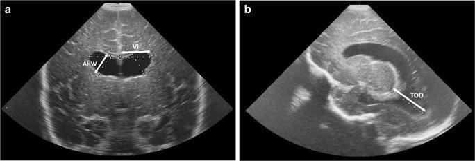
Cranial ultrasound (US) images of a preterm neonate demonstrating standardized linear measurements for assessing post-hemorrhagic ventricular dilatation. Image (a) is a coronal US scan showing measurements of the lateral ventricles: the Ventricular Index (VI), measured as the distance from the falx cerebri to the lateral wall of the anterior horn; and the Anterior Horn Width (AHW), measured as the maximum diagonal width of the anterior horn. Image (b) is a parasagittal US scan demonstrating the Thalamo-occipital Distance (TOD), measured from the posterior junction of the thalamus and choroid plexus to the outermost margin of the occipital horn. The images show significant ventricular enlargement and rounded morphology of the anterior horns, characteristic of hydrocephalus following germinal matrix-intraventricular hemorrhage (GM-IVH). These standardized 2D measurements are critical clinical tools in neonatal neurology for monitoring ventricular volume changes and determining the need for neurosurgical intervention, such as ventricular tapping or reservoir placement.
🧠 Neonatology Grand Rounds: Intraventricular Hemorrhage in Preterm Infants
DM Neonatology Teaching Session | Neonatal Neurology Series
SECTION 1: WHY IVH OCCURS IN PRETERM INFANTS
Germinal Matrix: The Anatomical Achilles' Heel
- Located beneath the ependyma of the lateral ventricles, predominantly at the head of the caudate nucleus at the caudothalamic groove (the landmark you look for on every neurosonogram)
- At 24 weeks, GM is at the frontal horn roof; by 28 weeks it has partly involuted; by 34 weeks it is nearly gone
- The GM vessels are thin-walled, lack a basement membrane, lack smooth muscle and pericyte support - essentially naked capillaries
- Blood drains via the internal cerebral veins which join at the vein of Galen - a venous "U-turn" that makes them susceptible to back-pressure
- The arterial supply comes from the lenticulostriate, choroidal, and recurrent arteries of Heubner
Why Extremely Preterm Infants Are at Highest Risk
| Gestational Age | IVH Incidence (All Grades) | Severe IVH (Grade III-IV) |
|---|---|---|
| <24 weeks | ~50-60% | ~25-30% |
| 24-26 weeks | ~35-45% | ~15-20% |
| 27-29 weeks | ~20-30% | ~8-12% |
| 30-32 weeks | ~10-15% | ~3-5% |
| >32 weeks | ~2-5% | ~1-2% |
- Vascular fragility - Thin-walled, unsupported GM vessels rupture easily
- Pressure-passive circulation - Impaired cerebrovascular autoregulation means systemic BP fluctuations directly translate to cerebral perfusion swings. There is no buffer.
- Coagulopathy - Immature coagulation factors + increased fibrinolytic activity = slower hemostasis once bleeding starts
Risk Factor Map
| Category | High-Impact Risk Factors | Lower-Impact / Modifiable |
|---|---|---|
| Antenatal | No antenatal steroids, chorioamnionitis, no in-utero transfer | PPROM >18 hrs, maternal fever |
| Perinatal | Precipitate/traumatic delivery, severe birth asphyxia, emergency CS without steroid cover | Delayed cord clamping NOT done |
| Postnatal - Respiratory | RDS with hypoxia/hypercarbia, pneumothorax, high PIP/MAP, rapid ventilator weaning | Surfactant not given early |
| Postnatal - Hemodynamic | Hypotension, rapid volume boluses, hypertonic saline/NaHCO3, Patent Ductus Arteriosus (large shunt) | Vasopressors causing BP spikes |
| Postnatal - Handling | Vigorous suctioning, painful procedures without sedation, rapid position changes | Head rotation/neck flexion |
| Other | Low platelet count, DIC, twin-to-twin transfusion | Polycythemia |
SECTION 2: PAPILE GRADING (I-IV)
The Grading System - and Why You Need to Know Its Limits
Ultrasound Features of Each Grade
| Grade | What You See on Ultrasound | Memory Hook | Approx. Incidence in <32 wk |
|---|---|---|---|
| I | Echogenic focus CONFINED to caudothalamic groove, anterior to the choroid plexus; ventricles normal size; no IVH | "GM only - don't panic" | ~40% of all IVH |
| II | Echogenic blood IN the ventricle (usually filling <50% of ventricle); NO ventricular dilatation; may see layering in occipital horn | "Blood in but no big belly" | ~35% |
| III | Blood fills >50% of the ventricle WITH acute ventricular dilatation (ballooning of frontal horn); may be bilateral | "Blood + Big ventricle" | ~15% |
| IV / PHI | Echogenic, triangular/fan-shaped periventricular parenchymal lesion pointing toward the ventricle; SEPARATE from the choroid plexus; almost always ipsilateral to the larger IVH | "Parenchyma on fire - this is PHI, NOT blood going into brain" | ~10% |
The Most Important Misconception You Must Correct
"Grade IV = blood spreading into the brain parenchyma." This is WRONG.
- PHI is typically unilateral or asymmetric (unlike bilateral ischemic PVL)
- Its prognosis depends on SIZE and LOCATION - anterior/small lesions can have surprisingly good motor outcomes
- The hemisphere contralateral to PHI often has coexisting white matter injury - check both sides always
- Liquefaction of PHI leads to a porencephalic cyst (see Section 4)
Prognosis by Grade
| Grade | Neurodevelopment | Cerebral Palsy Risk | Cognitive Impairment | Epilepsy | Hydrocephalus |
|---|---|---|---|---|---|
| I | Near normal; slightly worse than controls | <5% | Subtle (10-15%); may affect learning | <5% | Rare |
| II | Near normal to mildly impaired | 5-10% | 15-25%; attention/executive function affected | 5-10% | Rare |
| III | Variable; ~35% adverse outcome | 20-35% (mostly diplegia) | 30-50%; significant if PHVD develops | 15-25% | 25-35% develop PHVD |
| PHI (IV) | Most adverse; ~70-80% abnormal | 60-80%; hemiplegia on contralateral side | 50-70% | 20-30% | 40-50% |
SECTION 3: NEUROSONOGRAM INTERPRETATION - THE STEPWISE BEDSIDE APPROACH
When to Scan: Standard Protocol
Day 1-3: All infants <32 weeks OR any infant with clinical concern
Day 7:   Repeat all infants <32 weeks
Day 28:  All infants <32 weeks (or at discharge if before Day 28)
Term-equivalent age (TEA): All infants <28 weeks
TRIGGERED: Any acute neurological change, hemodynamic collapse, pneumothorax
The 9-Step Systematic Neurosonogram Protocol
- This is your landmark. Every sagittal scan starts here.
- The CTG is where the caudate head meets the thalamus - it looks like a notch on the parasagittal view
- Normal: thin sliver of choroid plexus here. Abnormal: echogenic blob that is LARGER than expected, or a rounded lesion ANTERIOR to the choroid plexus = GMH
- Look at the frontal horns on coronal view - they should be slit-like in the normal preterm infant
- Ballooned, rounded frontal horns = acute ventricular distension
- Measure: Ventricular Index (VI), Anterior Horn Width (AHW), Thalamo-Occipital Distance (TOD) - see Section 7 for thresholds
- Fresh blood = uniformly echogenic (white)
- Resolving blood = heterogeneous (mixed white/grey)
- Old blood = central liquefaction with echogenic rim
- Clot that is brighter than or equal to choroid plexus = definitely blood
- If unsure: CHOROID PLEXUS RULE - normal choroid plexus is echogenic, but it is symmetric and has smooth, well-defined borders. IVH clots are asymmetric, irregular, and may protrude into the frontal horn
- Does blood fill <50% or >50% of the ventricle? (Grade II vs III distinction)
- Is there a clot at the foramen of Monro? (risk of obstruction + hydrocephalus)
- Is there blood in the 3rd or 4th ventricle? (communicating vs obstructive hydrocephalus risk)
- Acute dilatation: rounded frontal horns, increased VI on serial measurements
- PHVD definition: VI >97th centile for gestational age (Levene nomogram)
- Key question: Is the dilatation increasing between scans? Rate of increase matters.
- Scan the periventricular white matter on BOTH sagittal views systematically
- PHI = triangular, fan-shaped echogenic area in the frontoparietal white matter, pointing toward the lateral ventricle
- PHI is ALMOST ALWAYS IPSILATERAL to the larger IVH
- If you see bilateral parenchymal echogenicity = think PVL, not bilateral PHI
- Large unilateral PHI or large clot can cause midline shift
- Identify the falx, third ventricle, and cavum septum pellucidum on coronal view
- Midline shift in a neonate = significant mass effect = immediate neurosurgical discussion
- Periventricular flare: echodense white matter around the ventricles, especially in posterior areas
- If persisting >7-10 days, it is significant (early PVL)
- Cysts developing within a flare = cystic PVL (see Section 4)
- Always scan POSTERIOR and SUPERIOR white matter - PVL is often missed in the anterior horns
- Use the mastoid view. The normal cerebellum has symmetric, intermediate echogenicity
- Cerebellar hemorrhage = echogenic focus in one or both hemispheres
- Cerebellar hemorrhage in <32 weekers worsens prognosis even in low-grade IVH (PMID: 41916837)
- Check the 4th ventricle for blood
Red Flags Requiring Urgent Discussion
🔴 NEW FINDING - act within hours:
  - PHI (triangular parenchymal echogenicity adjacent to new Grade III IVH)
  - Rapidly progressive PHVD (VI increasing >1mm/day)
  - Bilateral PHI or bilateral severe PVL
  - Midline shift
  - Foramen of Monro obstruction (asymmetric dilatation)
  - Cerebellar hemorrhage >1cm

🟡 SAME DAY DISCUSSION:
  - New Grade III IVH
  - Persistent periventricular flare >2 weeks
  - Progressive ventricular enlargement crossing the 97th centile
  - New cyst formation in periventricular white matter
Doppler in Neonatal IVH - When Is It Useful?
- Resistance Index (RI) of the anterior cerebral artery (ACA): RI >0.85 suggests raised intracranial pressure. Not diagnostic but adds clinical context in PHVD.
- Diastolic flow absence or reversal: Suggests markedly raised ICP - urgent neurosurgical consult
- Doppler is an ADJUNCT, not a substitute for linear measurements in PHVD monitoring
- Useful when you suspect raised ICP clinically but measurements are borderline
SECTION 4: CYST FORMATION AFTER IVH - THE DEEP DIVE
Why Cysts Form
The Four Types You Must Distinguish
| Type | Location | Pathology | US Appearance | Prognosis | Timeline to Appear |
|---|---|---|---|---|---|
| Clot liquefaction / subependymal pseudocyst | Within germinal matrix or intraventricularly | Normal resorption of blood clot - NO parenchymal injury | Small, round, anechoic; at caudothalamic groove; may have thin echogenic rim | REASSURING - indicates healing | 2-4 weeks post-hemorrhage |
| Porencephalic cyst | White matter/cortex, communicates with lateral ventricle | Liquefaction of PHI (venous hemorrhagic infarction) - actual brain tissue DESTROYED | Large, irregular, anechoic, communicates with lateral ventricle; unilateral | Poor for motor on CONTRALATERAL side; better if small/anterior | 4-8 weeks post-PHI |
| Post-hemorrhagic cyst (subependymal) | At site of Grade I GMH in the germinal matrix | Resorption of subependymal blood; no parenchymal damage | Small (<1cm), round, near CTG; does NOT communicate with ventricle | Mostly reassuring; minor cognitive risk | 2-4 weeks |
| Periventricular leukomalacia (PVL) cysts | Bilateral periventricular white matter | Coagulative necrosis of oligodendrocyte precursors from ischemia/infection - DIFFERENT mechanism from IVH | Multiple small (<3mm) or large (>3mm) bilateral cysts in periventricular white matter; symmetric; NOT at CTG | Severe; high risk of spastic diplegia/quadriplegia and cognitive impairment | 3-8 weeks after hypoxic-ischemic insult |
The Critical Distinguishing Features at the Bedside
Question to ask yourself when you see a cyst:
  1. Where exactly is it? (CTG = reassuring; periventricular white matter = concerning)
  2. Does it communicate with the ventricle? (communicating = porencephaly = bad)
  3. Is it unilateral or bilateral? (bilateral cysts = PVL pattern = very bad)
  4. What preceded it? (prior PHI = porencephaly; prior flare = PVL; prior Grade I = pseudocyst)
  5. What is the size? (>1cm = significant; multiple small bilateral = PVL)
Typical Timeline of Cyst Evolution
Day 1-3:    GMH/IVH - echogenic, solid; maximum brightness
Day 5-10:   Clot becomes heterogeneous (partial liquefaction)
Day 10-21:  Central liquefaction begins; "target sign" (dark center, bright rim)
Day 14-28:  Subependymal pseudocyst forms (small, near CTG) if Grade I-II
Week 4-8:   PHI liquefies → porencephalic cyst if Grade IV/PHI
Week 3-8:   PVL flares → cystic PVL if there was separate white matter ischemia
Week 8-12:  Most cysts are at maximum size; some may be resorbing
Month 3-6:  Small pseudocysts often disappear; porencephalic cysts persist permanently
SCENARIO ANALYSIS: Grade III IVH Day 2 → Cyst on Day 10
The Clinical Scenario
Is Cyst Formation Reassuring or Concerning?
| Cyst Location on Day 10 | Interpretation | Action |
|---|---|---|
| Small, round cyst at the caudothalamic groove, <1cm, no ventricular communication | Reassuring - this is clot liquefaction within the GM, healing in progress | Continue serial scans |
| Irregular cyst in frontoparietal white matter ADJACENT to where Grade III IVH was (i.e., the PHI zone) | Concerning - porencephaly forming from PHI | Urgent counselling; MRI at TEA |
| Multiple bilateral small cysts in periventricular white matter (NOT at CTG) | Very Concerning - concurrent PVL (separate from the IVH) | Immediate counselling change |
| Cyst at CTG with communicating with ventricle | Moderate concern - evolving minor porencephaly | MRI at TEA; neurosurgery review |
Additional Ultrasound Findings That Determine Prognosis
- Ventricular size - Is PHVD developing? (VI measurement critical)
- Periventricular white matter - Any flares or cysts OUTSIDE the CTG zone?
- Is the cyst communicating with the lateral ventricle? (use Doppler to confirm if unsure)
- Contralateral hemisphere - Any parenchymal injury on the side without the IVH?
- Cerebellum - Any cerebellar hemorrhage (worsens prognosis independently)
- Bilateral vs unilateral - Bilateral findings = much worse prognosis
- Size of porencephalic cyst, if present - Posterior/large = motor cortex involved = hemiplegia likely; Anterior/small = better outcomes possible
Decision Algorithm for Day 10 Cyst After Grade III IVH
Grade III IVH (Day 2)
         ↓
Day 10: Cyst formation
         ↓
Where is the cyst?
    |                              |
At CTG only,                Periventricular WM
small, no communication     OR communicating OR large
    ↓                              ↓
REASSURING:                  CONCERNING:
Clot liquefaction            PHI → porencephaly
                             OR concurrent PVL
    ↓                              ↓
Continue weekly scans        MRI at TEA mandatory
Check for PHVD               Urgent family counselling
                             Neurodevelopment team referral
         ↓
Both pathways: Look for PHVD - measure VI on every scan
         ↓
If VI > 97th centile AND progressive: PHVD pathway (Section 7)
When to Reassure Parents
- The cyst is small (<1cm), round, located AT the caudothalamic groove
- It does NOT communicate with the lateral ventricle
- Ventricular size is stable or decreasing
- No periventricular white matter flares or cysts elsewhere
- Cerebellum appears normal
- The infant's neurological examination is improving (tone normalizing, sucking returning)
When to Be Worried
- The cyst is large, irregular, and communicates with the ventricle (porencephaly)
- New or progressive cysts appear in the periventricular white matter bilaterally (PVL)
- Ventricular size is increasing (PHVD developing)
- The neurological examination is worsening or not improving
- Seizures develop in the first 2-4 weeks
Follow-Up Scan Schedule After Grade III IVH with Cyst
Week 1-4 (NICU):    Weekly cranial ultrasound
                     Measure VI, AHW, TOD at each scan
                     Document cyst: size, location, communication
Week 4-8:           Fortnightly if stable; weekly if PHVD developing
Term-Equivalent Age: Brain MRI (MANDATORY in Grade III IVH)
3-6 months CA:      Follow-up cranial ultrasound or MRI if cyst still present
18-24 months:       Formal developmental assessment (Bayley-IV)
4-5 years:          Neuropsychological testing
- Subtle white matter injury not visible on ultrasound
- Impaired myelination in the corticospinal tracts
- Cortical injury / sulcation abnormalities
- Cerebellar volume loss
- Confirms presence/absence of porencephaly
SECTION 5: HOW PROGNOSIS CHANGES WITH ULTRASOUND EVOLUTION
| Serial Ultrasound Finding | Prognosis | Clinical Action |
|---|---|---|
| Blood decreases, no ventricular enlargement | Good - low risk of PHVD; neurodevelopment risk primarily from the grade of initial IVH | Weekly scans until discharge; developmental follow-up based on grade |
| Blood decreases but ventricles enlarge | PHVD developing - intermediate to poor depending on severity; shunt-dependent hydrocephalus in ~30-40% of progressive cases | Measure VI/AHW/TOD weekly; cross 97th centile = intervention discussion begins |
| Small localized cyst at CTG | Mostly reassuring (healing pseudocyst) - very low risk of structural deficit | Continue routine scans; reassure parents; MRI at TEA |
| Large porencephalic cyst (communicating) | Significant - unilateral hemiplegia on contralateral side in ~60-70%; cognitive impairment common; epilepsy in 20-30% | MRI at TEA mandatory; physiotherapy referral early; PERCH trial monitoring; counselling about hemiplegia |
| Diffuse cystic PVL (bilateral periventricular cysts) | Severe - spastic diplegia or quadriplegia in 60-80%; significant cognitive and learning impairment; epilepsy in 30-40% | Immediate family counselling; early intervention team; ophthalmology; orthopedics; epilepsy planning |
| Progressive hydrocephalus (PHVD requiring intervention) | Poor (if requiring VP shunt) - disability rate rises to ~60% in Grade III; ~70-80% in PHI with shunt | Neurosurgery involvement; Ommaya reservoir vs VP shunt decision; DRIFT trial context |
SECTION 6: COUNSELLING BY IVH GRADE
Grade I IVH Counselling
| Domain | Risk |
|---|---|
| Cerebral palsy | <5%; similar to matched preterms without IVH |
| Cognitive impairment | 10-15% minor deficits; mostly in attention/executive function |
| Epilepsy | <5% |
| Hydrocephalus | Rare (<2%) |
| Can prognosis improve on serial imaging? | Yes - if no PHVD develops and no co-occurring injury, outlook is good |
Grade II IVH Counselling
| Domain | Risk |
|---|---|
| Cerebral palsy | 5-10% |
| Cognitive impairment | 15-25%; attention, memory, learning difficulties |
| Epilepsy | 5-10% |
| Hydrocephalus | ~5% |
| Can prognosis improve on serial imaging? | Yes - if ventricles remain stable and no white matter injury |
Grade III IVH Counselling
| Domain | Risk |
|---|---|
| Cerebral palsy | 20-35%; predominantly spastic diplegia |
| Cognitive impairment | 30-50%; more likely with PHVD |
| Epilepsy | 15-25% |
| Hydrocephalus requiring surgery | 25-35% |
| Can prognosis improve on serial imaging? | Yes - if ventricles stabilize without PHVD, outcome can be significantly better than worst-case |
Grade IV (PHI) IVH Counselling
| Domain | Risk |
|---|---|
| Cerebral palsy | 60-80%; predominantly contralateral hemiplegia (if unilateral PHI) |
| Cognitive impairment | 50-70%; more severe if large bilateral injury |
| Epilepsy | 20-30% |
| Hydrocephalus requiring surgery | 40-50% |
| Can prognosis improve on serial imaging? | Partially - smaller/anterior PHI has better outcomes; bilateral PHI is worst; serial scans clarify extent |
SECTION 7: POST-HEMORRHAGIC VENTRICULAR DILATATION (PHVD)
The Mechanism
- Blood breakdown products + fibrin clots obstruct CSF flow at the aqueduct of Sylvius and/or basal cisterns
- Communicating (most common) or obstructive hydrocephalus develops
- Progressive ventricular enlargement compresses the periventricular white matter
- White matter ischemia adds to the existing injury
Monitoring Protocol - The Three Measurements
| Measurement | How to Measure | Normal | Action Threshold |
|---|---|---|---|
| Ventricular Index (VI) | Coronal view at level of foramen of Monro: falx to lateral wall of anterior horn | Age-adjusted (Levene nomogram): ~10-13mm at 28-32wk | >97th centile (Levene) = PHVD; >4mm above 97th = consider intervention |
| Anterior Horn Width (AHW) | Max width of anterior horn on coronal view (diagonal) | <3mm | >6mm = dilated |
| Thalamo-Occipital Distance (TOD) | Parasagittal: from posterior border of thalamus to outer wall of occipital horn | <25mm at 28-32 wk | >26mm with symptoms = intervention |
Thresholds for Intervention (2024-2026 Evidence)
Early intervention (before clinical/ultrasound threshold) leads to more procedures but does NOT improve survival without moderate-severe neurodevelopmental impairment compared to conservative management.
CLINICAL signs (any one = consider):
  - Tense, bulging anterior fontanelle
  - Splayed sutures
  - Rapidly increasing head circumference (>1.5cm/week)
  - Sunset sign (sunsetting of eyes)
  - Apneas/bradycardias increasing
  - Deteriorating neurological status

ULTRASOUND signs (with clinical symptoms):
  - VI >4mm above 97th centile on Levene nomogram
  - AHW >6mm with acute ventriculomegaly
  - TOD >26mm with symptoms
  - ACA Doppler: absent diastolic flow / raised RI >0.85
Intervention Ladder
| Intervention | When | Advantages | Limitations |
|---|---|---|---|
| Lumbar Puncture (LP) | First step in communicating PHVD; symptomatic | Bedside, no surgery, removes bloody CSF | Only works if CSF communicates freely; may need multiple LPs; not useful in obstructive hydrocephalus |
| Ventricular Reservoir (Ommaya/subcutaneous reservoir) | When LP fails or obstructive PHVD; infant too small for VP shunt (<2kg) | More reliable CSF drainage; no external line infection risk | Requires surgical placement; regular tapping needed |
| Ventriculosubgaleal Shunt (VSG) | Some centres use as temporizing in very small preterm | Drains under the scalp galeal layer | Can over-drain; slit ventricle syndrome; requires later removal/conversion |
| VP Shunt | Definitive treatment once infant is stable, infection-free, protein <1.5-2 g/L in CSF, weight >2kg | Permanent CSF diversion | ~40% blockage/revision rate in preterm; lifelong shunt dependence |
- VI crossing 4mm above 97th centile with clinical signs
- Rapid progression (>1mm/day) despite LP/reservoir
- Midline shift or compromised cerebral mantle
- Obstructive pattern not responding to LP
SECTION 8: CLINICAL CORRELATION - INTEGRATING THE WHOLE PICTURE
Never Interpret the Ultrasound in Isolation
| Clinical Parameter | What It Adds |
|---|---|
| Gestational age | <26 weeks: baseline high risk for all grades; adjust prognosis accordingly. 34 weeker with GMH: what extra insult occurred? |
| Neurological examination | Tone (hypotonia = diffuse injury), suck/swallow (brainstem), symmetry (unilateral PHI = early contralateral weakness), seizures, level of alertness |
| Head circumference trend | Plot on every visit. Falling centiles (before PHVD develops) = brain not growing = diffuse injury. Rising rapidly (>1.5cm/wk) = PHVD/hydrocephalus. |
| Seizures | Subclinical (amplitude-integrated EEG = aEEG is the standard in any Grade III-IV) - seizures suggest significant cortical injury; worsen prognosis |
| Fontanelle | Sunken = not high ICP; full/flat = normal; tense/bulging = raised ICP = intervene |
| MRI at TEA | MANDATORY in all Grade III-IV. Detects: cortical injury, impaired myelination, volume loss, cortical folding abnormalities, posterior fossa injury. MRI gives the most accurate neurodevelopmental prognosis. |
| Developmental follow-up | Bayley Scales of Infant Development (Bayley-IV) at 18-24 months corrected age; neuropsychological testing at 4-5 years; school-age follow-up for executive function |
The aEEG in IVH Management
- Apply aEEG monitoring in ALL infants with Grade III-IV IVH from diagnosis
- Look for: seizure burden, background pattern (burst-suppression = severe injury), sleep-wake cycling (absent = bad)
- Subclinical seizures can occur in up to 60% of Grade III-IV IVH - you will miss them without aEEG
- Treat seizures: first-line phenobarbitone, then consider levetiracetam
The Developmental Surveillance Framework
Discharge:     Cranial ultrasound report + MRI at TEA if Grade ≥III
               Written summary to developmental pediatrician
               Eye examination (ROP + neonatal visual impairment screen)
               Hearing screen

3 months CA:   Developmental assessment (physiotherapy, occupational therapy)
               Head circumference monitoring
               Neurological exam

6 months CA:   Bayley-IV screener
               Tone assessment (early spasticity diagnosis)
               EEG if seizures suspected

18-24 months:  Full Bayley-IV (cognitive, language, motor)
               Neurology review

4-5 years:     Neuropsychological testing
               School readiness assessment
               Ophthalmology (visual acuity, visual fields)
SECTION 9: HIGH-YIELD VIVA QUESTIONS (CONSULTANT LEVEL)
- The choroid plexus is symmetric bilaterally; IVH clots are often asymmetric
- The choroid plexus has smooth, well-defined margins; clots are irregular
- The choroid plexus does NOT extend into the frontal horn (anterior to the foramen of Monro); any echogenicity in the frontal horn = blood
- The choroid plexus is NOT present anterior to the caudothalamic groove; echogenicity anterior to CTG = GMH
- Serial scans are key: choroid plexus stays the same; blood changes (becomes heterogeneous, liquefies)
- If unsure: use the temporal/mastoid view which gives a different angle
SECTION 10: THE 15 BEDSIDE TAKEAWAYS EVERY NEONATOLOGIST MUST KNOW
REFERENCE SUMMARY
- Creasy & Resnik's Maternal-Fetal Medicine (Principles and Practice): IVH pathogenesis, grading, antenatal prevention
- Adams and Victor's Principles of Neurology (12th ed.): GMH anatomy, neonatal outcomes
- Grainger & Allison's Diagnostic Radiology: Ultrasound approach to PVL, PHI, cystic evolution
- Leijser LM et al. Long-term neurodevelopmental outcomes and follow-up of GMH-IVH in preterm infants. Semin Fetal Neonatal Med. 2026 (PMID: 41916837) - 2026 review; prognosis by grade, serial cUS + MRI role
- Cizmeci MN et al. Diagnostic neuroimaging and early detection of GMH-IVH. Semin Fetal Neonatal Med. 2026 (PMID: 41896077) - 2026 review; cUS + MRI diagnostic approach
- Blundell P et al. Intervention at early threshold for PHVD: systematic review and meta-analysis. World J Pediatr. 2024 (PMID: 39044109) - 7 RCTs; early vs conservative PHVD threshold
- Ghaderi M et al. Risk factors for IVH in preterm neonates: systematic review and meta-analysis. Eur J Med Res. 2025 (PMID: 40751269) - 2025 meta-analysis on risk factors