
Pulmonary edema defination, pathophysiology, clinical features, types and management
Reading File
Now let me search the medical library for pulmonary edema content across multiple angles simultaneously.
Finding Sources
Finding Sources
Searching PubMed
"pulmonary edema"[MeSH Terms] AND management
Excellent - I have multiple relevant sources. Now let me read the key sections from the most authoritative textbooks simultaneously.
Reading File
Reading File
Reading File
Now let me read the Rosen's Emergency Medicine section and the Murray & Nadel detailed pathophysiology section simultaneously.
Reading File
Reading File
Reading File
Excellent - I have rich content. Let me also get the Harrison's treatment section continuation and fetch the Guyton edema figure.
Reading File
Loading Image
I now have comprehensive content from multiple authoritative textbooks. Let me compile the full answer.
Pulmonary Edema
1. Definition
Pulmonary edema is the abnormal accumulation of fluid in the extravascular spaces of the lung - initially in the pulmonary interstitium and, when severe, spilling into the alveoli. Any factor that increases fluid filtration out of the pulmonary capillaries OR impedes pulmonary lymphatic function, causing interstitial fluid pressure to rise from the negative range into the positive range, results in pulmonary edema.
- Guyton and Hall Textbook of Medical Physiology, p. 509
- Robbins, Cotran & Kumar Pathologic Basis of Disease, p. 2187
2. Pathophysiology
Normal fluid balance in the lung is governed by the Starling forces:
| Force | Direction of fluid movement |
|---|---|
| Pulmonary capillary hydrostatic pressure (~7 mmHg) | Out of capillary |
| Plasma colloid osmotic pressure (~28 mmHg) | Into capillary |
| Interstitial oncotic pressure | Out of capillary |
| Lymphatic drainage | Removes excess fluid |
Pulmonary edema occurs when one or more of these forces is disrupted:
A. Increased Hydrostatic Pressure (Cardiogenic)
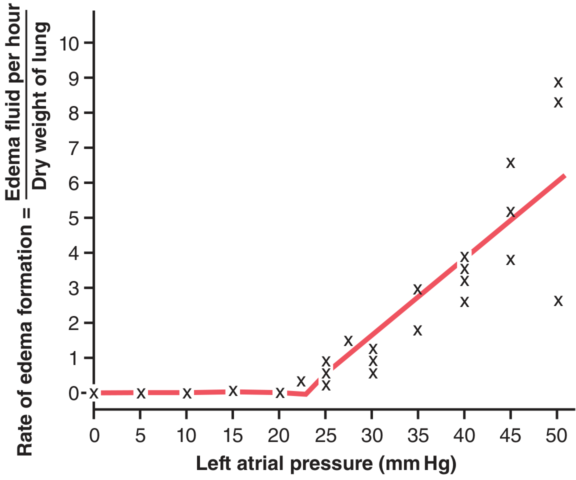
Left ventricular failure raises pulmonary venous pressure and capillary hydrostatic pressure. When capillary pressure exceeds the plasma colloid osmotic pressure (~25-28 mmHg), fluid pours into the interstitium and alveoli. The "safety factor" against pulmonary edema is ~21 mmHg (i.e., capillary pressure must rise from ~7 mmHg to >28 mmHg before significant edema occurs). As heart failure progresses, the Frank-Starling mechanism causes compensatory increases in vascular volume (preload), which further raises pulmonary venous pressure and accelerates edema formation exponentially.
- Guyton and Hall Textbook of Medical Physiology, p. 509
- Murray & Nadel's Textbook of Respiratory Medicine, p. 1508
Chronic compensation: In chronic conditions (e.g., mitral stenosis), pulmonary lymphatics can expand up to 10-fold, allowing patients to tolerate capillary pressures of 40-45 mmHg without lethal edema.
B. Increased Capillary Permeability (Non-Cardiogenic)
Direct or indirect injury to the alveolar-capillary membrane causes protein-rich fluid to leak into the alveoli. The Starling safety factors are overwhelmed even at normal hydrostatic pressures. This is the mechanism underlying ARDS (Acute Respiratory Distress Syndrome).
C. Decreased Oncotic Pressure
Hypoalbuminemia (nephrotic syndrome, liver disease, protein-losing enteropathy) reduces the force holding fluid in the capillary, tipping the balance toward filtration.
D. Lymphatic Obstruction
Impaired lymphatic drainage allows interstitial fluid to accumulate (rare; seen in malignant obstruction).
The graph below from Guyton shows how edema formation rate rises sharply once left atrial pressure exceeds ~25 mmHg:
Sequence of fluid accumulation:
- Fluid first enters the perivascular and peribronchial interstitium (interstitial edema)
- Then fills the alveolar walls
- Finally floods the alveoli (alveolar edema) - impairing gas exchange critically
3. Types of Pulmonary Edema
Classification (Robbins Pathologic Basis of Disease):
| Type | Mechanism | Common Causes |
|---|---|---|
| Hemodynamic (Cardiogenic) | Increased hydrostatic pressure | Left heart failure, mitral stenosis, volume overload, pulmonary vein obstruction |
| Hemodynamic - Low Oncotic | Decreased plasma oncotic pressure | Hypoalbuminemia, nephrotic syndrome, liver disease, protein-losing enteropathy |
| Hemodynamic - Lymphatic Obstruction | Impaired lymphatic drainage | Malignant obstruction (rare) |
| Increased Permeability (Non-Cardiogenic) | Alveolar-capillary injury - Direct | Pneumonia, inhaled gases (O2 toxicity, smoke), aspiration (gastric contents), radiation, lung trauma |
| Increased Permeability (Non-Cardiogenic) | Alveolar-capillary injury - Indirect | Sepsis, SIRS, burns, pancreatitis, blood transfusion (TRALI), drugs (bleomycin, amphotericin B, heroin, cocaine) |
| Neurogenic | Massive sympathetic discharge | Head injury, intracranial bleeding, abrupt naloxone reversal |
| High-Altitude | Hypoxic vasoconstriction + permeability | Rapid ascent to >2500 m |
Two Clinically Key Types:
1. Cardiogenic (Increased-Pressure) Pulmonary Edema
- PAWP (pulmonary artery wedge pressure) > 20 mmHg
- Fluid is a transudate (low protein)
- Histology: engorged alveolar capillaries, finely granular pale pink transudate, hemosiderin-laden macrophages ("heart failure cells"), brown induration in chronic cases
2. Non-Cardiogenic (Increased-Permeability) Pulmonary Edema / ARDS
- PAWP normal (<18 mmHg)
- Fluid is an exudate (protein-rich)
- Safety factors are lost; edema occurs even at normal pressures
- Resolves much more slowly than cardiogenic edema
- Robbins, Cotran & Kumar, p. 2189-2240
- Murray & Nadel's Textbook of Respiratory Medicine, p. 1501-1510
- Morgan and Mikhail's Clinical Anesthesiology (neurogenic type)
4. Clinical Features
Symptoms
- Dyspnea - initially on exertion, progressing to rest
- Paroxysmal nocturnal dyspnea (PND) - patient wakes gasping from sleep
- Orthopnea - breathlessness when lying flat (relieved by sitting up)
- Pink frothy sputum (blood-tinged alveolar exudate)
- Cough and wheezing ("cardiac asthma")
- Anxiety, diaphoresis, sense of drowning
Signs
| Sign | Mechanism |
|---|---|
| Tachypnea, tachycardia | Hypoxia + sympathetic activation |
| Severe hypoxemia (SpO2 ↓) | Alveolar flooding → V/Q mismatch + shunt |
| Bilateral basilar crackles/rales | Alveolar fluid |
| Wheezing | Peribronchial cuffing, airway compression |
| Hypertension | Endogenous catecholamine release (often present in acute cardiogenic) |
| Elevated JVP, S3 gallop, peripheral edema | In cardiogenic type |
"Crackles and wheezing due to alveolar flooding, increased airway fluid, and airway compression from peribronchial cuffing may be audible. Release of endogenous catecholamines often causes hypertension." - Harrison's Principles of Internal Medicine 22E, p. 2373
Investigations
- CXR: Bilateral "bat-wing" perihilar opacities, Kerley B lines, cardiomegaly (cardiogenic), pleural effusions
- ABG: Hypoxemia, respiratory alkalosis early; respiratory acidosis in severe/late cases
- BNP/NT-proBNP: Markedly elevated in cardiogenic edema - key differentiator
- ECG: Ischemia/MI pattern if acute MI is the trigger
- Echocardiography: Identifies LV systolic/diastolic dysfunction, valvular lesions
- Pulmonary Artery Catheter (PAC): PCWP >18-20 mmHg = cardiogenic; <18 mmHg = non-cardiogenic
5. Management
Management depends on etiology but several measures are applied immediately as it is a life-threatening emergency.
A. Immediate / Emergency Measures
| Measure | Details |
|---|---|
| Positioning | Sit patient upright, legs dangling - reduces venous return (preload) |
| Oxygen therapy | Target SpO2 ≥92%; high-flow nasal cannula preferred for hypoxemic failure with normal PaCO2 |
| Non-invasive ventilation (NIV) | CPAP or BiPAP - rests respiratory muscles, improves oxygenation and cardiac function, reduces intubation need |
| Intubation + PEEP | For refractory cases; PEEP decreases both preload and afterload, improves cardiac function, and keeps alveoli open |
B. Reduction of Preload
| Drug | Details |
|---|---|
| Furosemide (Loop diuretic) | First choice; also acts as a venodilator before diuresis; initial dose ≤0.5 mg/kg (increase to 1 mg/kg in renal impairment/chronic use) |
| Sublingual nitroglycerin | 0.4 mg × 3 every 5 min; first-line for acute cardiogenic pulmonary edema; reduces preload AND afterload rapidly |
| IV nitroglycerin | 5-10 µg/min if edema persists without hypotension |
| IV nitroprusside | 0.1-5 µg/kg/min; potent venous AND arterial dilator; use in hypertensive pulmonary edema (requires arterial line) |
| Morphine | 2-4 mg IV boluses; transient vasodilator, reduces anxiety and catecholamine levels; note: registry data suggests possible increased mortality - use cautiously |
| ACE inhibitors | Reduce preload and afterload; recommended in hypertensive patients; proven mortality benefit in acute MI with heart failure |
| Nesiritide | Recombinant BNP; potent arterial/venous vasodilator with diuretic effect; reserve for refractory cases |
C. Inotropic Support (when cardiogenic shock coexists)
- Dobutamine - beta-1 agonist, improves contractility
- Dopamine - at higher doses, vasopressor
- Milrinone - phosphodiesterase-3 inhibitor (inodilator); 50 µg/kg loading, then 0.25-0.75 µg/kg/min; stimulates contractility and causes vasodilation
D. Correct Precipitating Factors
- Arrhythmia control (e.g., rate control in AF)
- Revascularization for acute MI (emergent PCI)
- Antibiotics for infection
- Correct acidemia, anemia, electrolyte disturbances
E. Non-Cardiogenic Edema (ARDS)
- Lung-protective ventilation: tidal volume 6 mL/kg IBW, PEEP titration, prone positioning in severe cases
- Treat the underlying cause (sepsis, aspiration, etc.)
- Fluid restriction and conservative fluid strategy
F. Renal Replacement Therapy
- For refractory volume overload with: metabolic acidosis (pH <7.15-7.25), hypoxemia, or persistent hyperkalemia
- Continuous RRT preferred over intermittent hemodialysis when patient is hemodynamically unstable
- Harrison's Principles of Internal Medicine 22E, p. 2373-2374
- Murray & Nadel's Textbook of Respiratory Medicine, p. 1493-1510
- ROSEN's Emergency Medicine, p. 4855
Recent Evidence (2024-2026)
A 2025 systematic review and meta-analysis (PMID: 40506079) published in BMJ Open evaluated high-dose versus low-dose IV nitroglycerin in sympathetic crashing acute pulmonary edema, supporting its role in this specific phenotype. A 2026 meta-analysis of RCTs (PMID: 41643462) in Journal of Critical Care found lung ultrasound-guided decongestion improves outcomes in heart failure patients, reinforcing ultrasound as a monitoring tool during treatment.
Summary Table: Cardiogenic vs Non-Cardiogenic Pulmonary Edema
| Feature | Cardiogenic | Non-Cardiogenic (ARDS) |
|---|---|---|
| Mechanism | ↑ Hydrostatic pressure | ↑ Capillary permeability |
| PCWP | >18-20 mmHg | <18 mmHg |
| Fluid protein | Low (transudate) | High (exudate) |
| BNP | Markedly elevated | Usually normal/mildly elevated |
| CXR | Cardiomegaly, bilateral perihilar edema, Kerley B lines | Diffuse bilateral infiltrates, no cardiomegaly |
| Response to diuretics | Good | Poor |
| Resolution | Rapid (hours) with treatment | Slow (days-weeks) |
This is a shared conversation. Sign in to Orris to start your own chat.