I now have all content needed. Here are the complete, detailed MBBS notes:
🩺 MBBS Anatomy Notes: Superior Vena Cava · Inferior Vena Cava · Portal Vein
Branches, Tributaries & Anastomoses — Basics to Advanced
Primary source: Gray's Anatomy for Students · Gray's Anatomy for Students, Braunwald's Heart Disease, Schwartz's Principles of Surgery
═══════════════════════════════════════
PART 1: SUPERIOR VENA CAVA (SVC)
═══════════════════════════════════════
📌 What is the SVC?
The Superior Vena Cava (SVC) is a large, valveless vein that collects deoxygenated blood from the head, neck, both upper limbs, and thorax and returns it to the right atrium of the heart.
1.1 Formation & Course
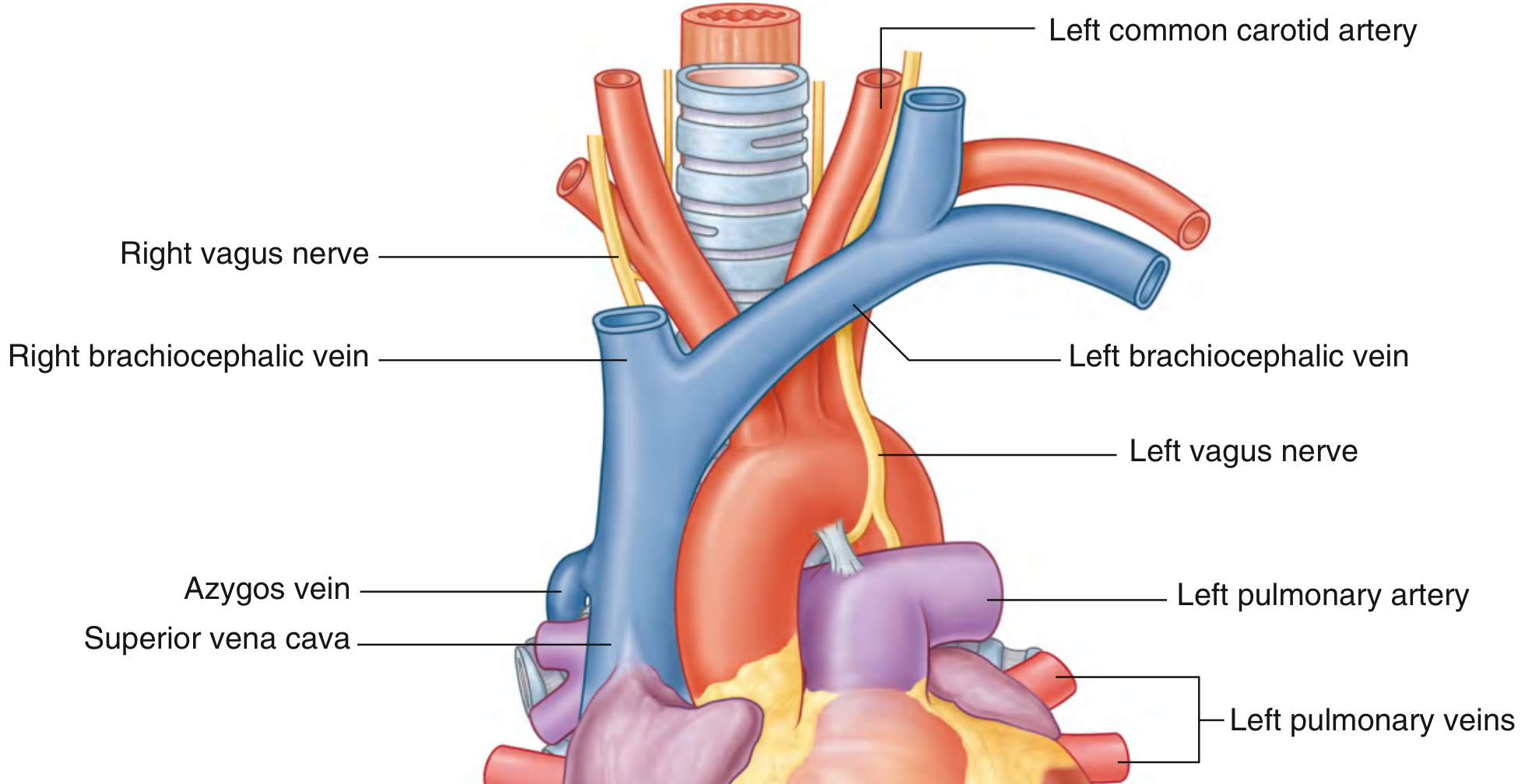
Fig. 3.91 — Superior Mediastinum with Thymus Removed, showing SVC formation from brachiocephalic veins. — Gray's Anatomy for Students
| Feature | Detail |
|---|
| Formed by | Union of right + left brachiocephalic veins |
| Level of origin | Posterior to lower edge of right 1st costal cartilage (near right sternal border) |
| Level of termination | Lower edge of right 3rd costal cartilage → right atrium |
| Length | ~7 cm |
| Location | Upper → superior mediastinum; Lower half → middle mediastinum (inside pericardial sac) |
| Valves | None (valveless) |
| CXR appearance | Forms right superolateral border of mediastinum |
⭐ Exam Pearl: The lower half of SVC is intrapericardial (middle mediastinum). The azygos vein opens into the SVC just before it enters the pericardial sac.
1.2 Tributaries of the SVC
The SVC has 2 main roots (the brachiocephalic veins) + the azygos vein + small mediastinal tributaries.
▶ ROOT 1: RIGHT BRACHIOCEPHALIC VEIN
- Formation: Junction of right internal jugular vein (IJV) + right subclavian vein, behind the right sternoclavicular joint (medial end of right clavicle)
- Course: Short (~2.5 cm), vertical descent
- Tributaries:
- Right vertebral vein
- Right 1st posterior intercostal vein
- Right internal thoracic vein
- Right inferior thyroid vein
- Thymic veins
▶ ROOT 2: LEFT BRACHIOCEPHALIC VEIN
- Formation: Junction of left IJV + left subclavian vein, behind medial end of left clavicle
- Course: ~6 cm, crosses obliquely to the right behind the manubrium, in a slightly inferior direction
- Tributaries:
- Left vertebral vein
- Left 1st posterior intercostal vein
- Left superior intercostal vein (drains 2nd–4th left intercostal spaces)
- Left inferior thyroid vein
- Left internal thoracic vein
- Thymic and pericardial veins
⚠️ Surgical note in children: In infants/children, the left brachiocephalic vein rises above the manubrium → less protected from injury during neck procedures.
▶ AZYGOS VEIN (Major tributary at T4)
The azygos vein opens into the posterior aspect of the SVC just before the SVC enters the pericardial sac (~T4 level). It is the crucial bridge between SVC and IVC territories.
▶ Other small tributaries
- Pericardial veins
- Mediastinal veins
1.3 Left Superior Intercostal Vein (Important!)
- Drains 2nd, 3rd (and sometimes 4th) posterior intercostal veins + left bronchial veins
- Passes over the left side of the aortic arch, lateral to left vagus nerve, medial to left phrenic nerve
- Opens into left brachiocephalic vein
- May connect inferiorly with the accessory hemiazygos vein
1.4 THE AZYGOS SYSTEM — The Bridge between SVC and IVC
The azygos system is the most important collateral pathway in the body. It connects the IVC below with the SVC above via posterior intercostal veins.
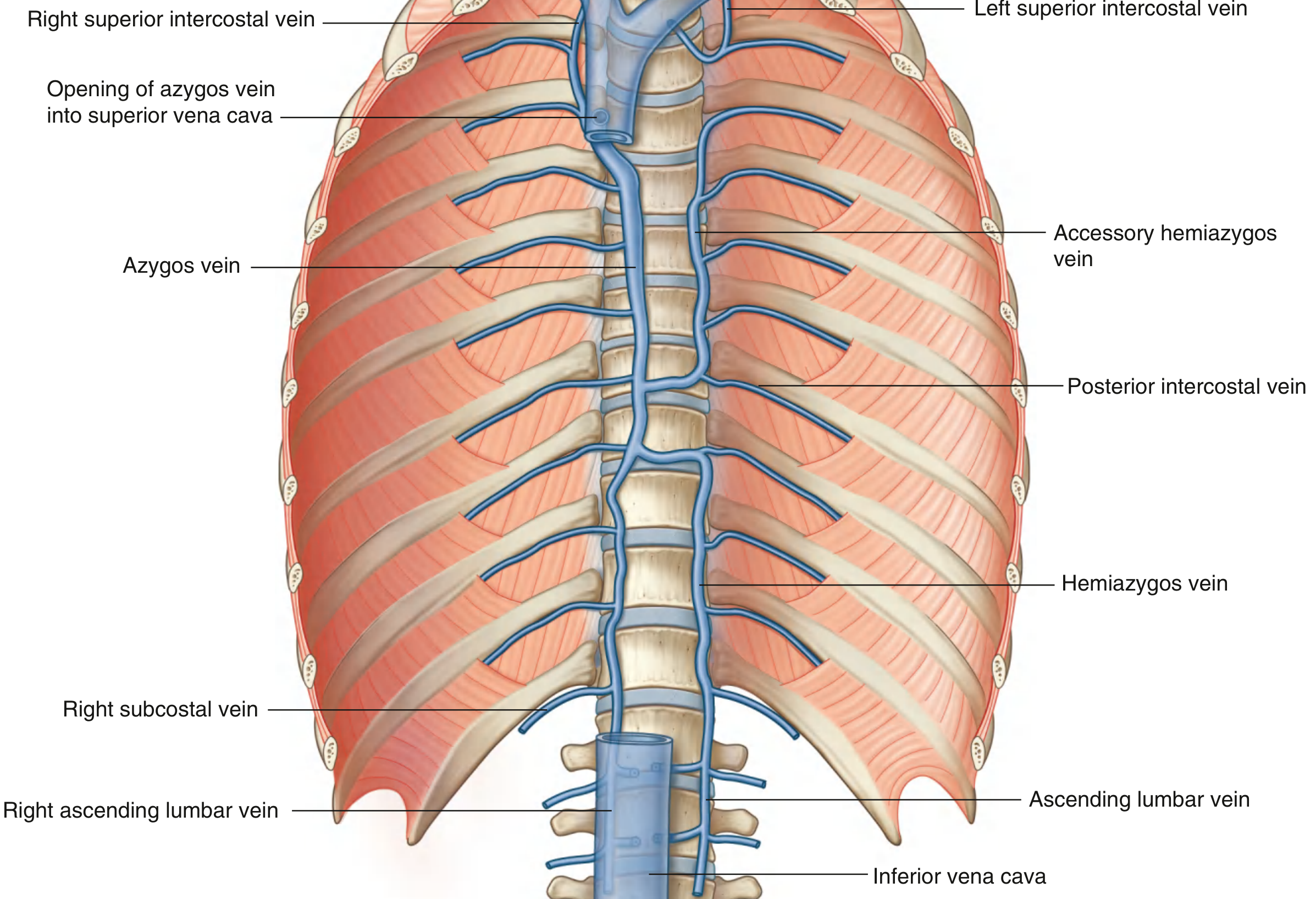
Fig. 3.105 — Azygos System of Veins — Gray's Anatomy for Students
AZYGOS VEIN (Right side — main trunk)
| Feature | Detail |
|---|
| Origin | Junction of right ascending lumbar vein + right subcostal vein at L1/L2 |
| Entry to thorax | Through aortic hiatus or posterior to right crus of diaphragm |
| Ascent | Right side of posterior mediastinum, to the right of thoracic duct |
| Terminates | Arches over root of right lung at T4 → enters SVC |
| Tributaries | Right superior intercostal vein (2nd–4th spaces); 5th–11th right posterior intercostal veins; hemiazygos vein; accessory hemiazygos vein; esophageal, mediastinal, pericardial, and right bronchial veins |
HEMIAZYGOS VEIN (Left side, inferior)
| Feature | Detail |
|---|
| Origin | Junction of left ascending lumbar vein + left subcostal vein; may connect to left renal vein |
| Entry to thorax | Through left crus of diaphragm |
| Ascent | Left posterior mediastinum to T9 |
| Termination | Crosses vertebral column (behind thoracic aorta, esophagus, thoracic duct) → azygos vein |
| Tributaries | Lowest 4–5 left posterior intercostal veins; esophageal veins; mediastinal veins |
ACCESSORY HEMIAZYGOS VEIN (Left side, superior)
| Feature | Detail |
|---|
| Course | Descends left side of posterior mediastinum to T8 |
| Termination | Crosses to join azygos vein or hemiazygos vein; connects superiorly to left superior intercostal vein |
| Tributaries | 4th–8th left posterior intercostal veins; left bronchial veins |
1.5 Relations of SVC
| Direction | Structure |
|---|
| Right | Right phrenic nerve, mediastinal pleura, right lung |
| Left | Ascending aorta |
| Posterior | Right pulmonary artery, right bronchus |
| Anterior | Thymus, pericardium |
1.6 Clinical: SVC Syndrome
| Feature | Detail |
|---|
| Cause | Obstruction of SVC — most commonly bronchogenic carcinoma (right upper lobe) or mediastinal lymphoma |
| Features | Facial/neck/arm edema, distended neck veins, dilated chest wall veins, dyspnea, headache, blurred vision |
| Collateral route | Azygos → hemiazygos → ascending lumbar veins → IVC |
| Emergency Rx | Radiotherapy, stenting, chemotherapy depending on cause |
═══════════════════════════════════════
PART 2: INFERIOR VENA CAVA (IVC)
═══════════════════════════════════════
📌 What is the IVC?
The IVC is the largest vein in the body. It returns blood from all structures below the diaphragm to the right atrium.
2.1 Formation & Course
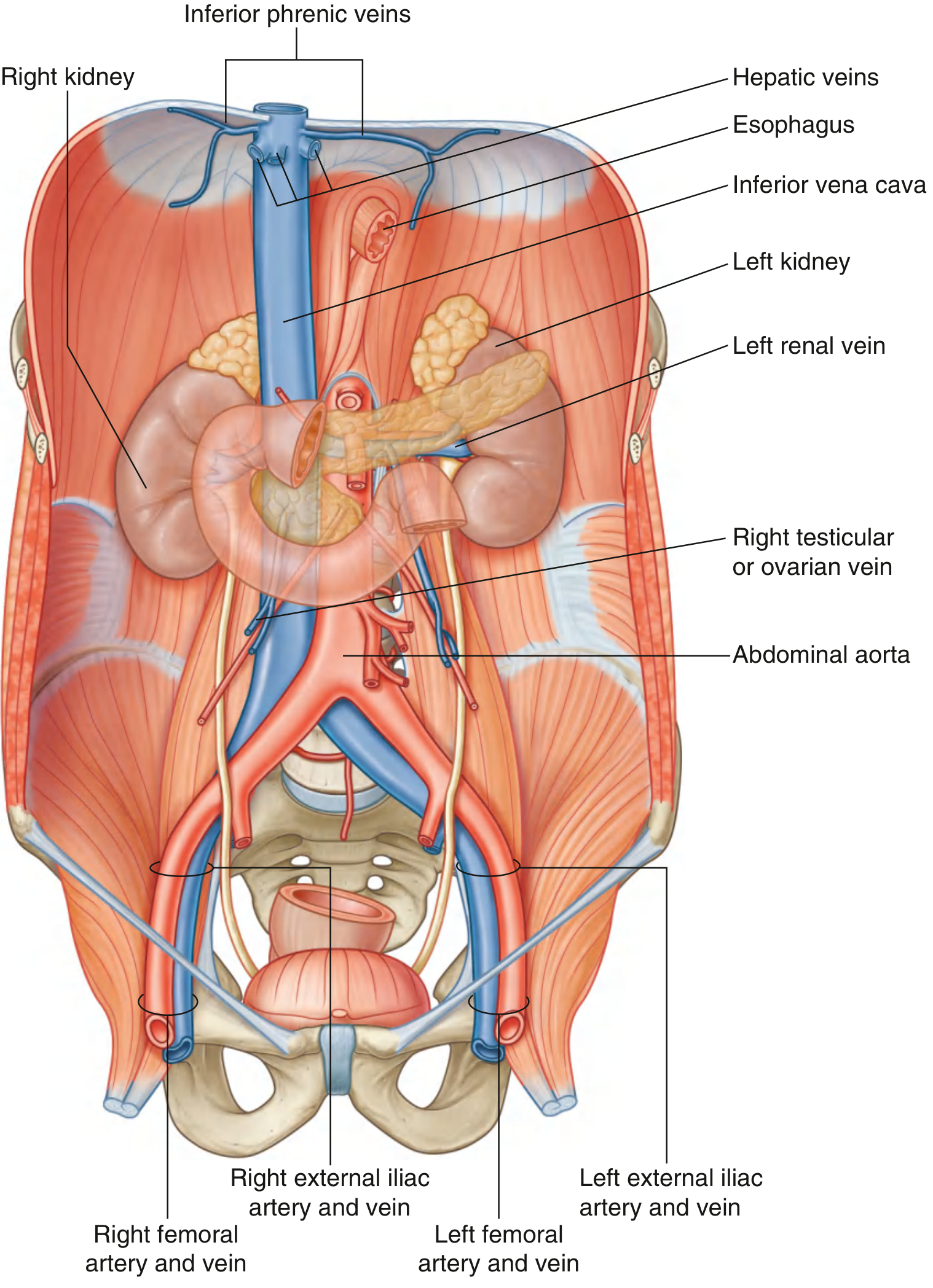
Fig. 4.169 — Inferior Vena Cava — Gray's Anatomy for Students
| Feature | Detail |
|---|
| Formation | Union of right + left common iliac veins |
| Level of formation | Vertebra LV, just to the right of midline |
| Course | Ascends on anterior surface of vertebral column, immediately to right of abdominal aorta |
| Exits abdomen | Pierces central tendon of diaphragm at T8 (IVC foramen) |
| Terminates | Right atrium of heart |
| Valves | None (in the main trunk) |
2.2 Tributaries of the IVC (from below upward)
| No. | Tributary | Level | Notes |
|---|
| 1 | Common iliac veins (R + L) | L5 | Unite to form IVC |
| 2 | Lumbar veins (3rd & 4th) | Various | Drain directly into IVC |
| 3 | Right testicular/ovarian vein | L2 | Drains directly; left side → left renal vein |
| 4 | Renal veins (R + L) | L1–L2 | Left renal vein longer (crosses aorta anteriorly) |
| 5 | Right suprarenal vein | — | Drains directly; left side → left renal vein |
| 6 | Inferior phrenic veins | T8 | Just below diaphragm |
| 7 | Hepatic veins (3: R, M, L) | T8–T9 | Enter IVC just below diaphragm |
🔴 Critical asymmetry — always asked in exams:
- Right gonadal vein → IVC directly
- Left gonadal vein → left renal vein → IVC
- Right suprarenal vein → IVC directly
- Left suprarenal vein → left renal vein → IVC
❌ What does NOT drain into IVC? Gut, spleen, pancreas, gallbladder — all go to the portal vein first.
2.3 Structures Crossing the ANTERIOR Surface of IVC
(From below upward — memorise for surgery/radiology)
- Right common iliac artery
- Root of mesentery
- Right testicular/ovarian artery
- Inferior part of duodenum (3rd part)
- Head of pancreas
- Superior part of duodenum (1st part)
- Bile duct
- Portal vein
- Liver (may completely surround IVC)
2.4 Lumbar Veins — The IVC–SVC Bridge
The lumbar veins are unique because they form the ascending lumbar veins, which link the IVC to the azygos/hemiazygos system:
| Lumbar Vein | Drainage |
|---|
| 5th lumbar vein | → iliolumbar vein → common iliac vein |
| 3rd & 4th lumbar veins | → directly into IVC |
| 1st & 2nd lumbar veins | → ascending lumbar veins |
| Ascending lumbar veins | Connect upward to azygos (right) and hemiazygos (left) in thorax |
📌 If IVC is blocked, the ascending lumbar veins enlarge and carry blood up to the azygos → SVC system — a critical collateral pathway.
2.5 Relations of IVC
| Region | Anterior | Posterior |
|---|
| Abdominal | Liver, portal vein, head of pancreas, duodenum, root of mesentery | Right crus of diaphragm, right renal artery, right suprarenal gland, right psoas major |
| Right lateral | Right ureter, right renal artery | — |
| Left | Abdominal aorta | — |
2.6 Clinical: IVC Obstruction
Causes: Thrombosis, tumour compression (RCC, HCC), retroperitoneal fibrosis
Collateral routes:
- Ascending lumbar veins (retroperitoneal) → azygos/hemiazygos → SVC
- Superficial epigastric veins → thoracoepigastric vein → lateral thoracic vein → axillary → SVC
- Paravertebral venous plexus
📌 In IVC obstruction — dilated superficial abdominal wall veins with flow going upward (toward SVC). Compare: in SVC obstruction, flow is downward. In portal hypertension (caput medusae), flow is away from umbilicus in all directions.
2.7 Clinical: IVC Filter
- Used in DVT patients when anticoagulation is contraindicated
- Inserted via femoral vein or via SVC → right atrium → IVC
- Traps emboli from lower limb/pelvic DVT before they cause pulmonary embolism
═══════════════════════════════════════
PART 3: PORTAL VEIN
═══════════════════════════════════════
📌 What is the Portal Vein?
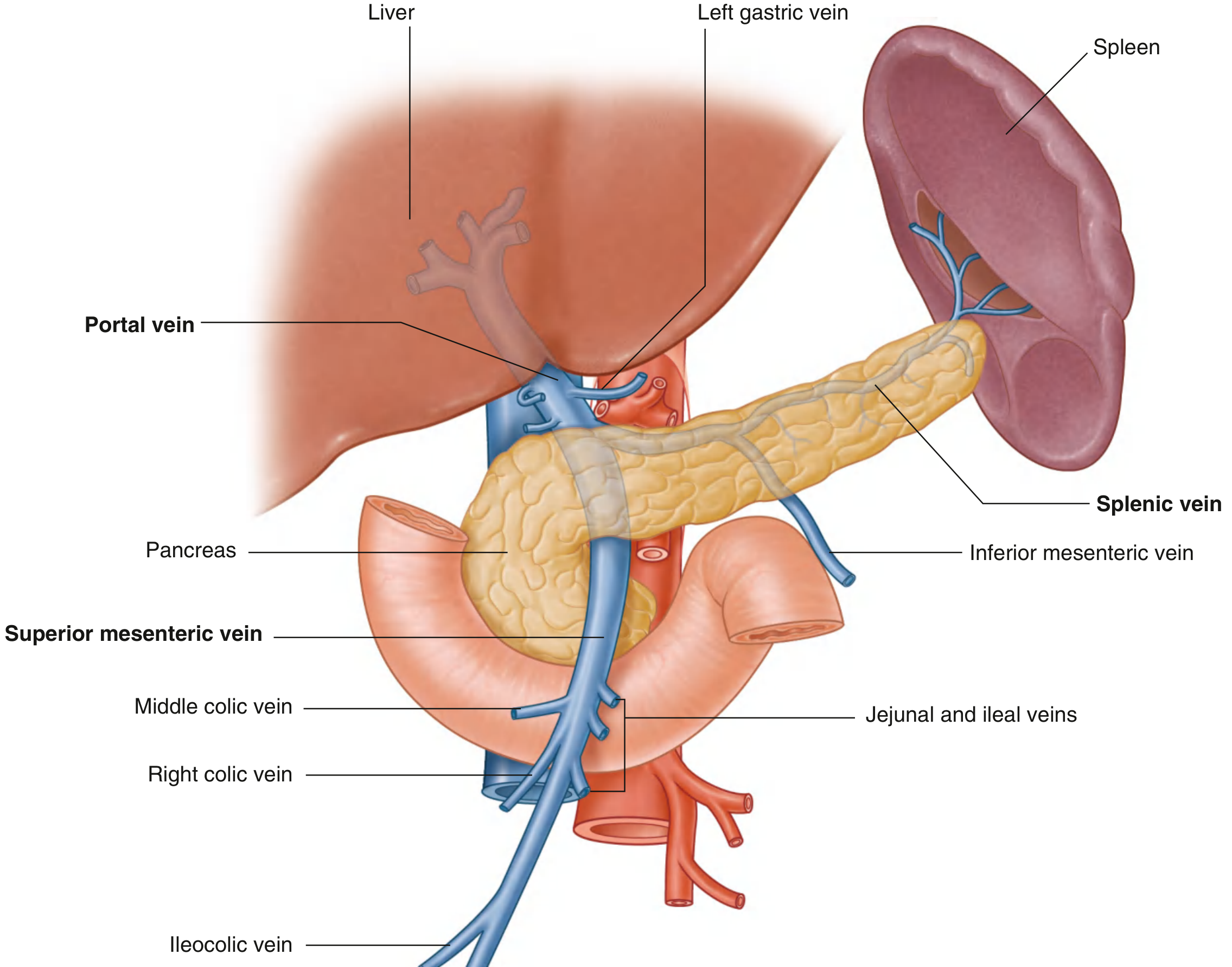
The portal vein is the final common pathway for venous blood from the spleen, pancreas, gallbladder, and abdominal gastrointestinal tract to the liver. It is a portal system — connecting two capillary beds (gut capillaries → liver sinusoids).
💡 Portal blood carries absorbed nutrients + toxins + bacteria from the gut to the liver for processing before entering systemic circulation.
3.1 Formation & Course
Fig. 4.132/133 — Portal Vein and Tributaries — Gray's Anatomy for Students
| Feature | Detail |
|---|
| Formation | Union of Superior Mesenteric Vein (SMV) + Splenic vein |
| Level | Posterior to neck of pancreas, at vertebra LII |
| Length | ~8 cm |
| Ascent | Passes posterior to superior part of duodenum → enters free edge of lesser omentum (hepatoduodenal ligament) |
| Contents of hepatoduodenal ligament | Portal vein (posterior), Bile duct (right anterior), Hepatic artery proper (left anterior) — "BPA" mnemonic |
| Omental foramen | Portal vein is anterior to it |
| Termination | Divides into right and left branches at porta hepatis → enter liver |
| Valves | None |
⭐ Exam Pearl: Portal vein is posterior in the hepatoduodenal ligament. Bile duct is right anterior, hepatic artery is left anterior — Together = "BPA" from right to left anteriorly (or: "Happy Americans Buy Porsches" = Hepatic Artery, Bile duct, Portal vein — right to left).
3.2 All Tributaries of the Portal Vein
The portal vein drains through 2 main roots + direct tributaries:
▶ ROOT 1: SUPERIOR MESENTERIC VEIN (SMV)
- Drains: Small intestine (jejunum, ileum), cecum, appendix, ascending colon, transverse colon
- Origin: Right iliac fossa — formed by junction of terminal ileal, cecal, and appendicular veins
- Course: Ascends in mesentery to the right of superior mesenteric artery (SMA)
- Unites with: Splenic vein posterior to neck of pancreas
Tributaries of SMV:
| Tributary | Drains |
|---|
| Jejunal veins | Jejunum |
| Ileal veins | Ileum |
| Ileocolic vein | Terminal ileum, cecum, appendix |
| Right colic vein | Ascending colon |
| Middle colic vein | Transverse colon |
| Right gastro-omental vein | Right part of greater curvature of stomach |
| Anterior and posterior inferior pancreaticoduodenal veins | Head of pancreas, duodenum |
▶ ROOT 2: SPLENIC VEIN
- Origin: Numerous vessels from hilum of spleen
- Course: Passes right through splenorenal ligament with splenic artery and tail of pancreas → runs along posterior surface of body of pancreas → unites with SMV
Tributaries of Splenic vein:
| Tributary | Drains |
|---|
| Short gastric veins | Fundus and left greater curvature of stomach |
| Left gastro-omental vein | Greater curvature of stomach (left part) |
| Pancreatic veins | Body and tail of pancreas |
| Inferior mesenteric vein (usually) | Rectum, sigmoid, descending colon, splenic flexure |
▶ INFERIOR MESENTERIC VEIN (IMV)
- Drains: Rectum (via superior rectal vein), sigmoid colon, descending colon, splenic flexure
- Begins as: Superior rectal vein
- Receives: Sigmoid veins, left colic vein
- Termination: Usually into splenic vein (occasionally at SMV–splenic junction or into SMV)
▶ DIRECT TRIBUTARIES of Portal Vein (small)
| Tributary | Drains |
|---|
| Right gastric vein | Lesser curvature of stomach (right) + lower esophagus |
| Left gastric vein (coronary vein) | Lesser curvature of stomach (left) + lower esophagus — ⭐ KEY in portal HTN |
| Cystic veins | Gallbladder |
| Para-umbilical veins | Run in falciform ligament with obliterated umbilical vein → connect to abdominal wall veins |
| Posterior superior pancreaticoduodenal vein | Usually empties directly into portal vein |
3.3 Portal Vein Diagram — Full System
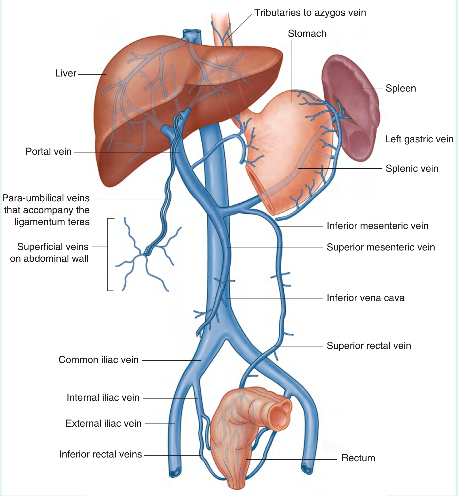
Fig. 4.134 — Portosystemic Anastomoses — Gray's Anatomy for Students
═══════════════════════════════════════
PART 4: PORTOSYSTEMIC ANASTOMOSES ⭐⭐⭐
═══════════════════════════════════════
📌 What are Portosystemic Anastomoses?
These are sites where the portal venous system communicates with the systemic venous system. Normally they carry minimal flow. In portal hypertension (portal pressure > 12 mmHg), they dilate massively to form varices — which can bleed catastrophically.
4.1 Three MAJOR Sites (Must Know for Exams)
| # | Site | Portal Side | Systemic Side | Varix Name | Clinical Risk |
|---|
| 1 | Gastroesophageal junction | Left gastric vein (coronary vein) + tributaries | Esophageal tributaries → azygos system → SVC | Esophageal varices | 💀 Life-threatening UGI bleed |
| 2 | Anorectal junction | Superior rectal vein (from IMV → portal) | Middle & inferior rectal veins → internal iliac → IVC | Anorectal varices | Lower GI bleed |
| 3 | Umbilicus (anterior abdominal wall) | Para-umbilical veins (along ligamentum teres/falciform ligament → left branch of portal vein) | Superficial + inferior epigastric veins → superior epigastric (SVC) + inferior epigastric (IVC) | Caput medusae | Cosmetic; massive varices rare |
4.2 Additional Anastomotic Sites
| Site | Portal vessel | Systemic vessel |
|---|
| Bare area of liver | Small portal branches in liver | Inferior phrenic veins → IVC |
| Retroperitoneum | Colic, mesenteric veins | Renal, gonadal, lumbar veins (Retzius veins) → IVC |
| Falciform ligament | Left portal vein branch | Inferior epigastric / internal thoracic veins |
4.3 Clinical Features of Portal Hypertension
Normal portal pressure: 5–10 mmHg
Portal hypertension: > 12 mmHg (clinically significant)
Classic triad:
- Splenomegaly (back-pressure → splenic venous congestion)
- Ascites (raised hydrostatic pressure + reduced albumin from liver failure)
- Portosystemic varices
Named varices:
- Esophageal varices — risk of torrential, potentially fatal haematemesis (most dangerous)
- Caput medusae — dilated veins radiating from umbilicus; flow is away from umbilicus in all directions
- Anorectal varices — bleeding per rectum (NOT hemorrhoids, which are anal cushions)
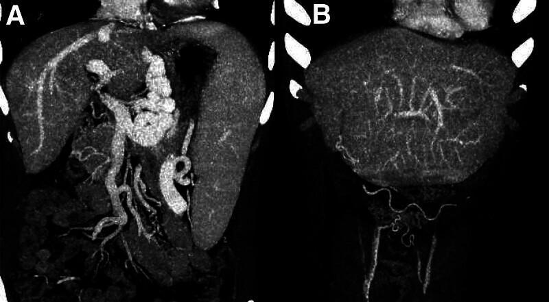
CT Portal Venography: (A) Dilated esophageal/gastric varices; (B) Caput medusae — recanalized umbilical vein with periumbilical varices in cirrhosis
4.4 Distinguishing Caput Medusae vs IVC Obstruction
| Feature | Caput Medusae (Portal HTN) | IVC Obstruction |
|---|
| Blood flow in abdominal veins | Away from umbilicus (all directions) | Upward (from groin toward chest) |
| Mechanism | Portal → para-umbilical → abdominal wall veins | Collateral bypass for blocked IVC |
| Umbilicus involved | Yes (epicentre) | No |
| Splenomegaly | Yes | No |
| Leg edema | Mild | Severe |
═══════════════════════════════════════
PART 5: COLLATERAL CIRCULATION SUMMARY
═══════════════════════════════════════
| Obstruction | Main Collaterals | Clinical Consequence |
|---|
| SVC obstruction | Azygos → hemiazygos → ascending lumbar → IVC | SVC syndrome (face/neck swelling, dilated chest veins) |
| IVC obstruction | Ascending lumbar → azygos/hemiazygos → SVC; or superficial epigastric veins | Leg edema, dilated abdominal veins (flow upward) |
| Portal hypertension | Para-umbilical; esophageal varices via left gastric → azygos; anorectal varices | Caput medusae, hematemesis, rectal bleeding |
═══════════════════════════════════════
PART 6: MNEMONICS & QUICK REVISION ⭐
═══════════════════════════════════════
IVC Tributaries (below → above):
"Come Let's Run, Really Super Incredible Heroes"
- Common iliac veins
- Lumbar veins
- Right gonadal vein (testicular/ovarian)
- Renal veins
- Suprarenal (right) vein
- Inferior phrenic veins
- Hepatic veins
Portal Vein Direct Tributaries:
"Right Gyms Cause Parachute Crashes"
- Right & left Gastric veins
- Cystic veins
- Para-umbilical veins
- (+ SMV and Splenic vein as main roots)
Hepatoduodenal ligament contents (right → left):
"Bill And Pam"
- Bile duct (right)
- A = hepatic Artery proper (left)
- Portal vein (posterior)
3 Portosystemic Anastomoses:
"GAU" (or "Good Anatomy Understanding")
- Gastroesophageal → esophageal varices
- Anorectal → anorectal varices
- Umbilicus → caput medusae
═══════════════════════════════════════
PART 7: HIGH-YIELD EXAM TABLE ⭐
═══════════════════════════════════════
| Question | Answer |
|---|
| SVC is formed by | R + L brachiocephalic veins |
| Level of SVC formation | Lower edge of right 1st costal cartilage |
| SVC terminates at | Lower edge of right 3rd costal cartilage → right atrium |
| Which half of SVC is intrapericardial? | Lower half |
| Azygos vein opens into SVC at | T4 (just before SVC enters pericardial sac) |
| Azygos vein formed by | Right ascending lumbar vein + right subcostal vein (L1/L2) |
| IVC formed by | R + L common iliac veins |
| Level of IVC formation | Vertebra LV |
| IVC exits abdomen at | T8 (central tendon of diaphragm) |
| Left gonadal vein drains into | Left renal vein (not IVC directly) |
| Left suprarenal vein drains into | Left renal vein |
| Right gonadal vein drains into | IVC directly |
| Portal vein formed by | SMV + Splenic vein |
| Level of portal vein formation | LII (posterior to neck of pancreas) |
| Portal vein length | ~8 cm |
| Portal vein position in hepatoduodenal ligament | Posterior |
| IMV usually drains into | Splenic vein |
| Major portosystemic anastomosis causing bleeding | Esophageal varices (left gastric → azygos) |
| Caput medusae flow direction | Away from umbilicus (radiates outward) |
| SVC syndrome most common cause | Bronchogenic carcinoma (right upper lobe) |
| Collateral in SVC obstruction | Azygos system |
| Collateral in IVC obstruction | Ascending lumbar veins |
| Normal portal pressure | 5–10 mmHg |
| Portal HTN threshold | > 12 mmHg |
| IVC filter used for | DVT with contraindication to anticoagulation |
═══════════════════════════════════════
PART 8: EMBRYOLOGY (Brief — MBBS Level)
═══════════════════════════════════════
| Structure | Embryological Origin |
|---|
| SVC | Right cardinal vein system (anterior cardinal vein) |
| IVC | Complex — four segments from multiple veins (posterior cardinal, subcardinal, supracardinal) |
| Portal vein | Vitelline (omphalomesenteric) veins |
| Ligamentum teres hepatis (round ligament) | Obliterated umbilical vein — carries para-umbilical veins in portal HTN |
| Ligamentum venosum | Obliterated ductus venosus (connected left portal branch → IVC in fetus) |
| Persistent left SVC | Failure of obliteration of left common cardinal vein |
Sources: Gray's Anatomy for Students (Mitchell, Drake, Vogl); Braunwald's Heart Disease; Schwartz's Principles of Surgery; Yamada's Textbook of GastroenterologyYour credits are exhausted. Your daily credits reset tomorrow, or you can purchase a top-up for more.