
Poland syndrome
Reading File
Finding Sources
Finding Sources
Searching PubMed
"Poland syndrome"
Reading File
Reading File
Poland Syndrome
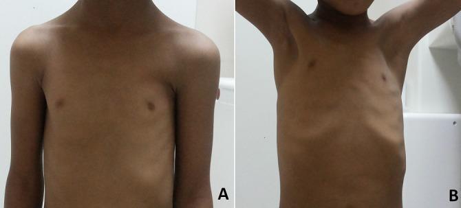
Poland syndrome is a rare congenital chest wall anomaly characterized by unilateral absence (agenesis or hypoplasia) of the pectoral muscles, most often the sternocostal head of pectoralis major, with a range of associated ipsilateral defects.
Epidemiology
- Incidence roughly 1 in 30,000 live births (Mulholland and Greenfield's Surgery, p. 4321).
- Right-sided predominance, and it occurs more often in males.
Clinical Features
- Core defect: absence or hypoplasia of the pectoralis major (sternocostal head) and often pectoralis minor.
- Associated chest wall findings: absence/deformity of costal cartilages of ribs 2-5, hypoplasia or absence of breast tissue and nipple (amastia/athelia), and rib anomalies.
- Limb findings: ipsilateral upper limb anomalies are common, especially syndactyly and brachydactyly (hand/digit shortening is typically unilateral) - Campbell's Operative Orthopaedics, p. 7283.
- Occasionally associated with renal and spinal lesions, and other syndromic overlaps (e.g., Möbius syndrome has been described in association).
- Most patients are asymptomatic from a respiratory standpoint, but if rib defects are extensive, paradoxical inward chest wall motion during inspiration (flail-chest-like physiology) or lung herniation can occur, sometimes requiring more urgent surgical repair - Sabiston Textbook of Surgery, p. 2067; Mulholland and Greenfield's Surgery, p. 4321.
Pathogenesis
The exact cause is unclear. The prevailing theory is an interruption or hypoplasia of the subclavian artery (or its branches, e.g., internal thoracic/intercostal arteries) during early fetal development (around the 6th week of gestation), causing hypoperfusion to the developing pectoral muscle and limb bud - resulting in the combined chest wall and ipsilateral limb defects (Sabiston Textbook of Surgery, p. 2067).
Imaging
On imaging, ipsilateral pectoral muscle changes are accompanied by hand/arm anomalies (particularly syndactyly), with or without absence of pectoralis minor, rib anomalies, and hypoplasia of breast and nipple (Grainger & Allison's Diagnostic Radiology, p. 1031).
Management
- Management is individualized and multidisciplinary (thoracic surgery, plastic surgery, sometimes orthopedics for limb defects).
- Surgery is ideally deferred until the patient has finished growing, unless urgent repair is needed for lung herniation or significant paradoxical chest wall motion.
- Reconstructive options include mesh placement over rib/cartilage defects and/or muscle flap transfer (e.g., latissimus dorsi flap) to restore chest wall contour, along with implant-based breast/soft tissue reconstruction in females with breast hypoplasia (Mulholland and Greenfield's Surgery, p. 4321 and references).
Notable Associations from Recent Literature
Recent case reports (not high-level evidence, so treat as observational associations rather than established causal links) have reported:
- Breast cancer occurring in patients with Poland syndrome, raising discussion of whether there is a true association or coincidence (PMID: 39365625, PMID: 36100049).
- Acute pericarditis in a Poland syndrome patient (PMID: 41426815) and esophageal cancer in another (PMID: 41403868) - both isolated case reports.
- A 2024 review, "Consultations for Poland Syndrome: The Essentials for a Thoracic Surgeon" (PMID: 39064607), reinforces the multidisciplinary referral pattern (breast surgery, plastic surgery, orthopedics, cardiology) that these patients often need.
These are case-level reports, so they don't change standard management, but they highlight the importance of monitoring for less common systemic associations in patients with Poland syndrome.
What are the surgical reconstruction options for Poland syndrome?
Finding Sources
Finding Sources
Searching PubMed
"Poland syndrome" reconstruction
Reading Article
PMID: 39818015
Surgical Reconstruction Options for Poland Syndrome
Reconstruction is individualized based on sex, severity of the skeletal/muscular defect, and desired chest/breast contour. There is no single gold-standard approach - a 2025 systematic review of 77 studies (Grande et al., JPRAS 2025, PMID: 39818015) found reconstructive strategy differs notably between men and women.
1. Skeletal/chest wall defect correction (done first if ribs/cartilage are missing)
- Mesh placement over the rib/cartilage defect to restore chest wall rigidity and prevent paradoxical motion or lung herniation - the standard approach when costal cartilage of ribs 2-5 is absent (Mulholland and Greenfield's Surgery, p. 4321).
- Rigid prosthesis/custom implants (e.g., ethacrylate or 3D-printed titanium/silicone chest wall prosthesis) have been described for more extensive bony defects (referenced in Mulholland and Greenfield's Surgery bibliography).
- Timing is ideally deferred until skeletal growth is complete, unless urgent repair is needed for flail-chest physiology or lung herniation.
2. Soft tissue / muscle contour restoration
- Flap-based reconstruction is the most common overall technique (about 60% of cases in the systematic review), with the latissimus dorsi myocutaneous flap being the dominant choice - it recreates the missing pectoralis bulk and anterior axillary fold in a single well-vascularized unit. Pedicled latissimus flaps can be tunneled anteriorly to fill the subclavicular/pectoral defect.
- Other flap options mentioned in older literature include pectoralis major transposition or free flaps for larger defects, though latissimus dorsi predominates.
3. Breast reconstruction (primarily in women)
- Tissue expander → implant reconstruction: used in about 22.5% of patients (all female in the review); roughly 80% of those who started with expanders went on to implant-based reconstruction.
- Hybrid reconstruction (flap + implant): the most common approach in women overall (about 34% of female cases) - typically combining a latissimus dorsi flap for muscle/soft-tissue coverage with a silicone implant to establish breast volume and symmetry.
- Custom/individualized silicone implants shaped to match the contralateral breast and chest wall contour (Ann Plast Surg 2007, cited in Mulholland and Greenfield's Surgery references).
4. Fat grafting
- Autologous fat grafting, alone or as an adjunct to flap/implant reconstruction, has become increasingly popular since the 1990s. It is used to fine-tune contour, camouflage step-off deformities, and reduce the volume of more invasive flap or implant procedures - often in staged sessions.
5. Sex-based patterns (from the 2025 systematic review)
- Men: flap reconstruction alone is most common (about 42% of cases), since the primary goal is restoring pectoral muscle bulk/contour rather than breast volume.
- Women: hybrid flap-plus-implant reconstruction predominates (about 34%), reflecting the added need to reconstruct breast shape and volume, often combined with contralateral symmetry procedures (e.g., mastopexy or reduction) if the unaffected breast is proportionally larger.
Associated hand/limb reconstruction (if present)
Ipsilateral syndactyly or brachydactyly, when present, is addressed separately by hand surgery - typically digit separation, commissure reconstruction, and resurfacing of the digit borders (Campbell's Operative Orthopaedics, p. 892) - and is generally staged independently from the chest/breast reconstruction.
Practical takeaway
A staged, individualized plan is standard: correct any skeletal instability first (mesh/prosthesis) if ribs are deficient, then restore soft-tissue contour with a latissimus dorsi flap, and finish breast volume/symmetry with implants and/or fat grafting as needed. Decision-making should involve a multidisciplinary team (thoracic surgery, plastic surgery, and hand surgery when limb anomalies coexist).
This is a shared conversation. Sign in to Orris to start your own chat.