
Secondary structure of protein in biochemistry
Reading File
Now let me search the medical library for secondary structure of protein content:
Finding Sources
Searching the Web
protein secondary structure alpha helix beta sheet biochemistry
Finding Sources
Finding Sources
Good - found Lippincott Biochemistry and Basic Medical Biochemistry. Let me read both:
Reading File
Reading File
Excellent - rich content from both Lippincott and Basic Medical Biochemistry. Let me read more of the Lippincott secondary structure section and continue reading the Basic Medical Biochemistry section:
Reading File
Reading File
I have rich content from both textbooks. Let me also fetch the key diagrams and read more of the beta sheet and other secondary structures section:
Reading File
Loading Image
Loading Image
Now let me get the beta sheet and turns/loops section from Lippincott, and fetch the beta sheet image:
Reading File
Reading File
I now have comprehensive content from both Lippincott and Basic Medical Biochemistry. Let me fetch one more key image - the alpha helix and beta sheet diagrams:
Loading Image
Loading Image
I have all the content I need. Let me now compile a comprehensive, well-structured answer.
Secondary Structure of Proteins
The secondary structure of a protein refers to recurring, localized structural patterns that form within short regions of a polypeptide chain. These structures arise when atoms of the peptide backbone form regular hydrogen bonds - they represent the second level of protein organization, sitting between the primary (linear amino acid sequence) and the tertiary (overall 3D fold) levels.
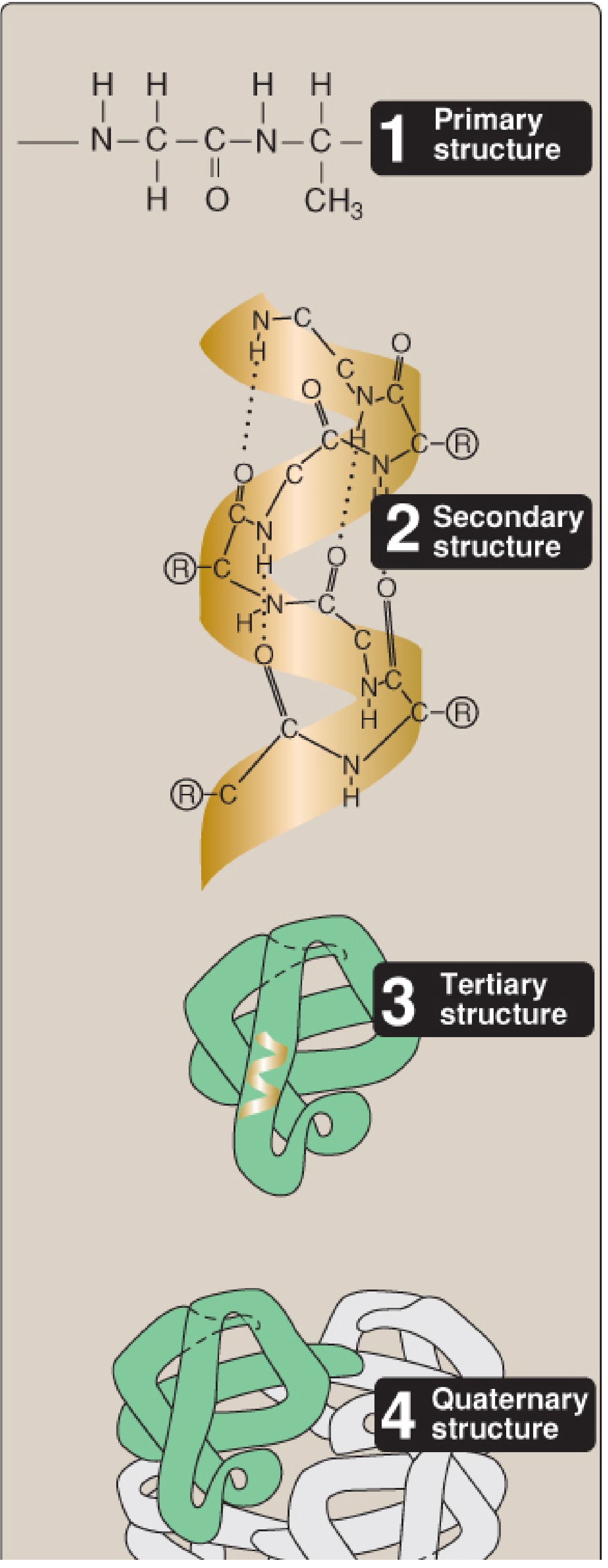
Fig. 2.1 - The four hierarchies of protein structure (Lippincott Biochemistry, 8th ed.)
Basis: The Peptide Backbone
Before secondary structure can form, there are geometric constraints on the polypeptide backbone. The peptide bond itself has partial double-bond character due to resonance, which keeps the carbonyl C and the amide N in the same plane (a rigid planar unit). Rotation is only allowed around the bonds connecting each alpha-carbon (Cα) to its neighboring atoms - described by torsion angles phi (φ) and psi (ψ). Steric clashes between side chains restrict which angles are permissible, which in turn limits the secondary structures that can form.
- Biochemistry, Lippincott 8th ed., p. 51
- Basic Medical Biochemistry - A Clinical Approach, 6e, p. 181
The Two Major Regular Secondary Structures
1. The α-Helix
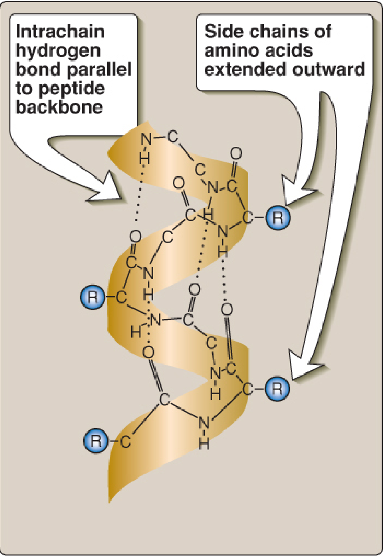
Fig. 2.6 - Structure of an α-helix (Lippincott Biochemistry, 8th ed.)
The α-helix is the most common secondary structure element. Key features:
| Property | Detail |
|---|---|
| Shape | Right-handed spiral (clockwise when viewed from N to C terminus) |
| Amino acids per turn | 3.6 residues per full turn |
| Hydrogen bonds | Each C=O (carbonyl oxygen) of one peptide bond forms an H-bond with the N-H of the residue 4 positions ahead in the chain |
| H-bond orientation | Parallel to the helix axis (backbone) |
| Side chains | Project outward and backward from the central axis, avoiding steric clash |
| Stabilization | All peptide bonds (except first and last) are involved in H-bonding |
The α-helix is found in diverse proteins: the near-entirely helical keratins (hair, skin, nails) and globular proteins like myoglobin.
Amphipathic helices: The arrangement of R groups can create a helix with one polar face and one nonpolar face - important for membrane-spanning and lipid-interacting proteins.
Amino acids that disrupt the α-helix:
-
Proline - its rigid secondary amino group (the N is part of a ring) cannot adopt the required geometry; it inserts a kink and is called a "helix breaker"
-
Glycine - too much conformational flexibility
-
Amino acids with bulky R groups (e.g., tryptophan), charged R groups (e.g., glutamate), or β-carbon branching (e.g., valine) are disfavored
-
Lippincott Biochemistry, 8th ed., p. 60-62
-
Basic Medical Biochemistry, 6e, p. 182
2. The β-Sheet (β-Pleated Sheet)
The β-sheet is the second major regular secondary structure. All peptide bond components participate in hydrogen bonding.
Key features:
- Formed by two or more polypeptide strands (β-strands) aligned laterally
- Stabilized by H-bonds between C=O and N-H groups of adjacent strands - these bonds are perpendicular to the polypeptide backbone (contrast with the α-helix where they are parallel)
- The surface has a pleated (corrugated) appearance because successive α-carbons lie slightly above and below the plane of the sheet
- β-strands are almost fully extended (much more extended than in the helix)
- The sheet has a right-handed curl (twist) when viewed along the backbone
- R groups of adjacent residues project alternately above and below the plane of the sheet
Two types of β-sheets:
| Type | Description |
|---|---|
| Antiparallel | Adjacent strands run in opposite directions (alternating N-termini); H-bonds are more linear (stronger) |
| Parallel | Adjacent strands run in the same direction; H-bonds are slightly distorted |
Just as with the α-helix, the positioning of R groups can make β-sheets amphipathic (with polar and nonpolar faces).
- Lippincott Biochemistry, 8th ed., p. 63-64
Non-Regular Secondary Structures
3. β-Bends (β-Turns / Reverse Turns)
- Allow the polypeptide chain to reverse direction, giving proteins their compact globular shape
- Usually found on the surface of proteins, often containing charged residues
- Connect successive strands of antiparallel β-sheets
- Typically composed of 4 amino acids
- Common residues: Proline (causes kink) and Glycine (smallest R group, allows tight turns)
- Stabilized by an H-bond between the C=O of residue 1 and the N-H of residue 4
4. Loops and Coils
About half of an average globular protein is organized into repetitive (regular) structures (α-helices and β-sheets). The rest consists of loops and coils - segments that connect helices and sheets.
Important points:
-
These are NOT random - they have specific, reproducible conformations stabilized by H-bonds dictated by the primary sequence
-
They should never be called "random coils" (that term is reserved for denatured proteins)
-
They are generally more flexible than helices or β-sheets
-
They often form hinge regions that allow protein segments to move during ligand binding or folding
-
Named loops include Ω-loops (omega loops)
-
Lippincott Biochemistry, 8th ed., p. 65
-
Basic Medical Biochemistry, 6e, p. 187
Summary Comparison Table
| Feature | α-Helix | β-Sheet | β-Bend |
|---|---|---|---|
| Shape | Right-handed coil | Extended, pleated strands | 4-residue hairpin turn |
| H-bond direction | Parallel to helix axis | Perpendicular to strand | Between residues 1 and 4 |
| H-bond linkage | i to i+4 within one chain | Between separate strands | Within the turn |
| Side chain orientation | Outward from helix | Alternate above/below plane | Variable |
| Helix breaker | Proline, Gly, bulky/branched R groups | - | Pro, Gly common |
| Strand orientation | - | Parallel or antiparallel | - |
| Example proteins | Keratin, myoglobin | Silk fibroin, IgG | Connecting loops |
Stabilizing Forces in Secondary (and Higher) Structure
Secondary structure is maintained primarily by hydrogen bonds between backbone atoms. Overall protein conformation (tertiary/quaternary) is further stabilized by:
- Disulfide bonds (-S-S-) between cysteine residues (covalent)
- Hydrophobic interactions - nonpolar side chains cluster in the interior
- Ionic interactions - between oppositely charged R groups
- Van der Waals forces - nonspecific close-range attractions
- Hydrogen bonds involving side chains
- Lippincott Biochemistry, 8th ed., p. 70-72
- Basic Medical Biochemistry, 6e, p. 188
Clinical Relevance
Alterations in secondary/tertiary structure have major clinical consequences:
- Prion diseases (Creutzfeldt-Jakob disease, scrapie): normal PrP^C protein (predominantly α-helical) misfolds into PrP^Sc (predominantly β-sheet), which is protease-resistant and aggregates - causing neurodegeneration
- Sickle cell disease: a single amino acid substitution (Val for Glu at position 6 of β-globin) leads to abnormal hydrophobic interactions and polymerization under low oxygen conditions
- Amyloid diseases (Alzheimer's, type 2 diabetes): normally soluble proteins misfold into β-sheet-rich fibrils that deposit in tissues
Sources: Biochemistry, Lippincott Illustrated Reviews, 8th ed. | Basic Medical Biochemistry - A Clinical Approach, 6th ed.
This is a shared conversation. Sign in to Orris to start your own chat.