
Oxidase test and catalase test
Reading File
Now let me search the medical library for content on oxidase and catalase tests in parallel.
Finding Sources
Finding Sources
Good results. Now let me read the detailed sections from the best sources in parallel.
Reading File
Reading File
Reading File
Excellent content gathered. Let me also fetch the relevant images and get more detail on the oxidase test mechanism.
Loading Image
Loading Image
Loading Image
Reading File
I now have comprehensive information to provide a thorough answer. Here is the complete breakdown:
Oxidase Test and Catalase Test
Two foundational biochemical tests used in bacteriology for rapid identification and differentiation of organisms.
Catalase Test
Principle
The enzyme catalase catalyzes the decomposition of hydrogen peroxide (H₂O₂) into water and oxygen:
2 H₂O₂ → 2 H₂O + O₂
The liberated O₂ appears as visible gas bubbles, which is the positive result.
Procedure
- Transfer a bacterial colony to a clean glass slide
- Add 1-2 drops of 3% hydrogen peroxide
- Observe for immediate, sustained bubbling
| Result | Interpretation |
|---|---|
| Rapid, sustained effervescence (bubbles) | Catalase POSITIVE |
| No bubbles | Catalase NEGATIVE |
Important: Do not use bacteria taken from a blood agar plate - blood itself contains catalase and will cause a false positive.
Catalase-Positive Organisms
- Staphylococcus spp.
- Micrococcus spp.
- Bacillus spp.
- Listeria spp.
- Campylobacter spp.
Catalase-Negative Organisms
- Streptococcus spp.
- Enterococcus spp.
Primary Clinical Use
The most important use is differentiating Gram-positive cocci:
- Staphylococci = catalase POSITIVE
- Streptococci = catalase NEGATIVE
This is the first step in the identification algorithm shown below:
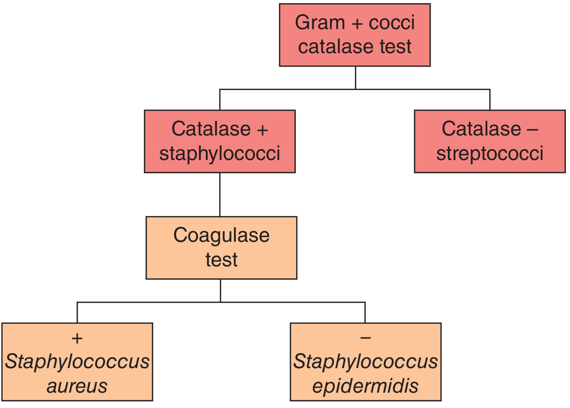
FIGURE: After the catalase test separates Staphylococci (+) from Streptococci (-), the coagulase test further differentiates S. aureus (coagulase +) from S. epidermidis (coagulase -). - Jawetz, Melnick & Adelberg's Medical Microbiology, 28e
The catalase test result in action (positive on left, negative on right):
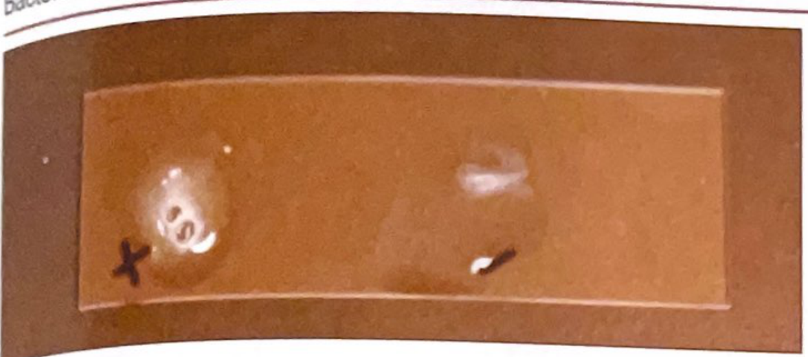
FIGURE: Rapid and sustained effervescence after H₂O₂ addition indicates catalase-positive organism (staphylococci). - Quick Compendium of Clinical Pathology, 5e
Oxidase Test
Principle
The test detects the presence of cytochrome c oxidase (the "c component" of the cytochrome-oxidase complex), a terminal enzyme in the aerobic respiratory chain. The reagent (tetramethyl-p-phenylenediamine, TMPD) acts as an artificial electron acceptor - it changes from colorless to purple/dark blue when oxidized by cytochrome c oxidase.
Cytochrome c oxidase transfers electrons from reduced cytochrome c to molecular oxygen.
Procedure (Spot Test - most common)
- Place filter paper moistened with oxidase reagent (or use an oxidase disk/swab)
- Smear a colony onto the reagent using a platinum loop (NOT nichrome/iron - these give false positives)
- Observe for color change within 10-60 seconds
| Result | Interpretation |
|---|---|
| Purple/dark blue color within 10-60 seconds | Oxidase POSITIVE |
| No color change | Oxidase NEGATIVE |
Positive oxidase test - purple/blue color on swab:
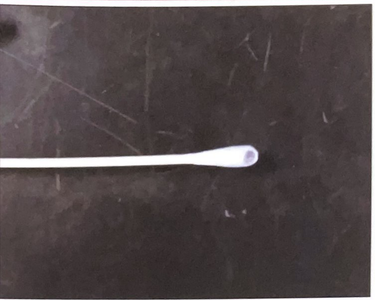
FIGURE: Positive oxidase test denoted by purple/blue color on the swab tip. - Quick Compendium of Clinical Pathology, 5e
Oxidase-Positive Organisms (key examples)
| Organism | Clinical Relevance |
|---|---|
| Pseudomonas aeruginosa | Nosocomial infections, CF lung disease |
| Neisseria spp. | Meningitis, gonorrhea |
| Haemophilus spp. (some) | Meningitis, epiglottitis |
| Vibrio spp. | Cholera, gastroenteritis |
| Campylobacter spp. | Gastroenteritis |
| Helicobacter pylori | Peptic ulcer disease |
| Aeromonas spp. | GI infections |
| Brucella spp. | Brucellosis |
| Moraxella catarrhalis | Respiratory infections |
Oxidase-Negative Organisms (key examples)
- All Enterobacteriaceae (E. coli, Klebsiella, Salmonella, Shigella, Proteus)
- Staphylococcus spp.
- Acinetobacter spp.
Primary Clinical Use
Rapidly differentiates Gram-negative rods:
- Pseudomonas aeruginosa = oxidase POSITIVE
- Escherichia coli (and all Enterobacteriaceae) = oxidase NEGATIVE
This is very useful when screening stool/wound cultures - most normal gut flora (Enterobacteriaceae) are oxidase negative, so an oxidase-positive colony on a plate flags an important pathogen immediately.
Side-by-Side Comparison
| Feature | Catalase Test | Oxidase Test |
|---|---|---|
| Detects | Catalase enzyme | Cytochrome c oxidase enzyme |
| Reagent | 3% H₂O₂ | Tetramethyl-p-phenylenediamine (TMPD) |
| Positive result | Bubbles (O₂ released) | Purple/blue color |
| Primary use | Differentiate Staph (+) vs Strep (-) | Differentiate Pseudomonas (+) vs Enterobacteriaceae (-) |
| Key false positive | Blood on agar plate | Nichrome/iron loop |
| Organism groups | Gram-positive cocci | Gram-negative rods |
Clinical Identification Flow
- Gram stain: Is it Gram+ cocci? → Catalase test → Staph vs Strep
- Gram stain: Is it Gram- rod? → Oxidase test → Enterobacteriaceae (negative) vs non-fermenters like Pseudomonas (positive)
Sources: Jawetz, Melnick & Adelberg's Medical Microbiology 28e; Sherris & Ryan's Medical Microbiology 8e; Quick Compendium of Clinical Pathology 5e
This is a shared conversation. Sign in to Orris to start your own chat.