
Chronic colecystitis
"cholecystitis, chronic"[MeSH Terms] OR chronic cholecystitis
chronic cholecystitis gallbladder gallstones histology Rokitansky-Aschoff sinuses
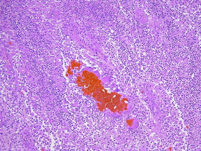
Imaging modality: bright-field light microscopy of a hematoxylin and eosin–stained gallbladder wall section, illustrating xanthogranulomatous inflammation. Anatomy: gallbladder wall with mucosa, submucosa, muscularis, and Rokitansky-Aschoff sinuses. Visual features: dense inflammatory infiltrate composed of lipid-laden foamy macrophages (xanthomatous histiocytes), plasma cells, lymphocytes, scattered neutrophils, and multinucleated giant cells; cholesterol clefts and bile pigments embedded within fibrotic stroma; focal ulceration and scattered calcifications are possible; perivascular and interstitial distribution with intralesional granulomas. Pathology: xanthogranulomatous cholecystitis (XC) characterized by foam cells, cholesterol crystallization, cholesterol clefts, foreign-body-type granulomas, and bile extravasation into subepithelial mesenchyme that provokes a vigorous inflammatory response; secondary bacterial superinfection may contribute. Diagnostic significance: essential to differentiate XC from gallbladder neoplasia; recognition prevents misdiagnosis of carcinoma and guides surgical management; histology may reflect chronic biliary obstruction and pigment deposition. Differential considerations: chronic cholecystitis without XC; cholesterolosis; adenomyomatosis; gallbladder adenocarcinoma or other gallbladder neoplasms with desmoplasia; infectious cholecystitis. Clinical correlation: commonly accompanies gallstones and chronic biliary disease; post-surgical histology confirms diagnosis and influences prognosis, follow-up, and patient counseling. Potential educational uses: pathology atlas references, diagnostic pattern recognition, and research into inflammatory gallbladder diseases. This image exemplifies XC histology for teaching, reference libraries, and computational indexing.
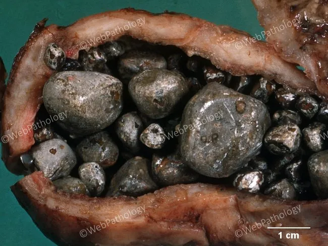
Gross pathology photograph of a cholecystectomy specimen showing chronic calculous cholecystitis. In this close-up, the gallbladder wall is visibly thickened and fibrotic, reflecting long-standing inflammation. The serosal surface is adherent in places, consistent with prior biliary inflammation and possible serosal adhesions. The lumen houses multiple intraluminal gallstones varying in size and calcific density. The stone-containing lumen contrasts with a dense, firm wall, and the mucosal lining is difficult to assess grossly due to distortion from fibrosis. Overall, the specimen demonstrates features of chronic lithiasic cholecystitis: persistent obstruction by gallstones leads to chronic inflammatory change, mural fibrosis, and sometimes loss of normal gallbladder pliability. The scale bar indicates 1 cm for size reference; stones are radiopaque on radiography and ultrasound correlate with echogenic foci and acoustic shadowing. Clinically, these findings explain recurrent biliary pain (biliary colic), potential episodes of acute inflammation, and surgical indication for cholecystectomy. Pathology correlation with microscopic histology would typically show dense fibrous wall, mononuclear infiltrates, and possible Rokitansky-Aschoff sinuses. This image is valuable for medical education, pathology training, and radiology-pathology correlation in gallbladder disease. The image therefore serves as a representative reference for teaching gross pathology features of chronic cholecystitis with calculi and facilitates recognition of similar patterns in biopsy specimens.
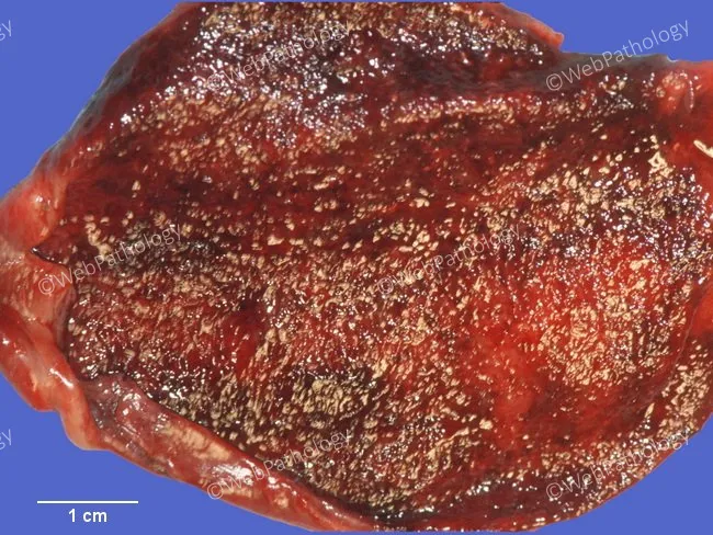
Gross pathology image of gallbladder from cholecystectomy demonstrating chronic acalculous cholecystitis. The gallbladder wall and mucosa are distended with pronounced congestion and patchy hemorrhages; surface shows a mottled reddish-brown to dark red appearance with areas of fibrous thickening. No radiopaque calculi are evident on this exterior view; the lumen mucosa is congested and irregular. Focal cholesterolosis is present as yellowish lipid-laden plaques on the mucosal surface. The image depicts a single external view of an intact gallbladder laid on a blue background with a centimeter scale bar for reference, scale approximately 1 cm. Notable features include chronic inflammatory thickening of gallbladder wall with mucosal congestion, submucosal hemorrhages, and cholesterolosis; absence of gallstones. These findings support a diagnosis of chronic acalculous cholecystitis with cholesterolosis. Clinically, this correlates with biliary colic or chronic right upper quadrant pain in the absence of cholelithiasis; histology would show mononuclear infiltration, fibrosis, and Rokitansky-Aschoff sinuses if examined microscopically. The image can be used for educational purposes in pathology, surgical education, and radiology correlation to illustrate gross appearance of acalculous cholecystitis and cholesterolosis. The image emphasizes mucosal changes rather than ductal stones and highlights the importance of gross assessment in forming preliminary diagnoses prior to histology. The specimen shows preserved bile ducts and no perforation.
Chronic cholecystitis
Definition and pathogenesis
- Association with stones: More than 90% of cases have cholelithiasis, so the at-risk population is the same as for gallstones.
- Role of stones: Stones may not directly start the inflammation. Chronic acalculous cholecystitis has similar symptoms and histology. Supersaturated bile seems to predispose to both chronic inflammation and stone formation.
- Bacteria: Organisms, usually E. coli and enterococci, are cultured from bile in about one-third of cases.
- Obstruction: Unlike acute calculous cholecystitis, outflow obstruction is not required. - Robbins, Cotran & Kumar Pathologic Basis of Disease, p. 812-813
- Mechanism with recurrent colic: Repeated, temporary cystic duct occlusion causes self-limited inflammation. This scars the gallbladder neck and cystic duct. Chronic cholecystitis therefore lies on a continuum with biliary colic. - Sabiston Textbook of Surgery, p. 1829
Pathology
- The wall is variably thickened and gray-white.
- The serosa may be dulled by subserosal fibrosis, with dense adhesions from prior acute inflammation.
- The lumen usually holds stones and green-yellow mucoid bile.
- The mucosa is generally preserved.
- In mild cases there are scattered lymphocytes, plasma cells and macrophages.
- In advanced cases there is subepithelial and subserosal fibrosis with mononuclear infiltrate.
- Rokitansky-Aschoff sinuses (mucosal outpouchings through the wall) may be prominent. - Robbins, Cotran & Kumar Pathologic Basis of Disease, p. 813
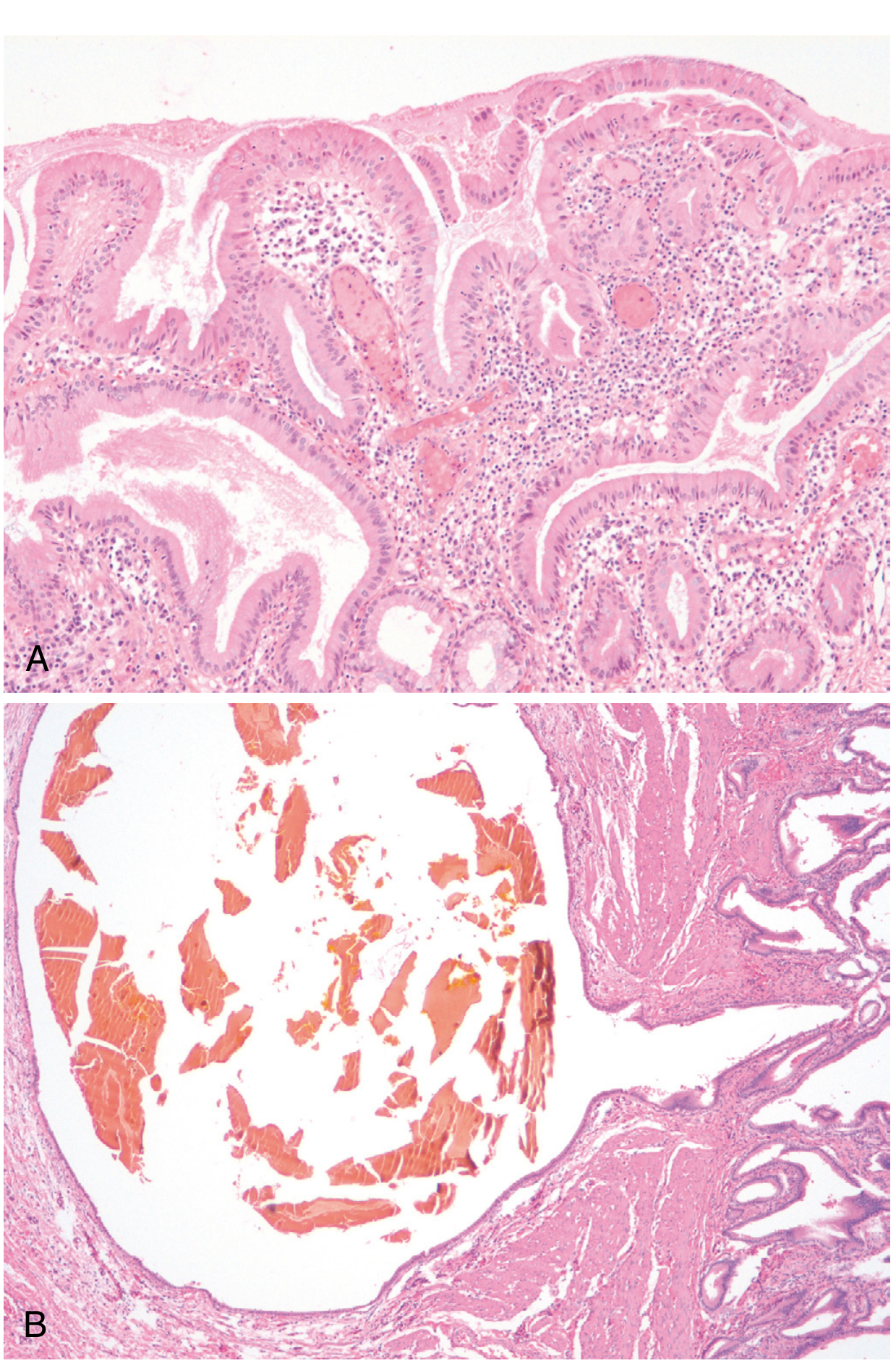
- Porcelain gallbladder: Extensive dystrophic wall calcification.
- Hyalinizing cholecystitis: The wall and mucosa are replaced by dense fibrosis, with or without calcification. It is associated with a higher incidence of gallbladder carcinoma.
- Xanthogranulomatous cholecystitis: The wall is massively thickened and nodular. It follows rupture of Rokitansky-Aschoff sinuses, with foamy xanthoma cells. It can mimic carcinoma.
- Hydrops: An atrophic, obstructed gallbladder containing only clear secretions.
- IgG4-related sclerosing cholecystitis: Rare.
Clinical features
- Recurrent steady epigastric or right upper quadrant pain, which may radiate to the scapula. Attacks last a few hours.
- Pain often follows a fatty meal (CCK release), although only about 50% of patients link it to food.
- Nausea, vomiting, bloating and fatty-food intolerance are common.
- Pain lasting more than 24 hours or with fever suggests acute cholecystitis.
- Murphy's sign is absent in simple biliary colic.
Diagnosis
- Clinical history of biliary-type pain.
- Transabdominal ultrasound is the first test. It shows stones, sludge or microlithiasis, polyps, porcelain gallbladder and CBD dilation. Wall thickening can also be seen.
- LFTs are usually normal, unlike in acute cholecystitis or choledocholithiasis.
- In acalculous disease with typical symptoms, HIDA scan with CCK (low ejection fraction) is used.
- Differential diagnosis includes peptic ulcer, gastritis, GERD, IBS and pancreatitis. - Sabiston Textbook of Surgery, p. 1830
Management
- Symptomatic disease: Elective laparoscopic cholecystectomy. It cures more than 90% of patients.
- Mild symptoms: Complications occur in about 1-3% per year. Observation with dietary and lifestyle changes is reasonable, especially if the patient declines surgery.
- Severe or recurrent symptoms: Complications occur in about 7% per year, so surgery is encouraged.
- Surgery on a diseased gallbladder: Fibrosis and adhesions raise the risk of difficult dissection. Use the critical view of safety to avoid bile duct injury.
- Prophylactic cholecystectomy: Consider it for porcelain gallbladder, because of carcinoma risk. - Sabiston Textbook of Surgery, p. 1830
Complications
- Acute-on-chronic cholecystitis
- Choledocholithiasis, cholangitis and gallstone pancreatitis
- Perforation, abscess, or rupture with peritonitis
- Mirizzi syndrome and cholecystoenteric fistula
- Gallbladder carcinoma, especially with porcelain gallbladder or hyalinizing cholecystitis
Recent evidence check
Chronic colecystitis acalculous
"acalculous cholecystitis" OR "biliary dyskinesia" cholecystectomy
Chronic acalculous cholecystitis (chronic acalculous gallbladder disease)
Definition and frequency
- It accounts for about 10% of patients with symptoms and histology of chronic cholecystitis.
- It is clinically and histologically indistinguishable from the calculous form, except that no stones are present.
- Symptoms usually resolve after cholecystectomy. The clinical challenge is to make the diagnosis before surgery, without invasive tests. - Yamada's Textbook of Gastroenterology, 7th ed, p. 2803
Pathogenesis
- Impaired gallbladder motility, with reduced CCK response or sphincter of Oddi dysfunction.
- Cystic duct dysfunction or narrowing.
- Supersaturated bile, microlithiasis or sludge missed on ultrasound.
- Chronic inflammation and fibrosis. Robbins notes that acalculous disease has histology similar to calculous disease, and that bile supersaturation predisposes to both. - Robbins, Cotran & Kumar Pathologic Basis of Disease, p. 812-813
Clinical presentation
- Recurrent biliary-type pain in the epigastrium or right upper quadrant, sometimes radiating to the scapula. Attacks last hours.
- Postprandial or fatty-food pain, with nausea, vomiting and bloating.
- Typically more often in young to middle-aged women, but it also occurs in children and adolescents.
- The physical exam and routine labs are usually normal between attacks.
Diagnosis
- Ultrasound shows no stones, sludge or microlithiasis.
- Exclude other causes:
- Upper endoscopy for peptic ulcer, gastritis and GERD.
- LFTs and lipase.
- Imaging for pancreatic, hepatic and CBD pathology.
- Consider sphincter of Oddi dysfunction, IBS, functional dyspepsia, and cardiac or abdominal wall causes.
- CCK cholescintigraphy (HIDA with sincalide) is the key test.
- A low gallbladder ejection fraction (GBEF) is diagnostic and predicts response to cholecystectomy.
- More than two dozen studies support this. In one randomized prospective study, over 91% of patients with a low GBEF who had surgery had resolution of symptoms and histologic chronic cholecystitis. Those with a low GBEF who were not operated on remained symptomatic. - Yamada's, p. 2803-2804
- The 60-minute sincalide infusion (0.02 μg/kg) has the least variability. The lower limit of normal is 38%. Three-minute infusions are highly variable and have a high false-positive rate. - Yamada's, p. 2804
- Cutoffs differ by source: below 35% (Grainger & Allison's), 35-40% (Fischer's Mastery of Surgery), and below 40% after 30 minutes of CCK (Maingot's).
- Poor contraction occurs in both chronic and acute disease. Good contraction rules out gallbladder disease. - Yamada's, p. 2803
Pathology
- Chronic inflammation, subepithelial and subserosal fibrosis, and Rokitansky-Aschoff sinuses. Some specimens also show cholesterolosis.
- Histology is the same as in calculous chronic cholecystitis.
Management
- Laparoscopic cholecystectomy is the treatment for patients with typical symptoms, a negative workup and a low GBEF. It is curative in the majority.
- Maingot's lists these criteria for surgery in biliary dyskinesia: typical biliary symptoms, EF below 40% on HIDA after 30 minutes of continuous CCK, and no pain for 12 months after cholecystectomy as the definition of a positive response. - Maingot's Abdominal Operations, Table 48-1
- If GBEF is normal and the workup is negative, consider a functional disorder and avoid surgery. Outcomes are less predictable.
- Children: There is no consensus on diagnostic criteria, and symptoms overlap with functional dyspepsia. Gallbladder dyskinesia was the most common indication for cholecystectomy in 58% of one pediatric cohort. - Sleisenger and Fordtran's GI and Liver Disease
Recent evidence (PubMed, last 3 years)
- Hyperkinetic biliary dyskinesia on HIDA: systematic review and meta-analysis, 2026 (PMID 42912575).
- Biliary dyskinesia in children: meta-analysis of 3,348 patients, 2026 (PMID 41308824).
- APSA pediatric evaluation and management: systematic review and practice guideline, 2024 (PMID 39227244).