
Is there any mechanism of hutinchon tooth and mulberry molar in syphilis and crireria
Reading File
Finding Sources
Finding Sources
Searching the Web
Hutchinson teeth mulberry molar syphilis mechanism pathogenesis criteria
Reading File
Reading File
Reading File
Reading Web Page
https://pmc.ncbi.nlm.nih.gov/articles/PMC4972761
Loading Image
Loading Image
Loading Image
Finding Sources
Finding Sources
Loading Image
I now have comprehensive information from multiple authoritative sources. Here is the complete answer:
Hutchinson's Teeth & Mulberry Molars in Congenital Syphilis
Background: Congenital Syphilis & Dental Involvement
These two dental signs are stigmata (permanent malformations) of late congenital syphilis - Treponema pallidum transmitted transplacentally from mother to fetus. They belong to a group of irreversible developmental defects caused during critical growth periods and persist throughout life, confirming the diagnosis.
Pathogenic Mechanism (Common to Both)
The core mechanism is syphilitic vasculitis affecting developing tooth buds in utero or in early infancy.
Step-by-step:
- Spirochetemia - T. pallidum enters fetal circulation transplacentally, causing widespread dissemination
- Vasculitis of the vasa vasorum - Treponemes trigger an obliterative endarteritis and periarteritis with a characteristic lymphoplasmacytic infiltrate surrounding small vessels
- Ischemic damage to the ameloblasts (enamel-forming cells) within developing tooth buds - because the blood supply is compromised during the critical window of tooth morphogenesis and enamel matrix deposition
- Enamel hypoplasia - the enamel covering (molar sheath) becomes thin and deformed; the underlying dentin and pulp remain structurally normal
- Permanent structural deformity - since enamel is formed only once and cannot regenerate, the damage is irreversible
"Peg-shaped notched central incisors (Hutchinson teeth) and mulberry molars, resulting from syphilis vasculitis in developing tooth buds." - Fitzpatrick's Dermatology
"Hutchinson teeth are small incisors shaped like a screwdriver or a peg, often with notches in the enamel." - Robbins & Cotran Pathologic Basis of Disease
Hutchinson's Teeth (Syphilitic Incisors)
Tooth affected: Permanent upper central incisors (secondary dentition - not the primary/deciduous teeth)
Timing: Become apparent when permanent teeth erupt (~age 6-8 years)
Morphology:
- The tooth is cylindrical (barrel-shaped) rather than the normal flat, fan shape
- The cutting (incisal) edge is narrower than the base - giving a peg or screwdriver shape
- A central notch may be present in the incisal edge (due to failure of the median tubercle to develop)
- Teeth may be widely spaced
- The enamel surface may appear pitted or hypoplastic
- Only the central upper incisors are affected (not all teeth)
Clinical photo (Andrews' Diseases of the Skin):
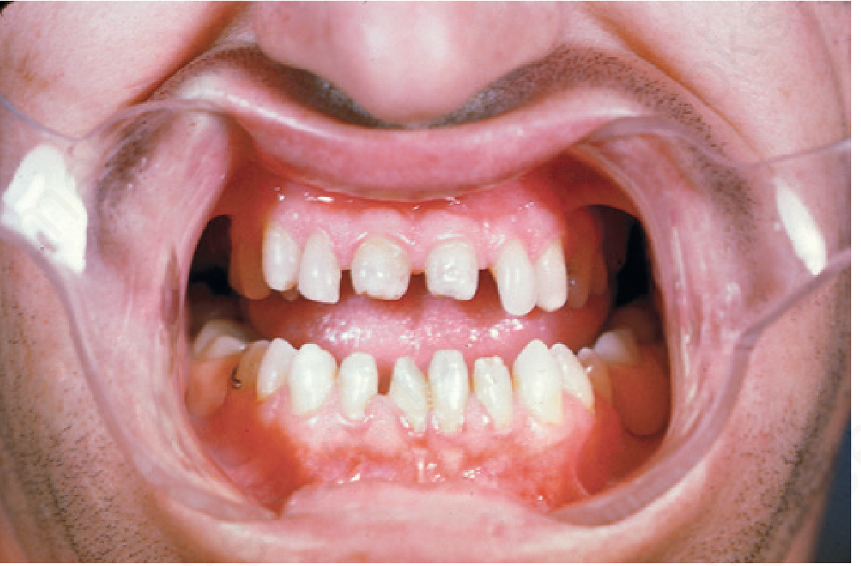
Hutchinson's teeth in congenital syphilis (Andrews' Diseases of the Skin)
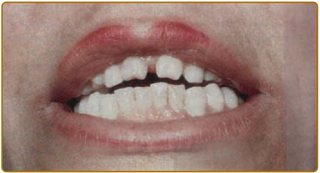
Close-up showing notched, peg-shaped central incisors - Fig. 170-36 from Fitzpatrick's Dermatology
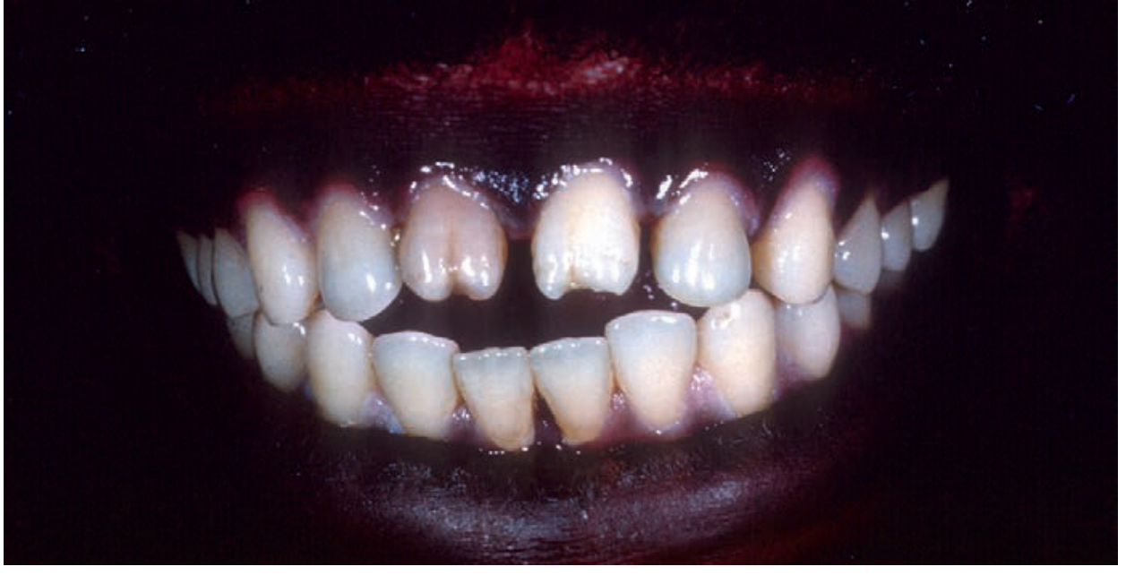
Another example of Hutchinson's incisors - congenital syphilis (Andrews' Clinical Atlas)
Mulberry Molars (Fournier/Moon Molars)
Tooth affected: Usually the permanent first molar (also known as the 6-year molar)
Timing: Erupts around age 6 years
Morphology:
- The mulberry molar is a hyperplastic tooth (excessive but abnormal tissue formation)
- Its occlusal (biting) surface is flat with multiple small rounded knobs - representing abortive (failed) cusps
- The appearance resembles the surface of a mulberry fruit - hence the name
- Just above the gum line the tooth looks near-normal; the deformity is only apparent at the cusp/occlusal surface
- The tooth is diminished in size in all dimensions ("stumpy")
- Enamel covering is thin and deformed; underlying dentin and pulp are normal
"The mulberry molar, usually the first molar and appearing about age 6 years, is a hyperplastic tooth; its flat occlusal surface is covered with a group of little knobs representing abortive cusps." - Andrews' Diseases of the Skin
"Congenital syphilis produces tapering, notched (Hutchinson's) incisors and finely nodular (mulberry) molar crowns." - Harrison's Principles of Internal Medicine 22E
Comparison Table
| Feature | Hutchinson's Teeth | Mulberry Molar |
|---|---|---|
| Tooth affected | Permanent upper central incisors | Permanent first molars |
| Age of appearance | ~6-8 years | ~6 years |
| Shape | Peg/screwdriver-shaped, narrow incisal edge | Flat top, multiple small rounded cusps |
| Surface | Notch in centre of incisal edge | Knob-like abortive cusps (mulberry-like) |
| Type of change | Hypoplasia (reduced/defective) | Hyperplastic but deformed |
| Enamel | Thin, hypoplastic | Thin and deformed |
| Dentin/Pulp | Normal | Normal |
| Mechanism | Vasculitis - ischemic damage to ameloblasts | Same - vasculitis of tooth bud |
Hutchinson's Triad (Classic Diagnostic Criteria)
Sir Jonathan Hutchinson originally described three findings that together are pathognomonic for late congenital syphilis:
| Component | Description |
|---|---|
| 1. Hutchinson's teeth | Notched, peg-shaped upper permanent central incisors |
| 2. Interstitial keratitis | Diffuse corneal clouding without ulceration; pericorneal inflammation; may cause partial/complete corneal opacity; occurs in 20-50% of cases |
| 3. Eighth nerve deafness | Sensorineural hearing loss from meningovascular syphilis affecting CN VIII |
"Hutchinson emphasized the diagnostic importance of changes in the incisor teeth, opacities of the cornea, and eighth cranial nerve deafness, which have since become known as the Hutchinson triad." - Andrews' Diseases of the Skin
Note: Optic nerve atrophy can also occur secondary to meningovascular syphilis. Mulberry molars are an additional stigma but are not part of the classic triad.
Other Stigmata of Late Congenital Syphilis (Context)
These accompany Hutchinson's teeth and mulberry molars as persistent markers:
- Saddle nose - nasal cartilage destruction
- Saber shins - anterior tibial bowing from periostitis
- Frontal bossing (Olympian brow)
- Rhagades - perioral linear scars from early papular skin lesions
- Saddle nose (septal destruction)
- Higoumenakia sign - unilateral thickening of the inner clavicle
- Clutton joints - symmetric painless perisynovitis of knees
Sources: Robbins & Cotran Pathologic Basis of Disease; Fitzpatrick's Dermatology Vols. 1-2; Andrews' Diseases of the Skin; Harrison's Principles of Internal Medicine 22E; Andrews' Clinical Atlas 2e
This is a shared conversation. Sign in to Orris to start your own chat.