
Gall bladder stone
gallbladder stones cholelithiasis ultrasound types
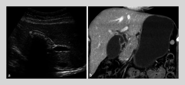
A side-by-side comparison of diagnostic imaging modalities for cholelithiasis. Panel (a) is a transabdominal ultrasonography of the right upper quadrant. It reveals multiple hyperechoic, irregular structures within the gallbladder lumen. These structures exhibit classic posterior acoustic shadowing, where ultrasound waves are reflected or absorbed, resulting in a dark, anechoic void distal to the stones. Panel (b) is a contrast-enhanced computed tomography (CT) scan in the axial or coronal plane of the same patient. The CT shows the gallbladder as a well-defined, hypoattenuating, fluid-filled structure located adjacent to the liver. In contrast to the ultrasound, the CT scan demonstrates no high-density (calcified) structures within the gallbladder lumen, illustrating the higher sensitivity of ultrasonography for detecting certain types of biliary stones compared to standard CT. The image serves as an educational comparison of imaging sensitivity in gastroenterology and radiology.
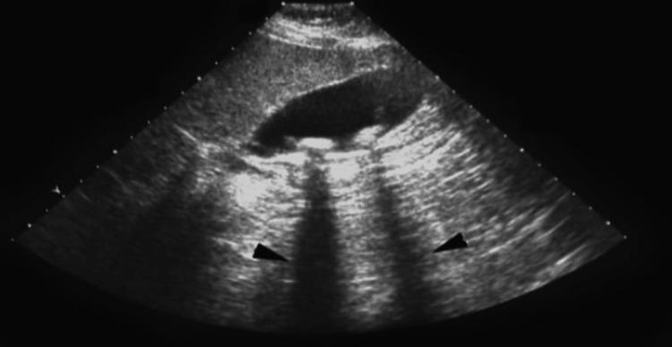
This abdominal ultrasound image displays a diagnostic view of the gallbladder containing cholelithiasis (gallstones). The gallbladder appears as a prominent, elongated, anechoic (black) sac-like structure. Inside the lumen, multiple hyperechoic (bright) foci are visible along the dependent wall, representing gallstones. Crucially, the image demonstrates the classic diagnostic sign of posterior acoustic shadowing, where dark, vertical hypoechoic bands extend inferiorly from the stones, indicated by black arrowheads. This phenomenon occurs as the dense stones attenuate the ultrasound beam, preventing sound waves from reaching deeper tissues. The surrounding liver parenchyma shows a normal, homogeneous intermediate echogenicity. This visual is typical for diagnosing symptomatic or asymptomatic gallstone disease in a clinical or preoperative setting.
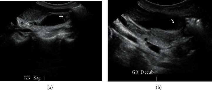
Diagnostic ultrasound images of the gallbladder in two planes: (a) sagittal view and (b) decubitus view. Both images reveal cholelithiasis, characterized by the presence of hyperechoic (bright) foci within the anechoic (fluid-filled) lumen of the gallbladder. White arrows in both frames highlight specific gallstones. The gallbladder wall appears thin and without significant distension, indicating cholelithiasis without acute cholecystitis. The liver parenchyma is visible adjacent to the gallbladder as a medium-echogenicity structure. These views are utilized in clinical practice to confirm the presence and mobility of stones, as the decubitus position helps redistribute luminal contents to differentiate mobile stones from stationary polyps. The image serves as an educational example of biliary imaging in the context of suspected gallstone pancreatitis or symptomatic cholelithiasis.
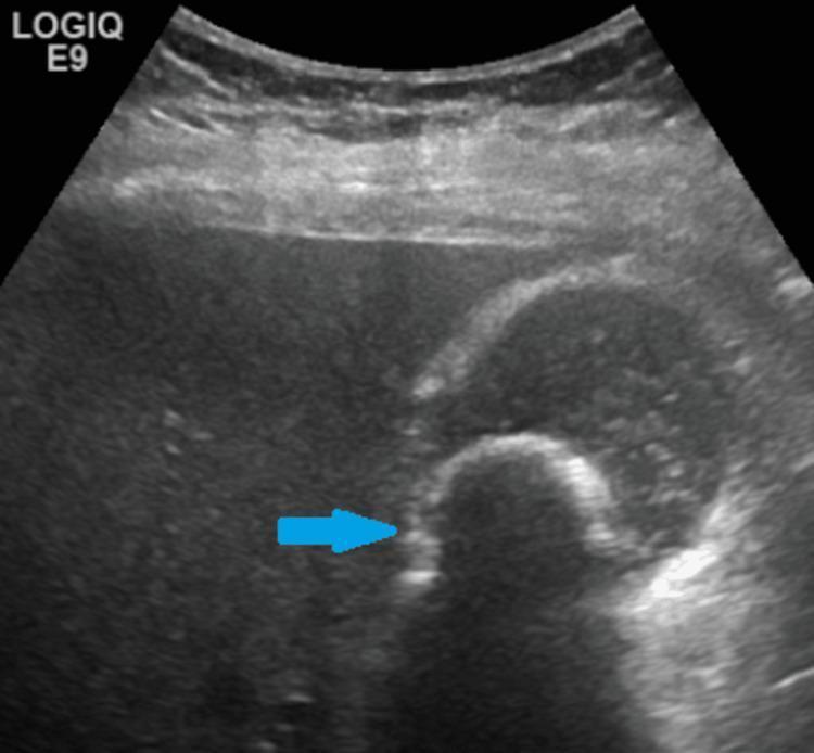
This abdominal ultrasound image displays a diagnostic view of the gallbladder containing a prominent 2.7-cm gallstone (cholelithiasis), indicated by a blue arrow. The gallstone appears as a curvilinear, highly hyperechoic structure within the gallbladder lumen. Characteristically, there is a clear, well-defined area of posterior acoustic shadowing distal to the calculus, resulting from the attenuation of ultrasound waves by the dense calcified object. The gallbladder itself appears as a sac-like structure with primarily anechoic (fluid-filled) contents, although scattered low-level internal echoes are present, suggesting the presence of biliary sludge. The surrounding liver parenchyma shows a uniform echotexture. This image is a classic representation used in radiology and gastroenterology for teaching the sonographic diagnosis of uncomplicated cholelithiasis, emphasizing the 'bright' appearance of stones and the diagnostic importance of acoustic shadowing.
"cholelithiasis"[MeSH Terms] AND "cholecystectomy"[MeSH Terms]
Gallbladder Stones (Cholelithiasis)
Epidemiology
- Robbins & Kumar Basic Pathology, p. 630
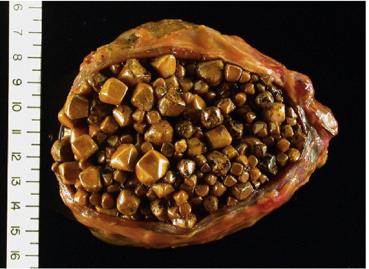
Types of Gallstones
1. Cholesterol Stones (80% in Western populations)
- Composed of crystalline cholesterol monohydrate
- Pure cholesterol stones are pale yellow and radiolucent
- Mixed stones (with calcium carbonate, phosphates, bilirubin) appear gray-white to black
- Usually ovoid and firm, often multiple with faceted surfaces
- As many as 20% may be radiopaque due to calcium carbonate content
- Arise exclusively in the gallbladder
2. Pigment Stones (predominant in non-Western populations)
- Composed of insoluble calcium bilirubinate salts
- Two subtypes:
- Black pigment stones: Small, numerous, found in sterile gallbladder bile; 50-75% radiopaque; associated with chronic hemolysis (sickle cell disease, hereditary spherocytosis)
- Brown pigment stones: Single or few, soft greasy/soaplike consistency; radiolucent; found in infected intra-hepatic or extrahepatic bile ducts
Risk Factors
| Cholesterol Stones | Pigment Stones |
|---|---|
| Advancing age | Chronic hemolysis (sickle cell anemia, hereditary spherocytosis) |
| Female sex (2x risk vs. males) | Biliary infection / liver flukes |
| Oral contraceptives | Ileal disease (Crohn disease), ileal resection, cystic fibrosis |
| Pregnancy | Cirrhosis |
| Obesity and insulin resistance | |
| Rapid weight reduction | |
| Gallbladder stasis | |
| Dyslipidemia | |
| Inborn disorders of bile acid metabolism |
Pathogenesis
Cholesterol Stones
- Gallbladder hypomotility (stasis) - promotes crystal nucleation
- Mucus hypersecretion - traps crystals and enhances aggregation into stones
Pigment Stones
- Chronic red cell hemolysis
- Biliary tract infections (bacteria deconjugate bilirubin)
- Cirrhosis (reduced bile salt synthesis)
- Crohn disease (altered enterohepatic cycling of bilirubin)
Clinical Features
- Biliary colic: Right upper quadrant (RUQ) or epigastric pain, often severe, constant or spasmodic. Classically precipitated by a fatty meal, which stimulates gallbladder contraction, pressing a stone against the outlet.
- Murphy's sign: Arrest of inspiration on palpation of the RUQ (seen in acute cholecystitis)
- Jaundice: When stones pass into the common bile duct (choledocholithiasis)
- Nausea, vomiting, bloating
Complications
| Complication | Mechanism |
|---|---|
| Acute calculous cholecystitis | Stone obstructs cystic duct (90% of cases); most common complication |
| Choledocholithiasis | Stone migrates to common bile duct |
| Obstructive jaundice / cholangitis | CBD obstruction + infection |
| Gallstone pancreatitis | Stone impacted at ampulla of Vater |
| Empyema | Pus-filled gallbladder |
| Gallbladder perforation | With peritonitis |
| Gallstone ileus | Rare; large stone erodes into small bowel causing intestinal obstruction |
| Gallbladder carcinoma | Long-term risk; gallstones are an important risk factor |
- Robbins & Kumar Basic Pathology, p. 631-632
Diagnosis
Imaging
| Modality | Details |
|---|---|
| Ultrasound (first-line) | Most sensitive and specific. Shows hyperechoic foci with posterior acoustic shadowing. Stones move with patient position. |
| CT scan | Good for complications; less sensitive for non-calcified stones (many are isodense with bile) |
| MRCP | Best for CBD stones and biliary tree evaluation |
| HIDA scan | Evaluates gallbladder function; failure to visualize = acute cholecystitis |
| Plain X-ray | Only ~15-20% of cholesterol stones are visible; useful for porcelain gallbladder |
| ERCP | Invasive; used therapeutically for CBD stones |
Management
Asymptomatic Gallstones ("Silent Stones")
- Generally watchful waiting - the annual risk of developing symptoms is only ~1-2%
- Exceptions where prophylactic cholecystectomy is considered:
- Porcelain gallbladder (risk of carcinoma - though this is debated)
- Very large stones (>3 cm)
- Patients with chronic hemolytic anemias about to undergo another surgery
- Immunocompromised patients
Symptomatic Gallstones
- Laparoscopic cholecystectomy is the definitive treatment - gold standard
- Advantages: shorter hospital stay, faster recovery, fewer complications vs. open surgery
- Most common non-obstetric surgical procedure
- Open cholecystectomy: Reserved for complex cases, failed laparoscopy
Special Scenarios
- Choledocholithiasis (CBD stones):
- Pre-operative or post-operative ERCP + sphincterotomy + stone extraction, followed by laparoscopic cholecystectomy
- A recent meta-analysis (Zhang et al., 2024) found one-stage intraoperative ERCP combined with laparoscopic cholecystectomy is comparable to the two-stage approach
- Pregnancy: Laparoscopic cholecystectomy preferred in the first and second trimester for symptomatic disease; delays increase recurrence risk (92% in first trimester, 64% in second trimester) - Sabiston Textbook of Surgery
- Gallstone ileus: Emergency surgery (enterolithotomy) +/- cholecystectomy
Non-Surgical Options (limited role)
- Ursodeoxycholic acid (UDCA): Dissolves small cholesterol stones in selected patients, but recurrence is high after stopping
- Extracorporeal shock wave lithotripsy (ESWL): Rarely used; reserved for poor surgical candidates
Gallstone Disease and Cancer Risk
Summary Table
| Feature | Cholesterol Stone | Black Pigment Stone | Brown Pigment Stone |
|---|---|---|---|
| Location | Gallbladder only | Gallbladder | Bile ducts |
| Color | Pale yellow to brown | Black | Brown/tan |
| Consistency | Hard | Hard, brittle | Soft, greasy |
| Radiopacity | Usually radiolucent (80%) | Radiopaque (50-75%) | Radiolucent |
| Number | Usually multiple | Multiple, small | Single or few |
| Key association | Obesity, female, OCP, pregnancy | Hemolysis, cirrhosis | Biliary infection |
All about gall bladder
gallbladder anatomy diagram biliary tract
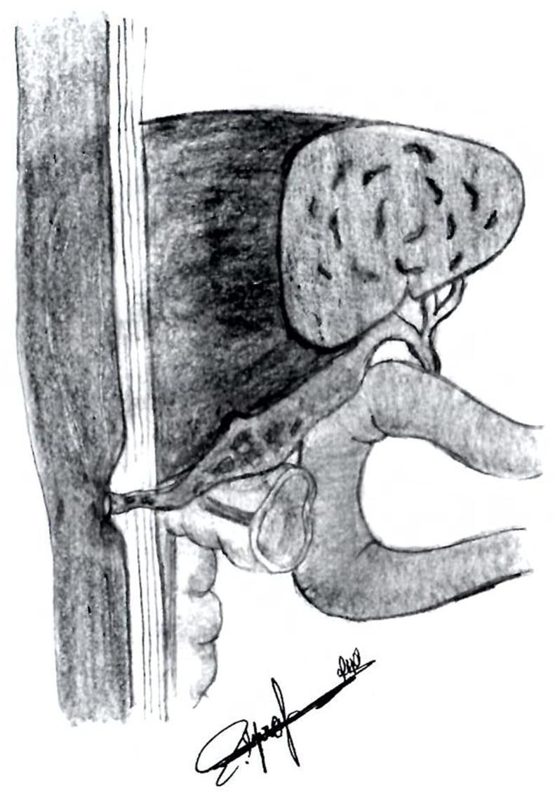
This medical illustration depicts a cholecystocutaneous fistula, a rare type of external biliary fistula. The grayscale diagram shows the anatomical relationship between the gallbladder, the biliary system, and the abdominal wall. The gallbladder is rendered with a textured, stippled surface, indicating potential inflammation or cholelithiasis, and is situated inferior to the liver. A pathological tubular tract (the fistula) is shown originating from the fundus or body of the gallbladder, extending anteriorly to penetrate through the muscular layers of the abdominal wall and the subcutaneous tissue, ultimately opening onto the skin surface. Surrounding structures, including the liver and segments of the intestine, provide anatomical context for the pathway of the tract. The illustration is designed to teach the pathophysiology of spontaneous or post-inflammatory biliary drainage to the exterior environment, emphasizing the abnormal connection between an internal organ and the cutaneous surface.
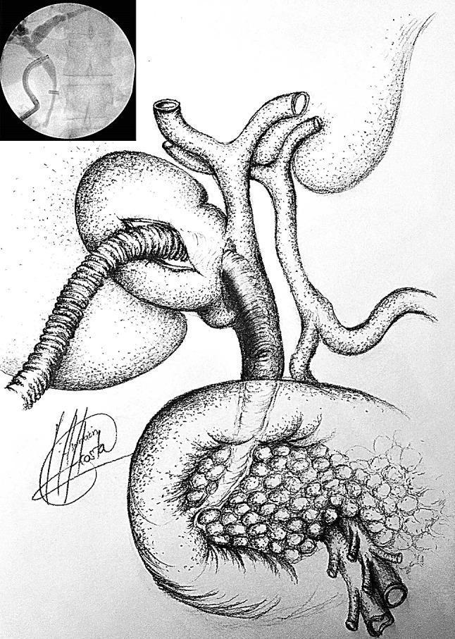
This composite educational image illustrates the surgical management of complex biliary disease, specifically Mirizzi syndrome with a cholecystobiliary fistula. The primary anatomical diagram depicts a gallbladder densely packed with multiple small, uniform stones (cholelithiasis). The biliary tree is shown with the right and left hepatic ducts joining into the common hepatic duct, which then becomes the common bile duct (CBD). A critical feature illustrated is a cholecystobiliary fistula—an abnormal connection between the gallbladder infundibulum and the CBD. A flexible medical device, representing a choledochoscope or cholangiocatheter, is shown entering through the gallbladder infundibulum and traversing the fistula to access the CBD for stone clearance. The upper-left inset contains a diagnostic fluoroscopic image from an intraoperative cholangiogram (IOC). This radiograph confirms the biliary anatomy, showing a radiopaque cholangiocatheter with a curved configuration positioned within the ductal system to assess patency and locate impacted stones. The image serves as a pedagogical tool for understanding the transinfundibular approach in laparoscopic biliary surgery when hilar inflammation is severe.
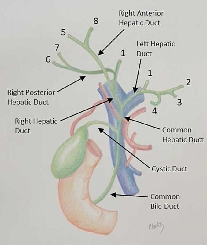
An anatomical diagram illustrating the human biliary tree and its relationship to adjacent hepatobiliary structures. The diagram depicts the biliary drainage system (colored green) beginning with the common bile duct, which receives the cystic duct from the gallbladder. Superior to this confluence is the common hepatic duct, which bifurcates into the right and left hepatic ducts. The right hepatic duct further branches into the right anterior (supplying segments 5 and 8) and right posterior (supplying segments 6 and 7) hepatic ducts. The left hepatic duct provides drainage for segments 1, 2, 3, and 4. Vascular landmarks are also shown: the portal vein (colored blue) is situated posterior to the common bile duct, and the right hepatic artery (colored red) is seen crossing posterior to the common hepatic duct. The gallbladder is positioned alongside the duodenum, with its cystic duct connecting to the main biliary channel. This diagram serves as a pedagogical tool for understanding segmental liver anatomy and surgical landmarks in the porta hepatis.
acute cholecystitis gallbladder histology
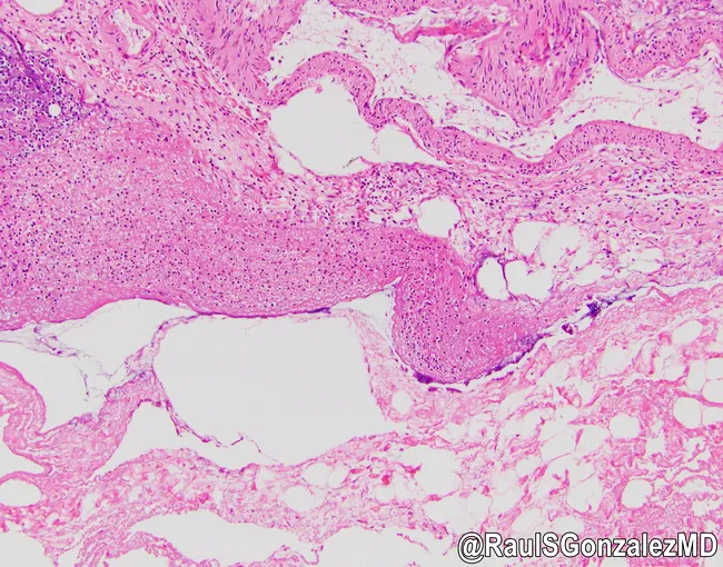
Histology of gallbladder wall illustrating acute emphysematous cholecystitis. The specimen is examined as a cross-sectional histologic slide prepared with Hematoxylin and Eosin stain and observed under light microscopy. The gallbladder wall shows transmural acute inflammation with dense neutrophilic infiltrate, mucosal ulceration, edema, and necrosis extending through the mucosa, muscularis, and serosa. Notably, intramural gas pockets create empty spaces within the wall, producing emphysematous changes. The overall architecture is disrupted by necrosis and inflammatory edema; vascular congestion and focal hemorrhage may be present. The image demonstrates gas-forming phenomenon within the gallbladder wall consistent with emphysematous cholecystitis, a surgical emergency with risk of gallbladder perforation, peritonitis, and sepsis. Clinically, this entity is associated with diabetic or elderly patients and correlates with rapid progression and high mortality if untreated. The histopathology complements radiologic findings of gas within the gallbladder and pericholecystic tissues. The data is relevant for education, pathology teaching, differential diagnosis between emphysematous and gangrenous cholecystitis, and research on gas-forming infections of the biliary tract. This representation aids learners in recognizing the hallmark correlate between microscopic gas spaces and tissue necrosis, guiding prompt surgical consultation, antibiotic therapy, and postoperative care. It also supports comparative studies of biliary tract infections in diabetic populations.
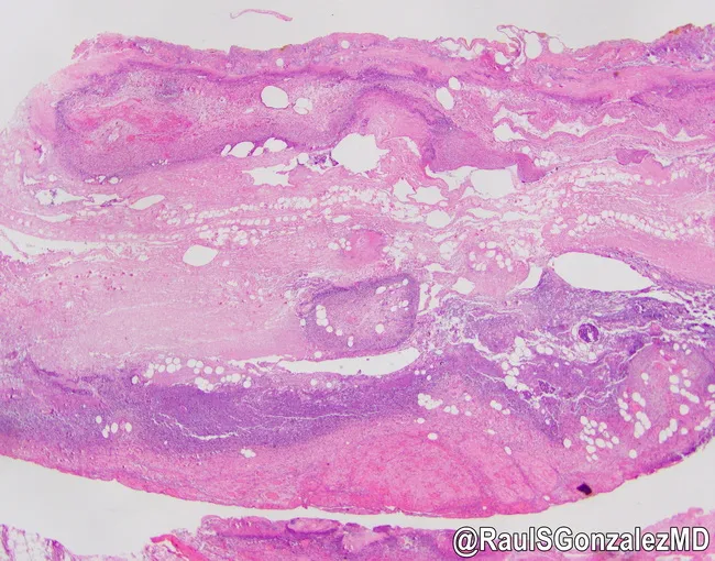
This image depicts a histopathologic section of the gallbladder wall stained with Hematoxylin and Eosin, viewed under brightfield light microscopy at low to moderate magnification. The specimen demonstrates acute emphysematous cholecystitis characterized by transmural inflammation with necrosis of the gallbladder wall. The mucosa is disrupted, and the muscularis shows dense neutrophilic infiltration and edema. Notably, numerous gas-filled spaces occupy the gallbladder wall and subserosal regions, producing empty-appearing lacunae that reflect intramural gas bubbles produced by gas-forming organisms. The surrounding tissue exhibits acute inflammatory exudate and focal necrosis, with hemorrhagic/serosanguinous components in the serosal surface. These features distinguish emphysematous cholecystitis from simple acute calculous cholecystitis and indicate a more fulminant infectious process. Clinically, this pathology corresponds to a high-risk surgical emergency with potential rapid progression to perforation and peritonitis if not promptly treated. Correlates with diabetes mellitus risk, elderly male patients, and poor tissue perfusion. The image is provided courtesy of Dr. Raul Gonzalez, with permission. Overall, the slide highlights the destructive inflammatory response, gas production within the gallbladder wall, and the necessity for urgent diagnosis and management in suspected cases of emphysematous cholecystitis. This histology correlates with radiologic gas patterns and supports urgent cholecystectomy planning in rapidly progressive cases.
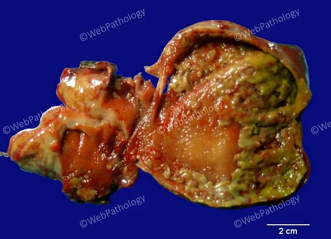
Gross pathology of a gallbladder with acute calculous cholecystitis. The specimen shows a distended, inflamed gallbladder wall with mucosal edema and erythema. The mucosa is congested and irregular, with focal necrosis and yellow-green exudate admixed with fibrinous material. Patchy greenish-yellow pseudomembranes overlying necrotic mucosa are evident, consistent with pseudomembranous cholecystitis. The serosa may be variably irritated, and adherent inflammatory adhesions can be seen along the gallbladder bed. The exterior surface demonstrates hyperemia; the wall may be thickened due to edema. A portion of the lumen is exposed, showing necrotic debris and exudate filling the lumen interface. The 2 cm scale bar provides context for dimensions; overall changes correlate with acute inflammatory insult, often precipitated by gallstone obstruction of the cystic duct. The appearance aligns with fulminant cholecystitis and may precede gangrenous transformation if infection progresses. These macroscopic findings are supported by typical histology of mucosal necrosis, neutrophilic infiltrate, edema, and fibrinous exudate on the surface. This image highlights hallmark features of pseudomembranous cholecystitis with inflammatory exudate forming pseudomembranes, mucosal ulceration, and mural necrosis within the gallbladder wall. Correlation with clinical history of gallstones, fever, RUQ pain, leukocytosis, and imaging findings reinforces diagnosis and guides emergent management for surgical intervention planning.
Make a surgical reference card for cholecystectomy indications and complications
Create a surgical reference PDF for cholecystectomy