
2 episodes of haemetemeis
Reading File
Finding Sources
Finding Sources
Searching PubMed
"upper gastrointestinal bleeding" AND management
Reading File
Reading File
Reading File
Reading File
Reading File
Loading Image
Finding Sources
Finding Sources
Reading File
Reading File
Here is a comprehensive clinical overview of haematemesis with two episodes:
Haematemesis - Clinical Overview
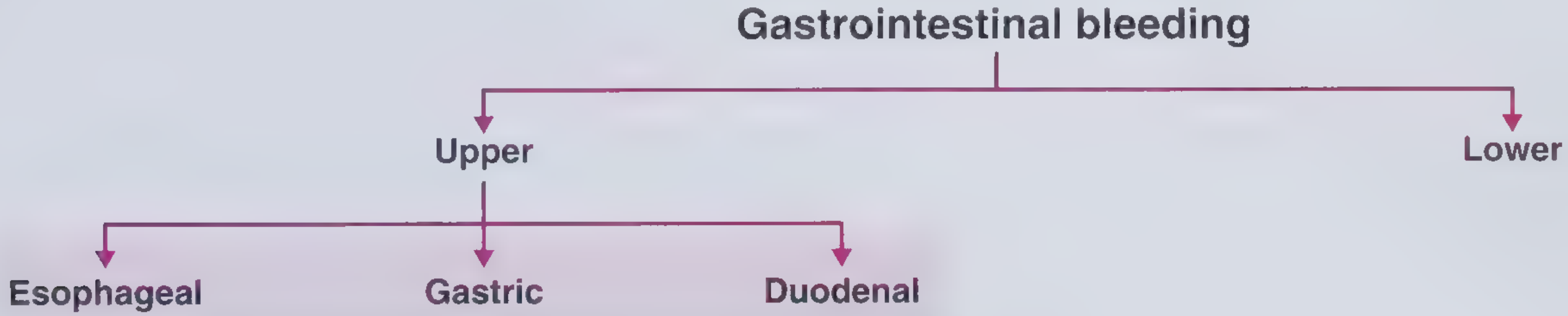
Haematemesis is the vomiting of blood, indicating an upper gastrointestinal (UGI) haemorrhage - i.e., a source proximal to the ligament of Treitz. Two episodes indicate active or recurrent bleeding requiring urgent evaluation.
Epidemiology
- Incidence: over 100 per 100,000/year in Western populations; increases with age
- In-hospital mortality: 5-10% overall; rises to 33% when bleeding is first observed in already-hospitalised patients
- Bleeding is self-limited in ~80% of patients; in the remaining 20% who continue to bleed or rebleed, mortality rises to 30-40%
- Strong association with NSAID use
Causes of Upper GI Bleeding
| Condition | Frequency (%) |
|---|---|
| Peptic ulcers (total) | ~40-60% |
| - Duodenal ulcer | 33 |
| - Gastric ulcer | 21 |
| - Oesophageal ulcer | 6 |
| Erosions (oesophageal, gastric, duodenal) | 26 |
| Oesophageal varices | 4-22% |
| Mallory-Weiss tear | 4 |
| Dieulafoy lesion | 3 |
| Tumour / Neoplasm | ~3 |
| Vascular lesions / Angioectasia | ~4 |
| No cause found | ~7 |
Bailey & Love's Short Practice of Surgery, p.1187; Sleisenger & Fordtran's GI and Liver Disease
Initial Assessment and Resuscitation
ABC approach first:
- IV access - large-bore x2; for severe bleeding add central venous pressure monitoring
- Urinary catheterisation - monitor urine output in haemodynamic compromise
- Cross-match blood - transfuse when >30% blood volume lost; use restrictive strategy (Hb threshold ~7-8 g/dL)
- Correct coagulopathy - especially in liver disease or anticoagulation therapy; use fresh-frozen plasma or concentrated clotting factors with haematology input
- NG lavage - red blood or coffee-ground aspirate confirms UGI source and aids endoscopy prep
- BUN:creatinine ratio - elevated ratio suggests UGI bleeding (digested blood raises BUN)
Note: There is no evidence for IV PPI administration prior to endoscopy to reduce rebleeding or mortality.
Risk Stratification
Glasgow-Blatchford Score (GBS) - Pre-endoscopy
Uses: BUN, Hb, systolic BP, HR, syncope, melaena, liver disease, heart failure
- GBS = 0: suitable for outpatient management
- GBS ≥ 6: ~50% likelihood of requiring intervention
Rockall Score - Pre- and Post-endoscopy
| Variable | Score 0 | Score 1 | Score 2 | Score 3 |
|---|---|---|---|---|
| Age | <60 | 60-79 | >80 | - |
| Shock | Pulse <100, BP >100 | Pulse >100 or BP <100 | Pulse >100, BP <100 | - |
| Comorbidities | None | - | CCF/CAD | Renal/liver failure, malignancy |
| Endoscopic signs | None/dark spot | - | Blood/clot/visible vessel | - |
| Diagnosis | Mallory-Weiss/no lesion | All others | UGI malignancy | - |
- Score 0-1: 0% mortality
- Score 5: ~10% mortality
- Score 8+: ~40% mortality
Bailey & Love's Short Practice of Surgery, p.1188; Current Surgical Therapy 14e
Investigation
- Upper GI endoscopy (EGD) is the investigation of choice
- Should be performed within 24 hours in most patients after haemodynamic resuscitation
- Immediately in severe/ongoing bleeding
- Consider prophylactic endotracheal intubation if active bleeding is severe (aspiration risk)
- Prokinetic (erythromycin or metoclopramide) before endoscopy improves visualisation by clearing blood
Treatment
Endoscopic (first-line)
Achieves haemostasis in approximately 70% of cases. Best evidence supports:
- Combination of adrenaline (epinephrine) injection + heater probe and/or clips
- Other methods: band ligation (varices), argon plasma coagulation, electrocoagulation, laser photocoagulation
- If rebleeding after first endoscopy: repeat endoscopic treatment before escalating
Medical
- PPI (proton pump inhibitor) after endoscopy reduces rebleeding risk
- Tranexamic acid (anti-fibrinolytic) - meta-analysis suggests possible reduction in overall mortality (recent 2025 systematic review [PMID: 40029534] confirms utility for acute GI bleeding)
- H2 antagonists: commonly used adjunct
Angiographic Embolisation
- Used when bleeding source cannot be identified endoscopically or after failed endoscopic treatment
- Low risk of ischaemia due to rich collateral supply of stomach and duodenum
- Rescue surgery after failed embolisation carries poor outcome
Surgical Treatment
Indicated when:
- Persistent or recurrent haemodynamic instability
- Transfusion requirement >6 units red cells
- Visible/spurting vessel or clot in ulcer base at endoscopy
- Frail/elderly patients should have earlier surgical intervention (paradoxically, higher operative risk if delayed)
Surgical options for bleeding peptic ulcer: oversewing alone; oversewing + vagotomy and drainage; vagotomy and antrectomy. Data suggest vagotomy + drainage has lower 30-day mortality (12%) vs. resection (23%) or oversewing alone (27%).
Special Considerations
- Variceal bleeding: suspected in cirrhosis/portal hypertension; managed with band ligation + octreotide/terlipressin + prophylactic antibiotics (norfloxacin/ceftriaxone); TIPS if refractory
- Mallory-Weiss tear: after forceful retching/vomiting; usually self-limiting; endoscopic treatment if persistent
- Coagulopathy: anticoagulation therapy and liver disease are the most important current causes; correct before/alongside endoscopy
Sources: Bailey & Love's Short Practice of Surgery 28th Ed.; Sleisenger & Fordtran's Gastrointestinal and Liver Disease; Current Surgical Therapy 14e; Frameworks for Internal Medicine. Recent evidence: Tranexamic acid in GI bleeding (2025 meta-analysis) | Endoscopic vs. medical management of peptic ulcers with adherent clots (2024 meta-analysis)
This is a shared conversation. Sign in to Orris to start your own chat.